. 2000 Aug;182(16):4578-86.
doi: 10.1128/JB.182.16.4578-4586.2000.

Vibrio fischeri lux genes play an important role in colonization and development of the host light organ

Affiliations

Vibrio fischeri lux genes play an important role in colonization and development of the host light organ

K L Visick et al. J Bacteriol. 2000 Aug.

Abstract

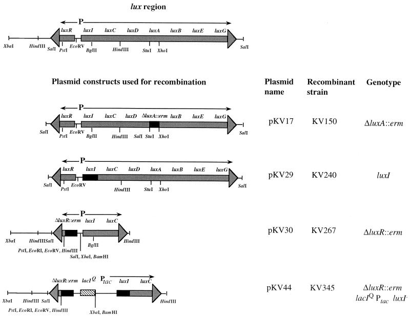
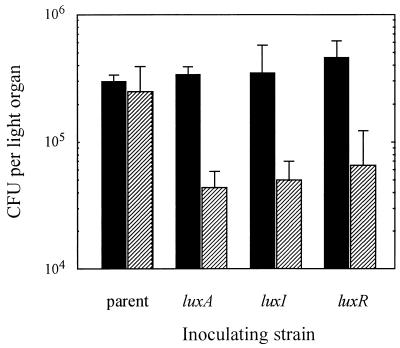
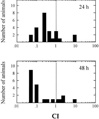
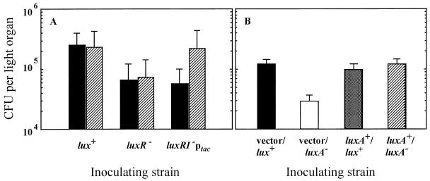
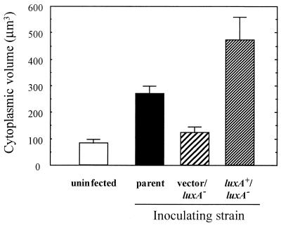
The bioluminescent bacterium Vibrio fischeri and juveniles of the squid Euprymna scolopes specifically recognize and respond to one another during the formation of a persistent colonization within the host's nascent light-emitting organ. The resulting fully developed light organ contains brightly luminescing bacteria and has undergone a bacterium-induced program of tissue differentiation, one component of which is a swelling of the epithelial cells that line the symbiont-containing crypts. While the luminescence (lux) genes of symbiotic V. fischeri have been shown to be highly induced within the crypts, the role of these genes in the initiation and persistence of the symbiosis has not been rigorously examined. We have constructed and examined three mutants (luxA, luxI, and luxR), defective in either luciferase enzymatic or regulatory proteins. All three are unable to induce normal luminescence levels in the host and, 2 days after initiating the association, had a three- to fourfold defect in the extent of colonization. Surprisingly, these lux mutants also were unable to induce swelling in the crypt epithelial cells. Complementing, in trans, the defect in light emission restored both normal colonization capability and induction of swelling. We hypothesize that a diminished level of oxygen consumption by a luciferase-deficient symbiotic population is responsible for the reduced fitness of lux mutants in the light organ crypts. This study is the first to show that the capacity for bioluminescence is critical for normal cell-cell interactions between a bacterium and its animal host and presents the first examples of V. fischeri genes that affect normal host tissue development.


Figures

FIG. 1
FIG. 1
Plasmids used for construction of lux mutants. The region of the chromosome containing the lux regulon is shown, with arrows demonstrating the direction of transcription of the two transcriptional units. Relevant restriction enzyme sites are indicated. Plasmid constructs that were used to make mutations in the chromosomal copy of the lux genes are also shown. Black boxes indicate the location of the mutation resulting from either a deletion and gene replacement with the erm gene or a frameshift mutation (see Materials and Methods); the box with diagonal stripes depicts the location of a lacIq/Ptac cassette.
FIG. 2
FIG. 2
Relative luminescence over time of newly hatched E. scolopes juveniles exposed to either the parent strain ESR1 (○), the luxA mutant strain KV150 (■), the luxI mutant strain KV240 (⧫), the luxR mutant strain KV267 (◊), or the luxR luxI Ptac strain KV345 (□). A subset of the squid exposed to KV345 (■) were treated with IPTG (see Materials and Methods) to induce luminescence genes. ESR1-exposed animals treated with IPTG (●) served as a control for IPTG effects on the association.
FIG. 3
FIG. 3
Symbiotic colonization levels achieved by lux mutant strains of V. fischeri and their parent, strain ESR1. The number of CFU present in the light organs of juvenile E. scolopes exposed to either lux mutant V. fischeri strains or the parent strain was determined at two times after inoculation, 24 h (black bars) or 48 h (striped bars). Each bar represents an average value obtained with at least four animals (standard error of the mean ranges are indicated). Similar results were obtained in three other independent trials.
FIG. 4
FIG. 4
Colonization of the E. scolopes light organ by a mixed inoculum of lux+ and luxA mutant V. fischeri cells. Forty-eight newly hatched E. scolopes juveniles were coinoculated with a 1:1 mixture of the luxA mutant (KV150) and its parent (ESR1). At two subsequent time points (24 and 48 h postinfection), the numbers of the two strains in the light organs of each of 24 animals were determined. The competitive index (CI) of the luxA mutants was calculated by dividing the number of KV150 mutant cells present in each organ by the number of ESR1 cells present. The number of animals with a given CI value is indicated by the bars. Symbiont populations of animals with a CI of <1 are dominated by lux+ cells.
FIG. 5
FIG. 5
Effect of colonization by lux mutants on host epithelial cell morphology. (A) TEMs illustrating the ultrastructural morphology of crypt epithelial cells 48 h after inoculation with wild-type and mutant strains of V. fischeri (v, bacterial cells). (a) Epithelial cells of uninoculated aposymbiotic animals have a narrow, columnar shape (n, nucleus). (b) Epithelial cells of animals exposed to V. fischeri strain ESR1 have become swollen and cuboidal. (c) Epithelial cells exposed to the V. fischeri luxA mutant have retained a narrow, columnar shape. (d) Epithelial cells exposed to a serine auxotrophic mutant, which is defective in colonizing the light organ at normal levels, nevertheless become swollen and cuboidal. (Bar = 10 μm) (B) Cytoplasmic volumes of light organ epithelial cells in E. scolopes infected by either lux+ or lux mutant V. fischeri. Uninfected (aposymbiotic) E. scolopes, and those that had been exposed to lux mutants or their lux+ parent, were fixed for TEM (Materials and Methods) after 48 h of symbiotic infection. The average cytoplasmic volume of the epithelial cells flanking the symbiont population was determined for each condition of inoculation. Measurements from at least 10 epithelial cells were used to determine the average cytoplasmic volume (error bars indicate 95% confidence limits). Similar results were obtained in two other experiments.
FIG. 6
FIG. 6
Complementation of the luminescence defect. (A) The average level of colonization of juvenile E. scolopes by the lux+ parent strain (ESR1), the luxR mutant (KV267), and the luxR luxI double mutant in which luxCDABE is under the control of Ptac (KV345) was determined after 46 h of infection. Throughout the course of the symbiotic infection, eight animals were exposed to IPTG (see Materials and Methods) (striped bars) and eight were not (black bars). (B) E. scolopes juveniles were exposed to one of four V. fischeri strains: a luxA mutant or its lux+ parent, carrying either a plasmid-borne copy of a complementing luxA+ gene or the parent vector alone. The number of CFU present in the light organs of 10 animals from each group was determined 48 h after inoculation. The bars represent the mean level of colonization (standard error of the mean ranges are indicated).
FIG. 7
FIG. 7
Cytoplasmic volumes of light organ epithelial cells in E. scolopes juveniles infected by a complemented lux mutant of V. fischeri. Uninfected (aposymbiotic) E. scolopes juveniles or those that had been exposed to either the lux+ parent, a luxA mutant carrying a luxA+-complementing plasmid, or the vector alone were fixed for TEM after 48 h of symbiotic infection. The average cytoplasmic volume of the epithelial cells flanking the symbiont population was determined for each condition of inoculation. Measurements from at least 10 epithelial cells were used to determine the average cytoplasmic volume (error bars indicate 95% confidence limits).

References

    1. Boettcher K J, Ruby E G. Depressed light emission by symbiotic Vibrio fischeri of the sepiolid squid Euprymna scolopes. J Bacteriol. 1990;172:3701–3706. - PMC - PubMed
    1. Boettcher K J, Ruby E G. Detection and quantification of Vibrio fischeri autoinducer from symbiotic squid light organs. J Bacteriol. 1995;177:1053–1058. - PMC - PubMed
    1. Boettcher K J, Ruby E G, McFall-Ngai M J. Bioluminescence in the symbiotic squid Euprymna scolopes is controlled by a daily biological rhythm. J Comp Physiol. 1996;179:65–73.
    1. Callahan S M, Dunlap P V. LuxR- and acyl-homoserine-lactone-controlled non-lux genes define a quorum-sensing regulon in Vibrio fischeri. J Bacteriol. 2000;182:2811–2822. - PMC - PubMed
    1. Chatterjee A, Cui Y, Liu Y, Dumenyo C K, Chatterjee A K. Inactivation of rmsA leads to overproduction of extracellular pectinases, cellulases, and proteases in Erwinia carotovora subsp. carotovora in the absence of the starvation/cell density-sensing signal, N-(3-oxohexanoyl)-l-homoserine lactone. Appl Environ Microbiol. 1995;61:1959–1967. - PMC - PubMed

Publication types

LinkOut - more resources