. 2000 Aug;182(16):4606-16.
doi: 10.1128/JB.182.16.4606-4616.2000.

sigma(BldN), an extracytoplasmic function RNA polymerase sigma factor required for aerial mycelium formation in Streptomyces coelicolor A3(2)

Affiliations

sigma(BldN), an extracytoplasmic function RNA polymerase sigma factor required for aerial mycelium formation in Streptomyces coelicolor A3(2)

M J Bibb et al. J Bacteriol. 2000 Aug.

Abstract

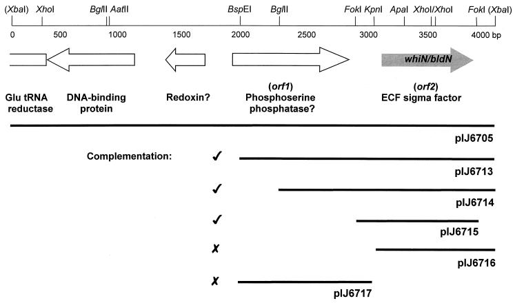
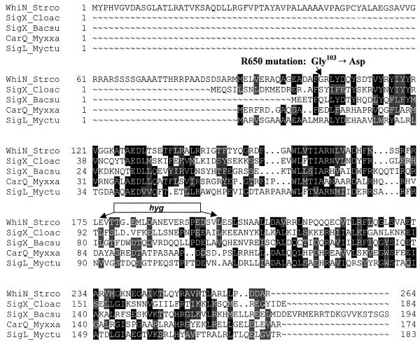
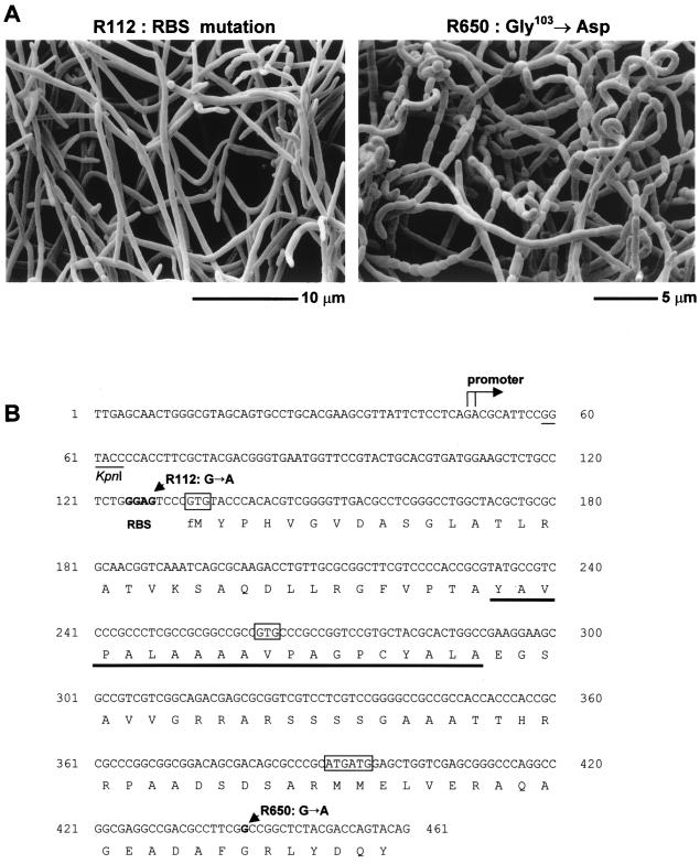
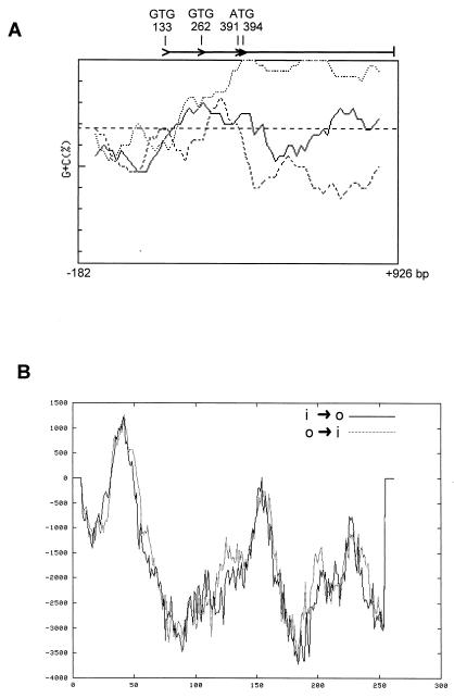
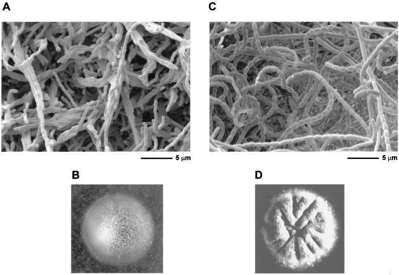
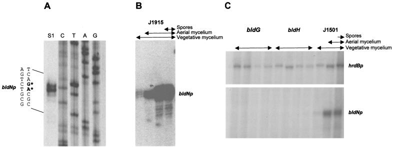
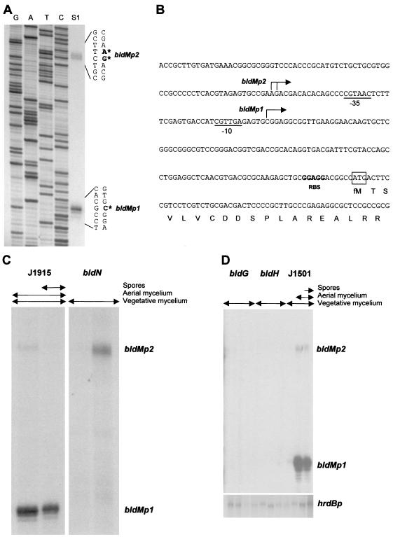
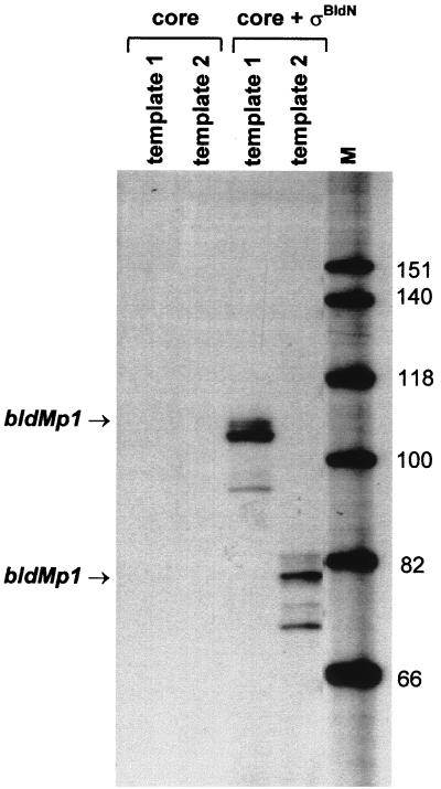
Sporulation mutants of Streptomyces coelicolor appear white because they are defective in the synthesis of the gray polyketide spore pigment, and such white (whi) mutants have been used to define 13 sporulation loci. whiN, one of five new whi loci identified in a recent screen of NTG (N-methyl-N'-nitro-N-nitrosoguanidine)-induced whi strains (N. J. Ryding et al., J. Bacteriol. 181:5419-5425, 1999), was defined by two mutants, R112 and R650. R650 produced frequent spores that were longer than those of the wild type. In contrast, R112 produced long, straight, undifferentiated hyphae, although rare spore chains were observed, sometimes showing highly irregular septum placement. Subcloning and sequencing showed that whiN encodes a member of the extracytoplasmic function subfamily of RNA polymerase sigma factors and that the sigma factor has an unusual N-terminal extension of approximately 86 residues that is not present in other sigma factors. A constructed whiN null mutant failed to form aerial mycelium (the "bald" phenotype) and, as a consequence, whiN was renamed bldN. This observation was not totally unexpected because, on some media, the R112 point mutant produced substantially less aerial mycelium than its parent, M145. The bldN null mutant did not fit simply into the extracellular signaling cascade proposed for S. coelicolor bld mutants. Expression of bldN was analyzed during colony development in wild-type and aerial mycelium-deficient bld strains. bldN was transcribed from a single promoter, bldNp. bldN transcription was developmentally regulated, commencing approximately at the time of aerial mycelium formation, and depended on bldG and bldH, but not on bldA, bldB, bldC, bldF, bldK, or bldJ or on bldN itself. Transcription from the p1 promoter of the response-regulator gene bldM depended on bldN in vivo, and the bldMp1 promoter was shown to be a direct biochemical target for sigma(BldN) holoenzyme in vitro.


Figures

FIG. 1
FIG. 1
Genetic organization of the 4.1-kb DNA fragment carrying whiN/bldN. The positions of the protein coding regions are indicated by arrows, and restriction sites referred to in the text are marked. The extent of the subclones described in the text and their ability to complement the whiN mutants R112 and R650 are shown below. XbaI sites are derived from the cloning vector, pIJ698. The DNA sequence of the lefthand 2-kb XbaI-BspEI fragment is taken from the ongoing S. coelicolor genome sequence (accession number AL079345).
FIG. 2
FIG. 2
Amino acid alignment of WhiN/BldN with other members of the ECF subfamily of sigma factors. The proteins and their corresponding amino acid sequence accession numbers are WhiN_Strco, S. coelicolor WhiN (CAB55345); SigX_Cloac, Clostridium acetobutylicum SigX (AAC12856); SigX_Bacsu, B. subtilis SigX (P35165); CarQ_Myxxa, M. xanthus CarQ (S39877) and SigL_Myctu, Mycobacterium tuberculosis SigL (CAA17502). The amino acid substitution carried by the whiN point mutant, R650, and the location of the hyg cassette (resulting from the replacement of a 66-bp XhoI fragment) in the whiN::hyg null mutant are shown.
FIG. 3
FIG. 3
(A) Scanning electron micrographs showing the phenotypes of the whiN/bldN point mutants, R112 and R650. (B) Nucleotide sequence of the promoter region and the 5′ end of the whiN/bldN gene showing the transcription start point, the putative ribosome binding site (RBS), the potential start codons (boxed), the nucleotide substitutions carried by the whiN point mutants, R112 and R650, and the KpnI site and the stretch of hydrophobic residues (underlined) discussed in the text.
FIG. 4
FIG. 4
(A) FRAME plot (2) of the whiN/bldN gene showing the unusual codon usage at the 5′ end. The GTG and ATG potential translation start codons are marked. The window size was 40 codons, and the step size was five codons. The sequence analyzed begins immediately after the stop codon of the upstream gene, and the numbering is that used in Fig. 3B. (B) TMPRED hydrophobicity plot of WhiN/BldN showing possible transmembrane helices. Predictions for both inside-to-outside helices (i → o) and outside-to-inside helices (o → i) are shown. Scores above 500 are considered significant.
FIG. 5
FIG. 5
Scanning electron micrographs and photographs showing the bald phenotype of the constructed bldN null mutant, J2177 (A and B), and its complementation by pIJ6715, the pSET152 derivative carrying bldN (see Fig. 1) (C and D).
FIG. 6
FIG. 6
Transcriptional analysis of bldN. (A) High-resolution S1 nuclease mapping of the 5′ end of the bldN transcript in S. coelicolor J1915. The most likely transcription start points are indicated by the asterisks. Lanes C, T, A, and G represent a dideoxy sequencing ladder generated using the same oligonucleotide that was used to make the S1 mapping probe. (B) S1 nuclease protection analysis of bldN transcription during development of S. coelicolor J1915 on R2YE solid medium. For each time point, the presence of vegetative mycelium, aerial mycelium, and spores, as judged by microscopic examination, is shown. (C) S1 nuclease protection analysis of bldN and hrdB transcription during development of S. coelicolor WC103 (bldG) and WC109 (bldH) and their congenic parent J1501 (bldG+ bldH+) on R2YE solid medium. For each time point, the presence of vegetative mycelium, aerial mycelium, and spores, as judged by microscopic examination, is shown.
FIG. 7
FIG. 7
Transcriptional analysis of bldM. (A) High-resolution S1 nuclease mapping of the 5′ ends of the bldMp1 and bldMp2 transcripts in S. coelicolor J1915. The most likely transcription start points are indicated by the asterisks. Lanes G, A, T, and C represent a dideoxy sequencing ladder generated using the same oligonucleotide that was used to make the S1 mapping probe. (B) Nucleotide sequence of the bldM promoter region showing the bldMp1 and bldMp2 transcription start points, the putative −10 and −35 sequences of the bldMp1 promoter, the putative ribosome binding site (RBS), and the start of the bldM coding sequence. (C) S1 nuclease protection analysis of bldM transcription during development of S. coelicolor J2177 (bldN) and its congenic parent J1915 (bldN+) on R2YE solid medium. For each time point, the presence of vegetative mycelium, aerial mycelium, and spores, as judged by microscopic examination, is shown. (D) S1 nuclease protection analysis of bldM and hrdB transcription during development of S. coelicolor WC103 (bldG) and WC109 (bldH) and their congenic parent J1501 (bldG+ bldH+) on R2YE solid medium. The RNA samples used were those described in Fig. 6C. For each time point, the presence of vegetative mycelium, aerial mycelium, and spores, as judged by microscopic examination, is shown.
FIG. 8
FIG. 8
In vitro transcription of the bldMp1 promoter by reconstituted EςBldN holoenzyme. Transcripts were generated from templates 1 and 2 (see Materials and Methods) using core RNA polymerase alone or core enzyme plus ςBldN. The expected sizes of the runoff transcripts from the bldMp1 promoter were 104 nucleotides (template 1) and 78 nucleotides (template 2). The size markers (M) were a 32P-end-labeled HinfI digest of ΦX174.

References

    1. Alper S, Dufour A, Garsin D A, Duncan L, Losick R. Role of adenosine nucleotides in the regulation of a stress-response transcription factor in Bacillus subtilis. J Mol Biol. 1996;260:165–177. - PubMed
    1. Bibb M J, Findlay P R, Johnson M W. The relationship between base composition and codon usage in bacterial genes and its use for the simple and reliable identification of protein-coding sequences. Gene. 1984;30:157–166. - PubMed
    1. Bierman M, Logan R, O'Brien K, Seno E T, Rao R N, Schoner B E. Plasmid cloning vectors for the conjugal transfer of DNA from Escherichia coli to Streptomyces spp. Gene. 1992;116:43–49. - PubMed
    1. Buttner M J, Fearnley I M, Bibb M J. The agarase gene (dagA) of Streptomyces coelicolor A3(2): nucleotide sequence and transcriptional analysis. Mol Gen Genet. 1987;209:101–109. - PubMed
    1. Buttner M J, Chater K F, Bibb M J. Cloning, disruption and transcriptional analysis of three RNA polymerase sigma factor genes of Streptomyces coelicolor A3(2) J Bacteriol. 1990;172:3367–3378. - PMC - PubMed

Publication types

MeSH terms

Substances

LinkOut - more resources