. 2000 Aug;20(16):5865-78.
doi: 10.1128/MCB.20.16.5865-5878.2000.

Regulation of c-SRC activity and function by the adapter protein CAS

Affiliations

Regulation of c-SRC activity and function by the adapter protein CAS

M R Burnham et al. Mol Cell Biol. 2000 Aug.

Abstract

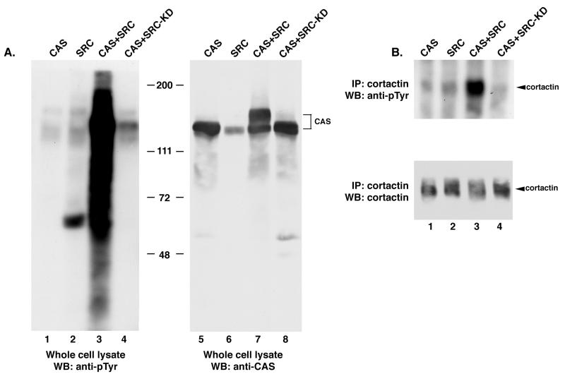
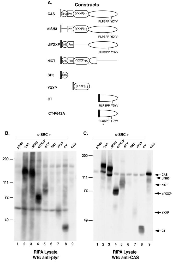
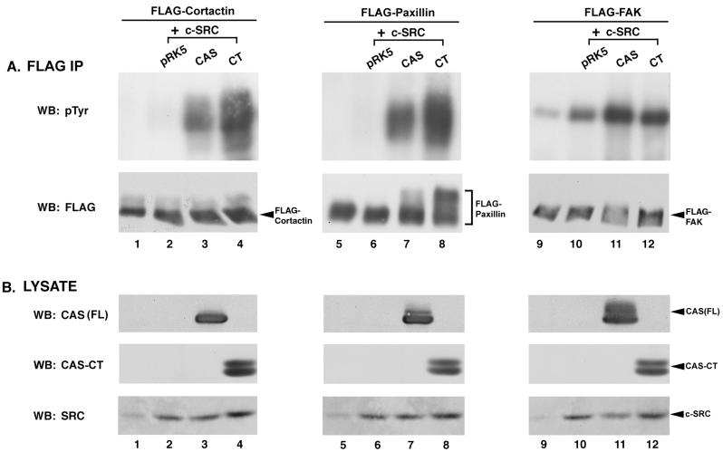
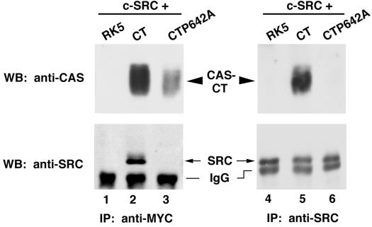
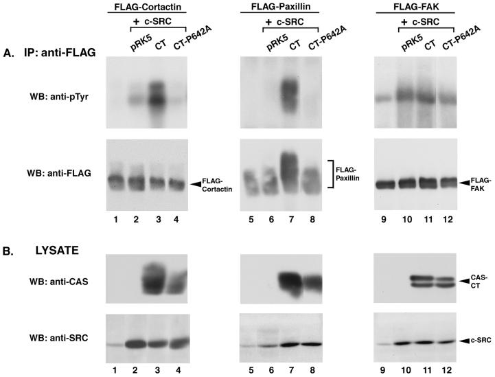
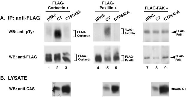
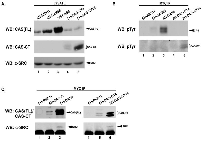
SRC family kinases play essential roles in a variety of cellular functions, including proliferation, survival, differentiation, and apoptosis. The activities of these kinases are regulated by intramolecular interactions and by heterologous binding partners that modulate the transition between active and inactive structural conformations. p130(CAS) (CAS) binds directly to both the SH2 and SH3 domains of c-SRC and therefore has the potential to structurally alter and activate this kinase. In this report, we demonstrate that overexpression of full-length CAS in COS-1 cells induces c-SRC-dependent tyrosine phosphorylation of multiple endogenous cellular proteins. A carboxy-terminal fragment of CAS (CAS-CT), which contains the c-SRC binding site, was sufficient to induce c-SRC-dependent protein tyrosine kinase activity, as measured by tyrosine phosphorylation of cortactin, paxillin, and, to a lesser extent, focal adhesion kinase. A single amino acid substitution located in the binding site for the SRC SH3 domain of CAS-CT disrupted CAS-CT's interaction with c-SRC and inhibited its ability to induce tyrosine phosphorylation of cortactin and paxillin. Murine C3H10T1/2 fibroblasts that expressed elevated levels of tyrosine phosphorylated CAS and c-SRC-CAS complexes exhibited an enhanced ability to form colonies in soft agar and to proliferate in the absence of serum or growth factors. CAS-CT fully substituted for CAS in mediating growth in soft agar but was less effective in promoting serum-independent growth. These data suggest that CAS plays an important role in regulating specific signaling pathways governing cell growth and/or survival, in part through its ability to interact with and modulate the activity of c-SRC.


Figures

FIG. 1
FIG. 1
Tyrosine phosphorylation of cellular proteins in COS-1 cells coexpressing CAS and c-SRC. COS-1 cells were transfected with a construct encoding CAS (lanes 1 and 5), SRC (lanes 2 and 6), both SRC and CAS (lanes 3 and 7), or a catalytically inactive SRC variant (SRC-KD) together with CAS (lanes 4 and 8). (A) Expression of CAS and catalytically active c-SRC induces PTK activity. Total cell protein (25 μg) was resolved by SDS–8% PAGE, transferred to nitrocellulose, and immunoblotted with anti-pTyr MAb 4G10 (lanes 1 to 4) or anti-CAS-F antiserum (lanes 5 to 8). (B) Expression of CAS and c-SRC induces tyrosine phosphorylation of cortactin. Cell lysate (350 μg) was incubated with the anti-cortactin MAb 4F11. The collected immune complexes were divided into two equal parts, separated by SDS–8% PAGE, and immunoblotted with either MAb 4G10 to determine pTyr levels (top blot) or MAb 4F11 to determine the amount of cortactin present in each immune complex (bottom blot). WB, Western blotting; IP, immunoprecipitation.
FIG. 2
FIG. 2
Induction of PTK activity by coexpression of c-SRC and CAS variants. (A) Schematic representation of Myc-tagged CAS and CAS variants. The circle represents the SH3 domain of CAS. The square represents a proline-rich region, and the oval designated (YXXP)15 represents the substrate-binding YXXP domain. The elongated oval represents the carboxy-terminal domain of CAS. RPLP642SPP and Y668DYV are the single-letter symbols for the amino acid residues contributing to the SRC binding sites located in the carboxy terminus of CAS (49). Single black lines indicate regions of the CAS protein that were deleted in each CAS variant. The asterisk indicates the site of the proline-to-alanine substitution in CAS-CTP642A. (B and C) Protein expression and phosphorylation in COS-1 cells coexpressing c-SRC and the indicated CAS variants. Total cell protein (25 μg) was resolved by SDS–8% PAGE, transferred to nitrocellulose, and immunoblotted either with anti-pTyr MAb 4G10 (B) or a mixture of polyclonal CAS-B and CAS-F antisera (C). WB, Western blot.
FIG. 3
FIG. 3
CAS and CAS-CT modulate c-SRC PTK activity toward specific SRC substrates. (A) Immunoblot analysis of FLAG immune complexes. One milligram of lysate from cells expressing the indicated proteins was incubated with anti-FLAG MAb M2-conjugated resin, and the collected immune complexes were divided into two equal parts. One half was immunoblotted with MAb 4G10 to determine pTyr levels of the indicated FLAG-tagged constructs (top blots), while the other half was immunoblotted with the anti-FLAG MAb M5 to verify that equal amounts of protein were present in the immune complexes (bottom blots). (B) Verification of recombinant protein expression. Total cell lysate (50 μg) from the indicated cells was separated by SDS–8% PAGE and immunoblotted with either CAS-F antiserum to determine expression levels of CAS-FL and CAS-CT (top blots) or the anti-SRC MAb 2-17 to determine levels of expression of c-SRC (bottom blots). IP, immunoprecipitation; WB, Western blotting.
FIG. 4
FIG. 4
Inhibition of the association between CAS-CT and c-SRC by a single amino acid substitution of CAS, proline642 to alanine (P642A). Cell lysate (500 μg) was incubated with either the anti-Myc MAb 9E10 (lanes 1 to 3) or the anti-SRC MAb EC10 (lanes 4 to 6), and the collected immune complexes were divided into two equal parts. Proteins were separated by SDS–8% PAGE and immunoblotted using either CAS-F antiserum (top blot) or the SRC MAb 2-17 (bottom blots). IP, immunoprecipitation; WB, Western blotting; IgG, immunoglobulin G.
FIG. 5
FIG. 5
Tyrosine phosphorylation of SRC substrates requires the SRC-binding site of CAS-CT. (A) Immunoblot analysis of FLAG immune complexes. One milligram of lysate from cells expressing the indicated proteins was incubated with anti-FLAG MAb M2-conjugated resin, and the collected immune complexes were divided into two equal parts. One half was immunoblotted with MAb 4G10 to determine pTyr levels of the indicated FLAG-tagged constructs (top blots), while the other half was immunoblotted with the anti-FLAG MAb M5 to verify that equal amounts of protein were present in the immune complexes (bottom blots). (B) Verification of recombinant protein expression. Fifty micrograms of total cell lysate from the indicated cells was separated by SDS–8% PAGE and immunoblotted with either CAS-F antiserum to determine expression levels of CAS-CT and CAS-CTP642A (top blots) or anti-SRC MAb 2-17 to determine levels of expression of c-SRC (bottom blots). IP, immunoprecipitation; WB, Western blotting.
FIG. 6
FIG. 6
Modulation of c-SRC activity by CAS-CT in C3H10T1/2-5H cells. (A) Immunoblot analysis of FLAG immune complexes. Total cell lysate (1 mg) was incubated with anti-FLAG MAb M2-conjugated resin to specifically immunoprecipitate the indicated FLAG-tagged constructs. Collected immune complexes were divided into two equal parts and immunoblotted using either the anti-pTyr MAb 4G10 (top blots) or the anti-FLAG MAb M5 to verify that equal amounts of FLAG-tagged protein were present in the immune complexes (bottom blots). (B) Verification of recombinant protein expression. Total cell protein (50 μg) from the indicated cells was separated by SDS–8% PAGE and immunoblotted with CAS-F antiserum. IP, immunoprecipitation; WB, Western blotting.
FIG. 7
FIG. 7
Association of c-SRC with CAS or CAS-CT in C3H10T1/2-5H clonal cell lines. (A) Protein expression in C3H10T1/2-5H stable cell lines. Total cell protein (50 μg) derived from representative C3H10T1/2-5H stable cell lines was resolved by SDS–8% PAGE, transferred to nitrocellulose, and immunoblotted using either CAS-F antiserum to determine relative levels of CAS or CAS-CT present in each cell line (top and middle blots) or anti-SRC MAb 2-17 to determine the amount of SRC in each cell line (bottom blot). The top two blots represent identical exposures of different regions of the same gel, allowing expression levels of CAS and CAS-CT to be directly compared. (B and C) CAS and CAS-CT are tyrosine phosphorylated and associate with c-SRC in cells expressing high levels of these proteins. Proteins from 1 mg of the indicated cell lysate were incubated with anti-Myc MAb 9E10 to selectively immunoprecipitate Myc-tagged CAS-FL or CAS-CT. Immune complexes were divided in half, resolved by SDS–8% PAGE, transferred to nitrocellulose, and immunoblotted with anti-pTyr MAb 4G10 (B), CAS-F antiserum (C, top blots), or anti-SRC MAb 2-17 (C, bottom blots). IP, immunoprecipitation; WB, Western blotting.
FIG. 8
FIG. 8
Biological activities of the c-SRC–CAS complex. (A) Serum-independent growth. The percentages of cells exhibiting nuclear staining of BrdU at 24, 48, and 72 h after serum withdrawal are presented for the indicated cell lines. The results for 5H-RK5 are representative of the combined results for two vector control cell lines. Values are averages (± standard deviations) of results of four independent experiments, and asterisks indicate a P value of ≤0.01. (B) Anchorage-independent growth. Colony number per 105 cells plated in soft agar is presented for each of the designated cell lines. Colonies were counted using EagleSight software, and the results presented are the averages of results of three independent experiments.

Similar articles

Cited by

References

    1. Abram C L, Courtneidge S A. Src family tyrosine kinases and growth factor signaling. Exp Cell Res. 2000;254:1–13. - PubMed
    1. Alexandropoulos K, Baltimore D. Coordinate activation of c-Src by SH3- and SH2-binding sites on a novel, p130CAS-related protein, Sin. Genes Dev. 1996;10:1341–1355. - PubMed
    1. Anderson R G W. The caveolae membrane system. Annu Rev Biochem. 1998;67:199–225. - PubMed
    1. Biscardi J S, Belsches A P, Parsons S J. Characterization of human epidermal growth factor receptor and c-Src interactions in human breast tumor cells. Mol Carcinog. 1998;21:261–272. - PubMed
    1. Biscardi J S, Tice D A, Parsons S J. c-Src, receptor tyrosine kinases, and human cancer. Adv Cancer Res. 1999;76:61–119. - PubMed

Publication types