. 2000 Aug;20(16):5908-16.
doi: 10.1128/MCB.20.16.5908-5916.2000.

TrkA immunoglobulin-like ligand binding domains inhibit spontaneous activation of the receptor

Affiliations

TrkA immunoglobulin-like ligand binding domains inhibit spontaneous activation of the receptor

J C Arevalo et al. Mol Cell Biol. 2000 Aug.

Abstract

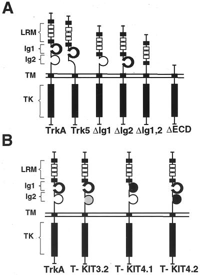
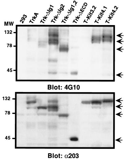
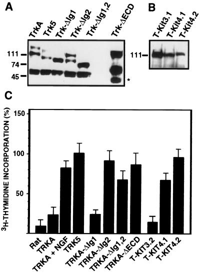
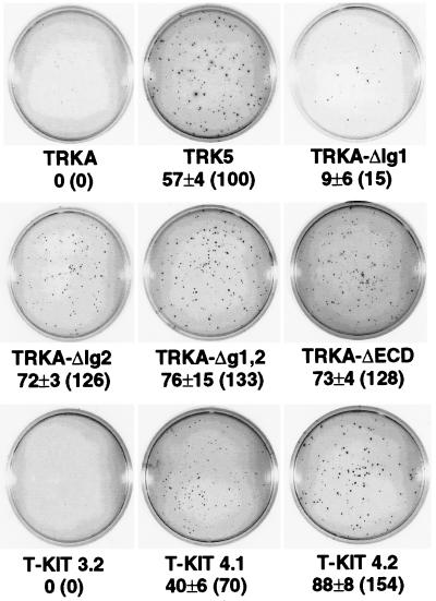
The extracellular region of the nerve growth factor (NGF) receptor, TrkA, contains two immunoglobulin (Ig)-like domains that are required for specific ligand binding. We have investigated the possible role of these two Ig-like domains in receptor dimerization and activation by using different mutants of the TrkA extracellular region. Deletions of each Ig-like domain, of both, and of the entire extracellular region were made. To probe the structural constraints on ligand-independent receptor dimerization, chimeric receptors were generated by swapping the Ig-like domains of the TrkA receptor for the third or fourth Ig-like domain of c-Kit. We also introduced single-amino-acid changes in conserved residues within the Ig-like domains of TrkA. Most of these TrkA variants did not bind NGF, and their expression in PC12nnr5 cells, which lack endogenous TrkA, promoted ligand-independent neurite outgrowth. Some TrkA mutant receptors induced malignant transformation of Rat-1 cells, as assessed by measuring proliferation in the absence of serum, anchorage-independent growth, and tumorigenesis in nude mice. These mutants exhibited constitutive phosphorylation and spontaneous dimerization consistent with their biological activities. Our data suggest that spontaneous dimerization of TrkA occurs when the structure of the Ig-like domains is altered, implying that the intact domains inhibit receptor dimerization in the absence of NGF.


Figures

FIG. 1
FIG. 1
Schematic representation of TrkA mutant receptors. (A) Mutants generated by deleting the Ig-like domains or the entire extracellular region. (B) TrkA/c-Kit chimeric receptors. TM, transmembrane domain; TK, tyrosine-kinase domain.
FIG. 2
FIG. 2
Ligand-independent neurite formation in PC12nnr5 cells transfected with TrkA mutant receptors. Neurite outgrowth was quantified 3 days after transfection by assessing the percentage of β-galactosidase-positive cells bearing neurites at least twice the length of their cell bodies. Results were normalized to the trk-5 oncogene response (set at 100%). Values were calculated from at least five independent experiments. Means and standard deviations (SD) are shown.
FIG. 3
FIG. 3
Ligand-independent dimerization of TrkA deletion mutants. (A) Schematic representation of HA- and Myc-tagged wild-type TrkA receptors. These epitopes were introduced in the same position within the different TrkA mutants. (B) Dimerization analysis in HEK293 cells transiently transfected with the following pairs of expression vectors: HA-TrkA and Myc-TrkA; HA-TrkA-ΔIg1 and Myc-TrkA-ΔIg1; HA-TrkA-ΔIg2 and Myc-TrkA-ΔIg2; and HA-TrkA-ΔIg1,2 and Myc-TrkA-ΔIg1,2. As a positive control, we used HA-TrkA- and Myc-TrkA-transfected cells treated with NGF (100 ng/ml) for 5 min. Two days after transfection, cells were lysed and immunoprecipitations (IP) were performed using the 12CA5 anti-HA antibody. Western blot was done with either 9E10 anti-Myc antibody (upper panel) or anti-203 pan-Trk antiserum (bottom panel). (C) Western blot of whole-cell extracts (40 μg of total protein) done with either 203 antiserum, 9E10, or 12CA5. Immunoreactive protein bands were detected by chemiluminescence. Sizes are shown in kilodaltons.
FIG. 4
FIG. 4
Tyrosine phosphorylation of TrkA mutant receptors in the absence of NGF. Expression vectors for the different mutants were transiently transfected into HEK293 cells. Two days after transfection, cells were lysed, and 50 μg of total protein extract was analyzed by Western blot using either 4G10 antiphosphotyrosine antibody (upper panel) or anti-203 pan-Trk antiserum (bottom panel). Sizes are shown in kilodaltons. Arrows indicate positions of the TrkA mutant receptors.
FIG. 5
FIG. 5
(A and B) Expression of mutant TrkA receptors in stably transfected Rat-1 cell lines. Immunoprecipitation and Western blot were performed on total cell extracts (2 mg of protein) using the 203 pan-Trk antiserum and HRP-labeled secondary anti-rabbit Ig antibody. Sizes are shown in kilodaltons. (C) [3H]thymidine incorporation by serum-starved Rat-1 cell lines stably expressing mutant TrkA receptors in the absence of NGF. Values are the means from at least five experiments performed with two independent clones of each mutant. Data are normalized to the [3H]thymidine incorporated by the cells in the presence of serum and expressed as a percentage of the incorporation measured in cells expressing the trk-5 oncogene (100%). Means and SD are shown.
FIG. 6
FIG. 6
Soft agar colony formation of Rat-1 cell lines expressing mutant TrkA receptors. Colonies were counted after 21 days in culture. Values are the means from at least five experiments performed with two independent clones of each mutant. The percentage of the foci formed by the trk-5-expressing clones (set at 100%) is shown in parentheses for each receptor.
FIG. 7
FIG. 7
(A) Schematic representation of point mutations affecting individual amino acid residues within the extracellular region of TrkA. (B) Expression and ligand-independent phosphorylation of mutant TrkA receptors in stably transfected Rat-1 cell lines. Immunoprecipitation (IP) was performed on total cell extracts (2 mg of protein) using the anti-203 pan-Trk antiserum, followed by Western blot with either anti-203 antiserum (upper panel) or 4G10 antiphosphotyrosine monoclonal antibody (lower panel). Expression of TrkA in PC-12 cells (2 mg of protein) is shown for comparison. Sizes are shown in kilodaltons. (C) [3H]thymidine incorporation by Rat-1 cell lines stably expressing mutant TrkA receptors in the absence of serum. Values are the means from at least five experiments performed with two independent clones of each mutant. Data are normalized relative to the [3H]thymidine incorporated by the cells in the presence of serum and expressed as a percentage of the incorporation measured in cells expressing the trk-5 oncogene (100%). Means and SD are shown.

References

    1. Ausubel F M, Brent R, Kingston R E, Moore D D, Seidman J G, Smith J A, Struhl K. Current protocols in molecular biology. New York, N.Y: John Wiley and Sons; 1995.
    1. Barbacid M. Neurotrophic factors and their receptors. Curr Opin Cell Biol. 1995;7:148–155. - PubMed
    1. Blechman J M, Lev S, Barg J, Eisenstein M, Vaks B, Vogel Z, Givol D, Yarden Y. The fourth immunoglobulin domain of the stem cell factor receptor couples ligand binding to signal transduction. Cell. 1995;80:103–113. - PubMed
    1. Brodeur G M, Maris J M, Yamashiro D J, Hogarty M D, White P S. Biology and genetics of human neuroblastomas. J Pediatr Hematol Oncol. 1997;19:93–101. - PubMed
    1. Canossa M, Rovelli G, Shooter E M. Transphosphorylation of the neurotrophin Trk receptors. J Biol Chem. 1996;271:5812–5818. - PubMed

Publication types

LinkOut - more resources