. 2000 Aug;20(16):5917-29.
doi: 10.1128/MCB.20.16.5917-5929.2000.

Receptor-like protein tyrosine phosphatase alpha homodimerizes on the cell surface

Affiliations

Receptor-like protein tyrosine phosphatase alpha homodimerizes on the cell surface

G Jiang et al. Mol Cell Biol. 2000 Aug.

Abstract

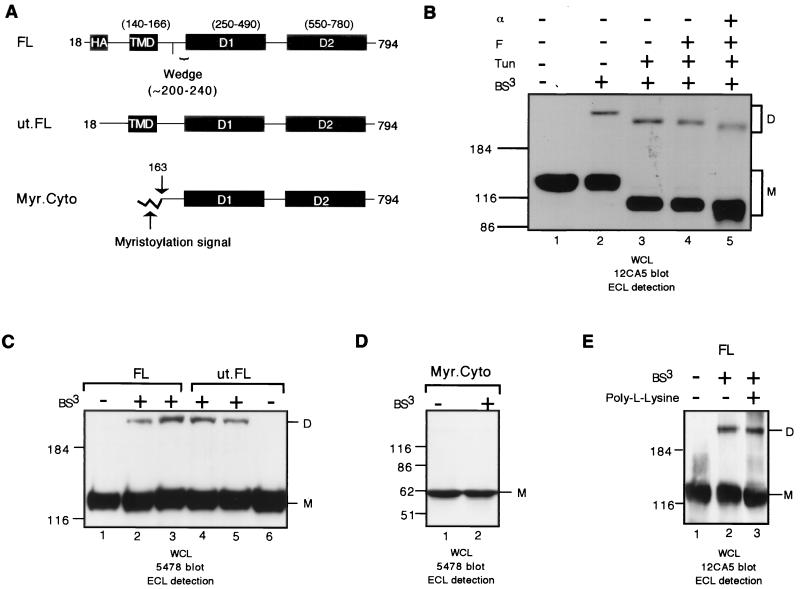
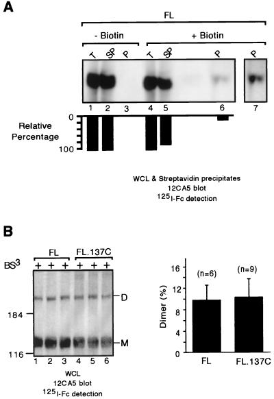
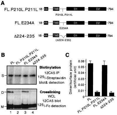
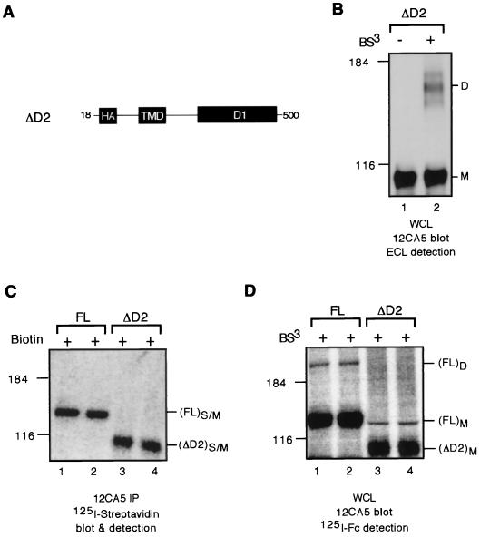
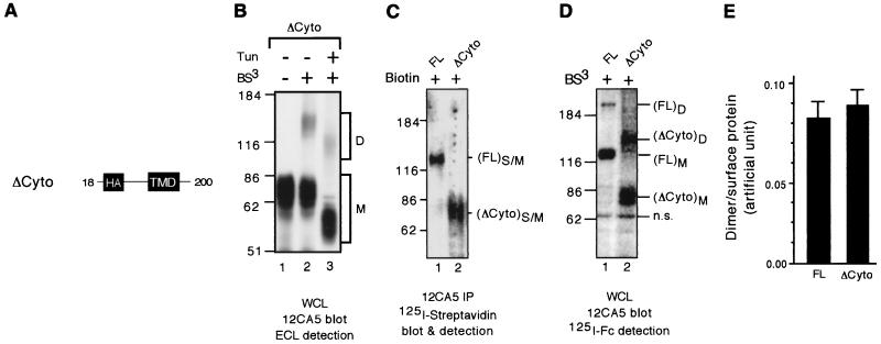
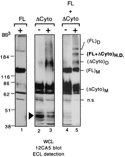
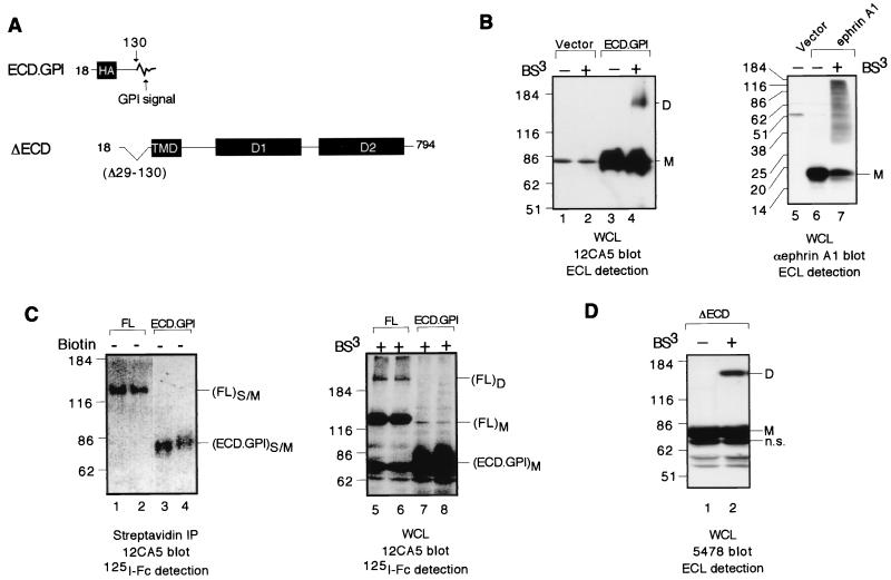
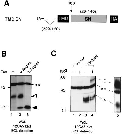
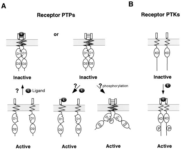
We reported previously that the N-terminal D1 catalytic domain of receptor protein-tyrosine phosphatase alpha (RPTPalpha) forms a symmetrical, inhibited dimer in a crystal structure, in which a helix-turn-helix wedge element from one monomer is inserted into the catalytic cleft of the other monomer. Previous functional studies also suggested that dimerization inhibits the biological activity of a CD45 chimeric RPTP and the catalytic activity of an isolated RPTPsigma D1 catalytic domain. Most recently, we have also shown that enforced dimerization inhibits the biological activity of full-length RPTPalpha in a wedge-dependent manner. The physiological significance of such inhibition is unknown, due to a lack of understanding of how RPTPalpha dimerization is regulated in vivo. In this study, we show that transiently expressed cell surface RPTPalpha exists predominantly as homodimers, suggesting that dimerization-mediated inhibition of RPTPalpha biological activity is likely to be physiologically relevant. Consistent with our published and unpublished crystallographic data, we show that mutations in the wedge region of D1 catalytic domain and deletion of the entire D2 catalytic domain independently reduced but did not abolish RPTPalpha homodimerization, suggesting that both domains are critically involved but that neither is essential for homodimerization. Finally, we also provide evidence that both the RPTPalpha extracellular domain and the transmembrane domain were independently able to homodimerize. These results lead us to propose a zipper model in which inactive RPTPalpha dimers are stabilized by multiple, relatively weak dimerization interfaces. Dimerization in this manner would provide a potential mechanism for negative regulation of RPTPalpha. Such RPTPalpha dimers could be activated by extracellular ligands or intracellular binding proteins that induce monomerization or by intracellular signaling events that induce an open conformation of the dimer.


Figures

FIG. 1
FIG. 1
RPTPα homodimerizes on the cell surface. (A) A schematic of RPTPα constructs used in this figure. Amino acids are numbered, and boundaries of the various structural domains, including the TMD, D1, and D2, are indicated according to the original (untagged) polypeptide (45). The boundary of the wedge region is indicated according to the D1 crystal structure (3). The construct FL contains an HA tag which was inserted between amino acids 19 and 20 and is exposed by signal peptide cleavage. The construct ut.FL corresponds to the full-length untagged RPTPα. The construct Myr.Cyto contains a myristoylation signal for membrane attachment. For details of vector construction, refer to Materials and Methods. Mock cross-linking and cross-linking on intact 293 cells transiently expressing FL (B and E), ut.FL (C), and Myr.Cyto (D) is shown. (E) Cells were cultured on plates without or with poly-l-lysine coating. Shown are the results of an immunoblotting analysis with anti-HA tag MAb 12CA5 on whole-cell lysates (WCL) using ECL detection (B and E) or polyclonal antiserum 5478 (C and D). BS3: −, mock cross-linking without BS3; +, cross-linking with BS3. Tun, transfected 293 cells were exposed to tunicamycin at 200 ng/ml; F, lysate was deglycosylated with N-glycosidase F in vitro; α, lysate was deglycosylated with endo-α-N-acetylgalactosaminidase in vitro. M, monomers; D, dimers. The positions of RPTPα monomer (M) and dimer (D) are indicated at the right side of the figure. The positions of molecular-weight markers (in kilodaltons) are indicated on the left side of the figure. Similar labels are used throughout the study.
FIG. 2
FIG. 2
RPTPα appears to exist on the cell surface predominantly as homodimers. (A) 293 cells transiently expressing FL were surface biotinylated or not biotinylated. Cells were lysed, and biotinylated proteins were precipitated with streptavidin beads and separated by SDS-PAGE, and the biotinylated FL was detected by immunoblotting using MAb 12CA5 followed by 125I-labeled sheep anti-mouse IgG F(ab′)2. The blot was quantified using a PhosphorImager as described in Materials and Methods. Top and bottom panels are the immunoblot and quantification, respectively. The loading for each of the lanes was standardized using an equivalent amount of whole-cell lysate. Lane 7 is a longer exposure of lane 6. + or − biotin, labeled or not labeled with biotin; WCL, total whole-cell lysate; SN, whole-cell lysate supernatant after streptavidin bead precipitation; P, streptavidin precipitate. (B) BS3 cross-linking was performed on 293 cells transiently transfected with either FL or FL.137C. Whole-cell lysates of cross-linked cells were separated by SDS-PAGE and probed using MAb 12CA5 followed by 125I-labeled sheep anti-mouse IgG F(ab′)2 and then quantified using a PhosphorImager as described in Materials and Methods. Shown are the results of an immunoblotting analysis. The right panel shows quantitation of the gel in left panel. n, number of replicates. Note that FL and FL.137C are similarly localized to the cell surface (20).
FIG. 3
FIG. 3
Mutations in the wedge diminish but do not abolish RPTPα oligomerization. (A) A schematic of RPTPα wedge mutant constructs, including point mutants FL.P210L.P211L and FL.E234A and deletion mutant Δ224-235. (B) For the top panel, transiently transfected 293 cells were biotinylated. Whole-cell lysates were immunoprecipitated with MAb 12CA5 to isolate the total RPTPα proteins, which were then subjected to SDS-PAGE and probed with 125I-labeled streptavidin to determine the levels of surface-expressed RPTPα protein. For the bottom panel, transiently transfected 293 cells were cross-linked with BS3. Whole-cell lysates were subjected to immunoblotting analysis with MAb 12CA5 followed by 125I-labeled sheep anti-mouse IgG F(ab′)2 to determine the levels of RPTPα dimers. The bands representing FL.P210L.P211L dimers and Δ224-235 dimers are faint but detectable by PhosphorImager analysis. Biotinylation and cross-linking were done on parallel dishes from the same transfection. All the constructs were expressed to a similar level on the cell surface. (C) Quantification of dimerization efficiency based on average of three replicates. The dimer/surface protein value is the ratio of the levels of RPTPα dimers over surface-expressed RPTPα, which were determined from the bottom and top portions of panel B, respectively, using a PhosphorImager. S, surface-expressed RPTPα (monomer).
FIG. 4
FIG. 4
Deletion of D2 diminishes but does not abolish RPTPα oligomerization. (A) A schematic of the D2 deletion mutant construct. (B) 293 cells transiently expressing ΔD2 protein were cross-linked or not cross-linked with BS3. Shown are the results of an immunoblotting analysis with anti-HA tag MAb 12CA5 on whole-cell lysates using ECL detection. (C) Transiently transfected 293 cells were biotinylated. Whole-cell lysates were immunoprecipitated with MAb 12CA5 to isolate the total RPTPα proteins, which were then subjected to SDS-PAGE and probed with 125I-labeled streptavidin to determine the levels of surface-expressed RPTPα protein. (D) Transiently transfected 293 cells were cross-linked with BS3. Whole-cell lysates were subjected to immunoblotting analysis using MAb 12CA5 followed by 125I-labeled sheep anti-mouse IgG F(ab′)2 to determine the levels of RPTPα dimers. Biotinylation (C) and cross-linking (D) were done on parallel dishes from the same transfection. Shown in panels C and D are images obtained via PhosphorImager analysis. S/M, surface-expressed monomeric proteins.
FIG. 5
FIG. 5
ΔCyto homodimerizes on the cell surface with high efficiency. (A) A schematic of the construct ΔCyto lacking the entire cytoplasmic domain. (B) 293 cells transiently transfected with FL or ΔCyto were treated or not treated with tunicamycin at 200 ng/ml and subsequently cross-linked with BS3. Whole-cell lysates were subjected to immunoblotting analysis with MAb 12CA5 using ECL detection. (C) Transiently transfected 293 cells were biotinylated. Whole-cell lysates were precipitated with streptavidin beads to isolate the total RPTPα proteins, which were then subjected to SDS-PAGE and probed with 125I-labeled streptavidin to determine the levels of surface-expressed RPTPα proteins. (D) Transiently transfected 293 cells were cross-linked with BS3. Whole-cell lysates were subjected to immunoblotting analysis using MAb 12CA5 followed by 125I-labeled sheep anti-mouse IgG F(ab′)2 to determine the levels of RPTPα dimers. Biotinylation (C) and cross-linking (D) were done on parallel dishes from the same transfection. Shown in panels C and D are images from PhosphorImager analysis. S/M, surface-expressed monomeric proteins. (E) Quantification of dimerization efficiency based on average of three replicates. The dimer/surface protein value is the ratio of the levels of RPTPα dimers over surface-expressed RPTPα, which were determined from panels C and D, respectively, using a PhosphorImager. n.s., nonspecific band.
FIG. 6
FIG. 6
RPTPα oligomers are homodimers. Mock cross-linking and cross-linking on 293 cells transiently transfected was done with the construct FL alone (lane 1), with ΔCyto alone (lanes 2 and 3), or with both FL and ΔCyto simultaneously (lanes 4 and 5). Whole-cell lysates were subjected to immunoblotting analysis with MAb 12CA5 using ECL detection. Note that a band corresponding to either partially glycosylated or degraded ΔCyto is present in the transfections of ΔCyto alone (lanes 2 and 3) but is virtually undetectable in the cotransfections (lanes 4 and 5). (FL+ΔCyto)H.D. is the cross-linked FL-ΔCyto heterodimer.
FIG. 7
FIG. 7
The ECD possesses relatively weak dimerization potential and is not required for the homodimerization of the full-length RPTPα. (A) A schematic of RPTPα constructs used in this figure. (B) Mock cross-linking and cross-linking on 293 cells transiently transfected with the construct ECD.GPI (lanes 1 to 4) or ephrin A1 (lanes 5 to 7). Shown are the results of immunoblotting analysis with MAb 12CA5 of whole-cell lysates using ECL detection. (C) In the left panel, transiently transfected 293 cells were biotinylated. Streptavidin-agarose beads were used to isolate the total biotinylated surface proteins, which were then subjected to immunoblotting analysis using MAb 12CA5 followed by 125I-labeled sheep anti-mouse IgG F(ab′)2 to determine the levels of surface-expressed RPTPα protein. In the right panel, transiently transfected 293 cells were cross-linked with BS3. Whole-cell lysates were subjected to immunoblotting analysis using MAb 12CA5 followed by 125I-sheep anti-mouse IgG F(ab′)2 to determine the levels of RPTPα dimers. n.s., nonspecific band. (D) Mock cross-linking and cross-linking on 293 cells transiently transfected with the construct ΔECD. Whole-cell lysates were subjected to immunoblotting analysis with anti-RPTPα serum 5478 using ECL detection.
FIG. 8
FIG. 8
The TMD of RPTPα is a potent dimerization domain. (A) A schematic of RPTPα constructs used in this figure. (B) 293 cells transiently transfected with TMD.SN were left untreated (lane 1) or were treated with tunicamycin at 200 ng/ml (lane 2) or 1,000 ng/ml (lane 3). Whole-cell lysates were subjected to immunoblotting analysis with MAb 12CA5 using ECL detection. Open arrow, most likely a N-glycosylated TMD.SN protein; closed arrow, most likely a nonglycosylated TMD.SN protein; n.s., a nonspecifically recognized band. (C) Mock cross-linking and cross-linking on intact cells by BS3 was performed on 293 cells transiently transfected with pSG5 vector alone or the TMD.SN construct. Whole-cell lysates were subjected to immunoblotting analysis with MAb 12CA5 using ECL detection.
FIG. 9
FIG. 9
A model for the regulation of RPTPs via dimerization. (A) Hypothetical model for regulation of RPTPs via dimerization. In the inactive state, RPTPs are dimerized via wedge-active site interaction in D1, interaction via the TMD, and interaction via the ECD. In the active state, the receptors are either monomers or dimers in which dimerization via D1 no longer occurs due to phosphorylation. Ligand binding can either destabilize or stabilize dimers. (B) Classical model of activation of receptor PTKs. Ligand binding leads to receptor dimerization, transautophosphorylation, and subsequent kinase activation. KD, kinase domain; L, ligand; P, phosphorylation.

Similar articles

Cited by

References

    1. Barnea G, Grumet M, Milev P, Silvennoinen O, Levy J B, Sap J, Schlessinger J. Receptor tyrosine phosphatase β is expressed in the form of proteoglycan and binds to the extracellular matrix protein tenascin. J Biol Chem. 1994;269:14349–14352. - PubMed
    1. Bhandari V, Lim K L, Pallen C J. Physical and functional interactions between receptor-like protein-tyrosine phosphatase α and p59fyn. J Biol Chem. 1998;273:8691–8698. - PubMed
    1. Bilwes A M, den Hertog J, Hunter T, Noel J P. Structural basis for inhibition of receptor protein-tyrosine phosphatase α by dimerization. Nature. 1996;382:555–559. - PubMed
    1. Buist, A., C. Blanchetot, L. G. Tertoolen, and J. den Hertog. Identification of p130Cas as an in vivo substrate of receptor protein-tyrosine phosphatase α. J. Biol. Chem., in press. - PubMed
    1. Buist A, Zhang Y L, Keng Y F, Wu L, Zhang Z Y, den Hertog J. Restoration of potent protein-tyrosine phosphatase activity into the membrane-distal domain of receptor protein-tyrosine phosphatase α. Biochemistry. 1999;38:914–922. - PubMed

Publication types

MeSH terms

Substances