. 2000 Aug;20(16):5974-85.
doi: 10.1128/MCB.20.16.5974-5985.2000.

Receptor isoforms mediate opposing proliferative effects through gbetagamma-activated p38 or Akt pathways

Affiliations

Receptor isoforms mediate opposing proliferative effects through gbetagamma-activated p38 or Akt pathways

L A Sellers et al. Mol Cell Biol. 2000 Aug.

Abstract

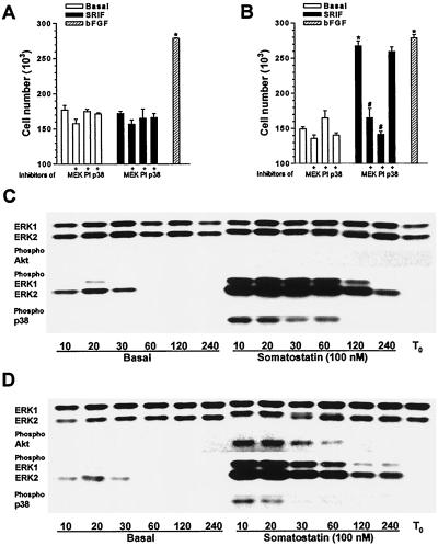
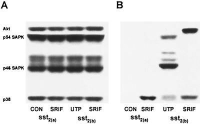
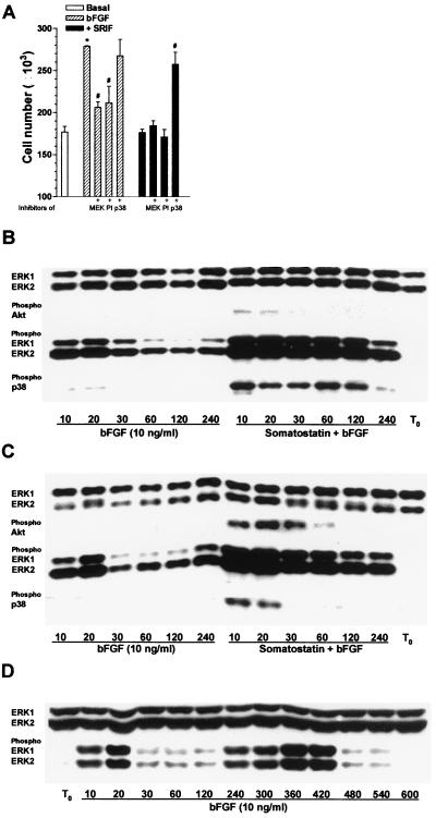
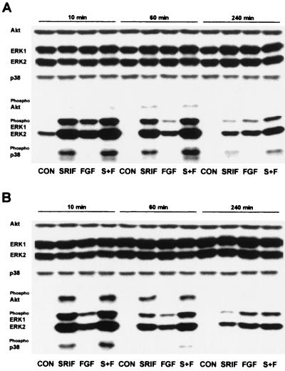
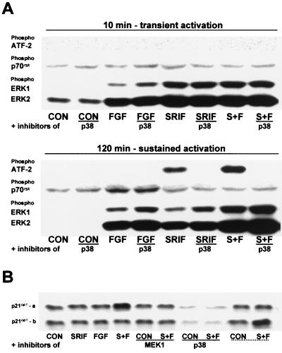
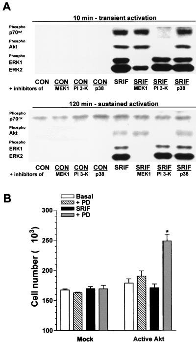
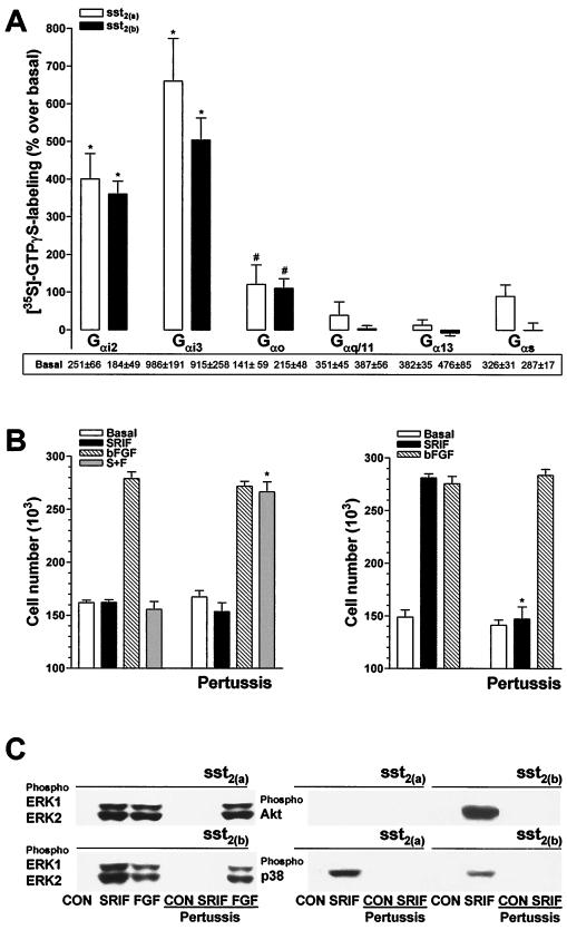
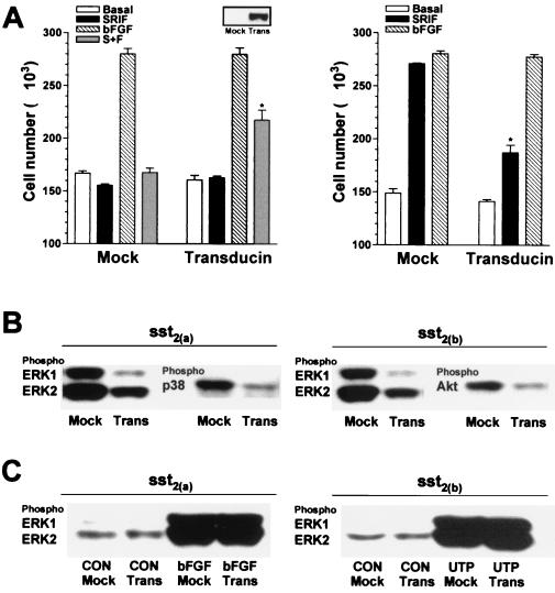
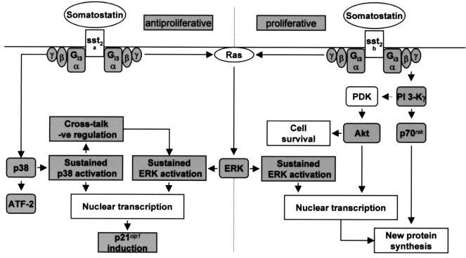
The opposing effects on proliferation mediated by G-protein-coupled receptor isoforms differing in their COOH termini could be correlated with the abilities of the receptors to differentially activate p38, implicated in apoptotic events, or phosphatidylinositol 3-kinase (PI 3-K), which provides a source of survival signals. These contrasting growth responses of the somatostatin sst(2) receptor isoforms, which couple to identical Galpha subunit pools (Galpha(i3) > Galpha(i2) >> Galpha(0)), were both inhibited following betagamma sequestration. The sst(2(a)) receptor-mediated ATF-2 activation and inhibition of proliferation induced by basic fibroblast growth factor (bFGF) were dependent on prolonged phosphorylation of p38. In contrast, cell proliferation and the associated transient phosphorylation of Akt and p70(rsk) induced by sst(2(b)) receptors were blocked by the PI 3-K inhibitor LY 294002. Stimulation with bFGF alone had no effect on the activity of either p38 or Akt but markedly enhanced p38 phosphorylation mediated by sst(2(a)) receptors, suggesting that a complex interplay exists between the transduction cascades activated by these distinct receptor types. In addition, although all receptors mediated a sustained activation of extracellular signal-regulated kinases (ERK1 and ERK2), induction of the tumor suppressor p21(cip1) was detected only following amplification of ERK and p38 phosphorylation by concomitant bFGF and sst(2(a)) receptor activation. Expression of constitutively active Akt in the presence of a p38 inhibitor enabled a proliferative response to be detected in sst(2(a)) receptor-expressing cells. These findings demonstrate that the duration of activation and a critical balance between the mitogen-activated protein kinase and PI 3-K pathways are important for controlling cell proliferation and that the COOH termini of the sst(2) receptor isoforms may determine the selection of appropriate betagamma-pairings necessary for interaction with distinct kinase cascades.


Figures

FIG. 1
FIG. 1
Effect of somatostatin on cell proliferation and the phosphorylation status of MAP kinases and Akt in CHO-K1 cells recombinantly expressing sst2(a) or sst2(b) receptors. (A and B) The number of CHOsst2(a) (A) and CHOsst2(b) (B) cells harvested from a single coverslip following incubation with 100 nM somatostatin (SRIF; solid histograms) in the presence or absence of the MEK1 inhibitor PD 98059 (20 μM) (MEK), the PI 3-K inhibitor LY 294002 (100 μM) (PI), or the p38 inhibitor PD 169316 (10 μM) (p38) 24 h after application to partially denuded cell monolayers is shown. Open histograms indicate basal repopulation; hatched histograms indicate the induced increase in cell number obtained with bFGF (10 ng/ml). Groups labeled with an asterisk are significantly different from basal (P < 0.001), and those labeled with a pound sign are significantly different from that incubated in the presence of somatostatin (P < 0.01). (C and D) Changes induced in the phosphorylation status of ERK1, ERK2, p38, and Akt during initial processes in the repopulation of partially denuded monolayers of CHOsst2(a) (C) or CHOsst2(b) (D) cells, as determined by Western analysis. Whole-cell extracts were prepared from confluent monolayers immediately following denudation (T0) and after incubation with incomplete medium (Basal) or 100 nM somatostatin for the times shown (in minutes). The consistency of protein loading was substantiated by determining the immunoreactivity of samples with phosphorylation state-independent anti-ERK antibodies. Phosphorylation changes were demonstrated by detection with an antibody to ERK1 and ERK2 that recognizes only the doubly phosphorylated (at Thr202 and Tyr204) and hence active forms. Similarly, p38 activation was assessed using an antibody specific for the doubly phosphorylated form at residues Thr180 and Tyr182 within the TGY sequence. The phosphospecific antibody for Akt recognizes this kinase only when phosphorylated at Ser473, which was shown to correlate with Akt activation. Cross-reactivity of the phosphospecific antibodies was not observed in this study.
FIG. 2
FIG. 2
Changes in the phosphorylation status of SAPKs, p38, and Akt in CHO-K1 cells recombinantly expressing sst2(a) or sst2(b) receptors. Whole-cell extracts were prepared from partially denuded confluent monolayers after incubation for 10 min with incomplete medium (CON), 100 nM somatostatin (SRIF), or 100 nM UTP and analyzed by Western blotting. (A) The consistency of protein loading was substantiated by determining the immunoreactivity of samples with phosphorylation state-independent antibodies to Akt, p38, and the SAPKs. (B) Phosphorylation changes were demonstrated by detection with an antibody to p38 and Akt that recognizes only the phosphorylated and hence active forms. Similarly, SAPK activation was assessed using an antibody specific for the doubly phosphorylated forms of all SAPK isoforms at residues Thr183 and Tyr185 within the TPY sequence.
FIG. 3
FIG. 3
Effect of somatostatin on bFGF-induced cell proliferation in CHOsst2(a) cells and the phosphorylation changes observed in MAP kinases and Akt following concomitant activation of bFGF and either sst2(a) or sst2(b) receptor isoforms. (A) The mean number of CHOsst2(a) cells harvested from a single partially denuded coverslip incubated for 24 h with bFGF (10 ng/ml) (hatched histograms) and the effect of coapplication with 20 μM PD 98059 (MEK), 100 μM LY 294002 (PI), or 10 μM PD 169316 (p38) is shown. Solid histograms show the effect of 100 nM somatostatin (SRIF) and bFGF on CHOsst2(a) cell proliferation with or without the kinase inhibitors present. The group labeled with an asterisk is significantly different (P < 0.001) from basal (open histogram), and those labeled with a pound sign are significantly different from incubation with bFGF (P < 0.01) or with bFGF in the presence of somatostatin (P < 0.001). (B and C) The time dependency of ERK1 and ERK2 phosphorylation induced by bFGF and that evoked by the combined effect of bFGF (10 ng/ml) and somatostatin (100 nM) in both CHOsst2(a) (B) and CHOsst2(b) (C) cells. Whole-cell extracts prepared from confluent monolayers immediately following partial denudation (T0) and after incubation for the times shown (in minutes) were analyzed by Western blotting. Detection of phosphorylated ERK1 and ERK2 as well as Akt and p38 is shown together with the expression levels of ERK1 and ERK2. (D) An extended time course showing the biphasic activation of ERK1 and ERK2 following incubation of partially denuded monolayers of CHO-K1 cells for the times shown (in minutes) with bFGF (10 ng/ml).
FIG. 4
FIG. 4
Comparison of the changes induced by somatostatin and bFGF in the phosphorylation status of ERK1, ERK2, p38, and Akt during initial processes in the repopulation of partially denuded monolayers of CHOsst2(a) (A) or CHOsst2(b) (B) cells. Analysis at 10, 60, and 240 min following partial denudation of confluent monolayers was determined by Western detection with antibodies specific to Akt, ERK1, ERK2, and p38 or those recognizing the phosphorylated and thus active forms. The immunoreactivity obtained with phosphoindependent antibodies shows that expression of the kinases remained unaffected by the various treatments or between the time points examined. Whole-cell protein extracts were prepared from partially denuded monolayers incubated in the presence of incomplete medium (CON), 100 nM somatostatin (SRIF), 10 ng of bFGF per ml (FGF), or somatostatin in the presence of bFGF (S+F).
FIG. 5
FIG. 5
Effect of p38 inhibition on the phosphorylation of ATF-2, p70rsk, ERK1, and ERK2 and the induction of p21cip1 following somatostatin and bFGF application to CHOsst2(a) cells. (A) The effect of 10 μM PD 169316 (p38) on the phosphorylation of ATF-2, p70rsk, ERK1, and ERK2 induced by 10 ng of bFGF per ml (FGF), 100 nM somatostatin (SRIF), or somatostatin in the presence of bFGF (S+F) at both 10 and 120 min following partial denudation of confluent monolayers of CHOsst2(a) cells is shown. Control samples incubated in incomplete medium (CON), with or without the p38 inhibitor at both time points are also shown. Detection was made by Western analysis using phosphospecific antibodies. Transcriptionally active ATF-2 requires phosphorylation of both Thr69 and Thr71, and the antibody used recognizes only this doubly phosphorylated form. The activity of p70rsk is controlled by multiple phosphorylation events. Ser411, Thr421, and Ser424 lie within a Ser-Pro-rich region located in the pseudosubstrate domain, and the antibody used detects the kinase when either Thr421 or Ser424 is phosphorylated. (B) Induction of the cell cycle inhibitor p21cip1 following activation of either sst2(a) (top panel) or sst2(b) (bottom panel) receptors. Immediately postdenudation, cell monolayers were incubated in the presence of incomplete medium (CON), 100 nM somatostatin (SRIF), 10 ng of bFGF per ml (FGF), or somatostatin and bFGF (S+F) with and without 20 μM PD 98059 (MEK1) or 10 μM PD 169316 (p38). Whole-cell protein extracts were prepared 24 h later and analyzed by an anti-p21cip1 antibody following separation on 15% polyacrylamide gels. For comparison, the last two lanes of each panel (underscored with dotted line) show the immunoreactivity obtained from the alternative cell line (i.e., the top panel shows samples from CHOsst2(b) cells and the bottom panel shows samples from CHOsst2(a) cells). Western detection was also performed with an anti-β-actin antibody to demonstrate consistency of protein loading (data not shown).
FIG. 6
FIG. 6
Effect of protein kinase inhibitors on the somatostatin-induced phosphorylation of p70rsk, Akt, ERK1, and ERK2 in CHOsst2(b) cells and the ability of constitutively active Akt to evoke a proliferative activity in CHOsst2(a) cells. (A) The effect of coincubation for 10 and 120 min with 20 μM PD 98059 (MEK1), 100 μM LY 294002 (PI 3-K), or 10 μM PD 169316 (p38) on the phosphorylation of p70rsk, Akt, ERK1, and ERK2 induced by 100 nM somatostatin (SRIF) in partially denuded CHOsst2(b) cell monolayers. The effect of the inhibitors in the presence of incomplete medium (CON) is also shown. Detection was performed by Western analysis using phosphospecific antibodies. (B) Effect of transient expression of tagged Akt1 with c-src-derived residues required for myristylation on the number of cells determined 24 h following application of 100 nM somatostatin (SRIF; solid histograms) in the absence or presence of 10 μM PD 169316 (PD; shaded histograms) to partially denuded CHOsst2(a) monolayers. The cell counts obtained following incubation in the presence of incomplete media (Basal; open histograms) and with PD 169316 present (PD; hatched histograms) are shown, and the effect of transfection with the empty plasmid is represented by the histograms labeled Mock. Values are expressed as the mean cell number harvested from a single coverslip (from separate transfections, four replicates). The group labeled with an asterisk is significantly different from that of mock-transfected cells (P < 0.01).
FIG. 7
FIG. 7
Involvement of G-protein α subunits in the mediation of the proliferative and phosphorylation effects induced by somatostatin in CHOsst2(a) and CHOsst2(b) cells. (A) Membrane preparations from either sst2(a) (open histograms) or sst2(b) (solid histograms) receptor-expressing cells were preincubated (2 min at 30°C) in the presence or absence of 300 nM somatostatin and then incubated with 2 nM [35S]GTPγS (2 min at 30°C). Subsequent immunoprecipitation of G-protein α subunits was performed using the polyclonal antibodies EC/2 (specific for Gαi3), AS/7 (cross-reacting with Gαi1 and Gαi2), K-20 (for Gαs), K-20 (for Gα0), A-20 (for Gα13), and C-19 (for Gαq/11). Results are expressed as the percent stimulation over basal values, shown as cpm under each histogram. Gαi1 could not be detected by Western analysis in CHO-K1 cells. Groups labeled with an asterisk (P < 0.001) or with a pound sign (P < 0.05) are significantly different from basal values (n = 3 or 4). (B) The effect of pertussis toxin pretreatment (100 ng/ml for 18 h) on cell proliferation induced by 100 nM somatostatin (SRIF; solid histograms), 10 ng of bFGF per ml (hatched histograms), or somatostatin in the presence of bFGF (S+F; shaded histograms), determined 24 h following application to partially denuded monolayers of either CHOsst2(a) (left) or CHOsst2(b) (right) cells. Basal proliferation in the presence of incomplete medium is shown by the open histograms. Values are expressed as the mean cell number harvested from a single coverslip (n = 3, three replicates). Groups labeled with an asterisk are significantly different from the respective treatment group for cells not pretreated with pertussis toxin (P < 0.001). (C) The effect of pertussis toxin pretreatment (100 ng/ml for 18 h) on the phosphorylation of Akt, ERK1, ERK2, and p38 in CHOsst2(a) and CHOsst2(b) cells. Western detection was performed using phosphospecific antibodies of samples prepared from whole-cell extracts of partially denuded monolayers incubated for 10 min in the presence of incomplete medium (CON), 100 nM somatostatin (SRIF), or 10 ng of bFGF per ml (FGF). Samples from cells that had been previously incubated with the toxin are underscored.
FIG. 8
FIG. 8
Involvement of βγ subunits in the mediation of the proliferative and phosphorylation effects induced by somatostatin in CHOsst2(a) and CHOsst2(b) cells. (A) The effect of transient expression of the βγ sequestrant transducin on cell proliferation induced by 100 nM somatostatin (SRIF; solid histograms), 10 ng of bFGF per ml (hatched histograms), or somatostatin in the presence of bFGF (S+F; shaded histograms), determined 24 h following application to partially denuded monolayers of either CHOsst2(a) (left graph) or CHOsst2(b) (right graph) cells is shown. The effect of transfection with the empty plasmid, pCDNA3, is represented by the histograms labeled Mock, and open histograms show basal repopulation. Values are expressed as the mean cell number harvested from a single coverslip (from two separate transfections, three replicates). Groups labeled with an asterisk are significantly different from the respective treatment group for cells without transducin expression (P < 0.05). Cell samples extracted immediately prior to partial denudation that had been transfected 48 h previously with either pCDNA3 (Mock) or pCDNA3 incorporating transducin cDNA (Trans) were analyzed by Western detection using an appropriate antibody to confirm expression of transducin (inset). (B) The effect of βγ sequestration on somatostatin-induced phosphorylation of ERK1, ERK2, and p38 in CHOsst2(a) cells or of ERK1, ERK2, and Akt in CHOsst2(b) cells. Western detection was performed using phosphospecific antibodies of samples prepared from whole-cell extracts of partially denuded monolayers incubated in the presence of 100 nM somatostatin for 10 min. Samples from cells transfected with the empty plasmid 48 h prior to partial denudation are labeled Mock, and those from cells expressing transducin are labeled Trans. (C) The effect of βγ sequestration on the phosphorylation of ERK1 and ERK2 stimulated by 10 ng of bFGF per ml in CHOsst2(a) cells or by 100 nM UTP in CHOsst2(b) cells following incubation for 10 min immediately after partial denudation of confluent monolayers. Western detection was performed using phosphospecific anti-ERK antibodies of samples prepared from whole cells that had been either transfected with the empty plasmid 48 h prior to partial denudation (Mock) or transfected to overexpress transducin (Trans).
FIG. 9
FIG. 9
The transduction pathways activated by recombinant somatostatin sst2(a) or sst2(b) receptor isoforms and proposed mechanism which determines the proliferative fate of the host cell. G-protein-coupled receptors and receptor tyrosine kinases stimulate mitogenesis in part via ERKs, which are members of the MAP kinase family. It is becoming evident that many of the same intermediates as those utilized by the receptor tyrosine kinases are involved in the mechanism of ERK activation by G-protein-coupled receptors. For example, ERK1 and ERK2 are regulated by Gi-coupled receptors through a Ras-dependent pathway by stimulating the recruitment of the guanine nucleotide exchange factor, SOS, into a plasma membrane-associated signaling complex, where it activates Ras by catalyzing GTP-for-GDP exchange. This recruitment is the consequence of receptor-mediated stimulation of tyrosine protein kinases, which phosphorylate adapter proteins including Shc, followed by the Grb-2-mediated docking of SOS to the plasma membrane. Considerable evidence supports the role of the Src family kinases in the Gi-mediated stimulation of ERK1 and ERK2 through a mechanism dependent on Gβγ release. Activation of the other MAP kinase pathways, such as p38, also involves a cascade of kinases downstream from Ras family members such as Rac, but little is known about the mechanism by which G-protein-coupled receptors activate these alternative signaling pathways. The proliferative response mediated via the sst2(b) receptor requires a sustained activation of ERK1 and ERK2 that is βγ sensitive. However, the proliferative effect is additionally dependent on a parallel and distinct PI 3-K pathway, which is also mediated by βγ release. Substrates of PI 3-K such as p70rsk, important for protein synthesis and cell cycle progression, and Akt, which affords protection against apoptotic processes, are both phosphorylated by activated sst2(b) receptors. The sustained activation of ERK mediated by the sst2(a) receptor, together with a βγ-dependent prolonged activation of p38, which activates the transcription factor ATF-2, is required to inhibit the cell growth induced by bFGF. Amplification of these MAP kinase cascades by the cooperative effects of the bFGF and sst2(a) receptors enables the induction of p21cip1, which interacts with cyclin-dependent kinases associated with cyclins A, D1, D2, D3, and E to inhibit cyclin-dependent kinase activity and thus block cell cycle progression.

Similar articles

Cited by

References

    1. Alessi D R, Cohen P. Mechanism of activation and function of protein kinase B. Curr Opin Genet Dev. 1998;8:55–62. - PubMed
    1. Berra E, Municio M M, Sanz L, Frutos S, Diaz-Meco M T, Moscat J. Positioning atypical protein kinase C isoforms in the UV-induced apoptotic signaling cascade. Mol Cell Biol. 1997;17:4346–4354. - PMC - PubMed
    1. Brevini T A L, Bianchi R, Motta M. Direct inhibitory effect of somatostatin on the growth of the human prostatic cancer cell line LNCaP: possible mechanism of action. J Clin Endocrinol Metab. 1993;77:626–631. - PubMed
    1. Brunet A, Bonni A, Zigmond M J, Lin M Z, Juo P, Hu L S, Anderson M J, Arden K C, Blenis J, Greenberg M E. Akt promotes cell survival by phosphorylating and inhibiting a Forkhead transcription factor. Cell. 1999;96:857–868. - PubMed
    1. Burgering B M, Coffer P J. Protein kinase B (c-Akt) in phosphatidylinositol-3-OH kinase signal transduction. Nature. 1995;376:599–602. - PubMed

MeSH terms

Substances