. 2000 Aug;20(16):5998-6007.
doi: 10.1128/MCB.20.16.5998-6007.2000.

Separation of membrane trafficking and actin remodeling functions of ARF6 with an effector domain mutant

Affiliations

Separation of membrane trafficking and actin remodeling functions of ARF6 with an effector domain mutant

O Al-Awar et al. Mol Cell Biol. 2000 Aug.

Abstract

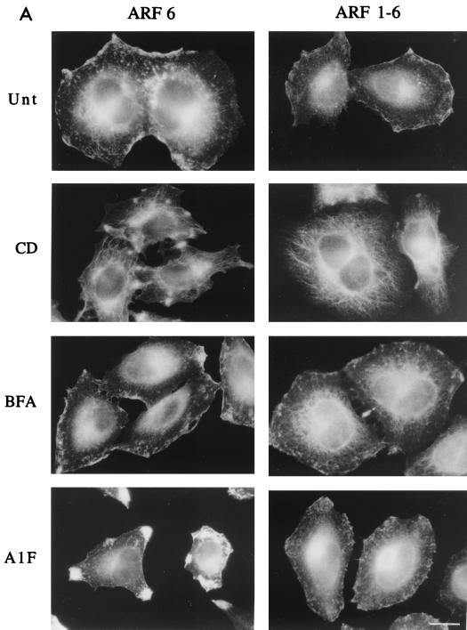
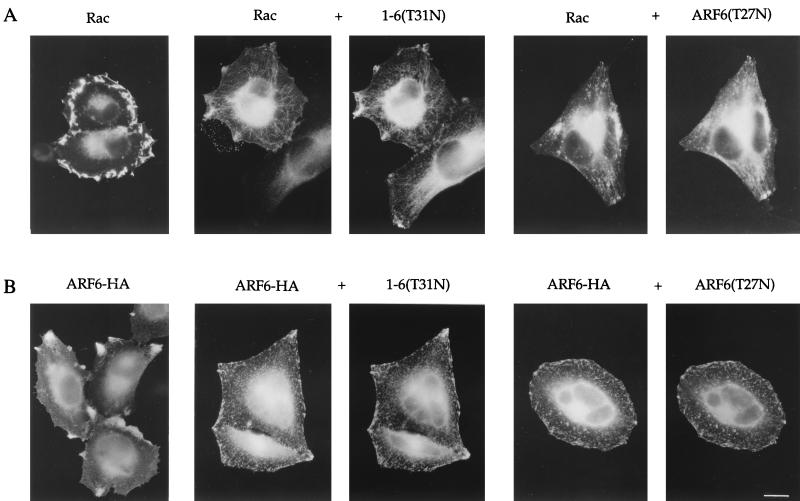
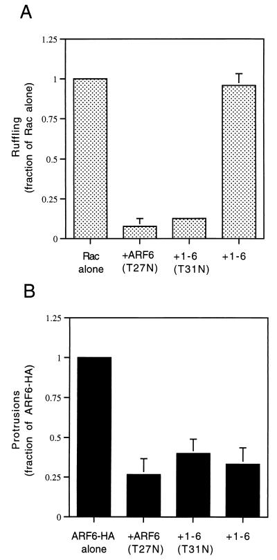
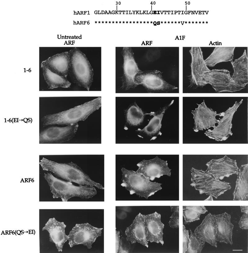
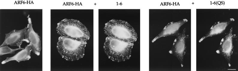
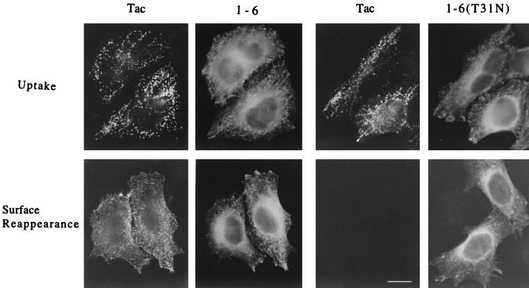
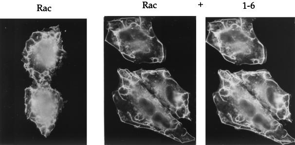
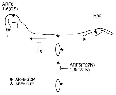
The ADP-ribosylation factor 6 (ARF6) GTPase has a dual function in cells, regulating membrane traffic and organizing cortical actin. ARF6 activation is required for recycling of the endosomal membrane back to the plasma membrane (PM) and also for ruffling at the PM induced by Rac. Additionally, ARF6 at the PM induces the formation of actin-containing protrusions. To identify sequences in ARF6 that are necessary for these distinct functions, we examined the behavior of a chimeric protein of ARF1 and ARF6. The 1-6 chimera (with the amino half of ARF1 and the carboxyl half of ARF6) localized like ARF6 in HeLa cells and moved between the endosome and PM, but it did not form protrusions, an ARF6 effector function. Two residues in the amino-terminal half of ARF6, Q37 and S38, when substituted into the 1-6 chimera allowed protrusion formation, whereas removal of these residues from ARF6 resulted in an inability to form protrusions. Interestingly, expression of 1-6 in cells selectively inhibited protrusions induced by wild-type ARF6 but had no effect on ARF6-regulated membrane movement or Rac-induced ruffling. Thus, we have uncoupled two functions of ARF6, one involved in membrane trafficking, which is necessary for Rac ruffling, and another involved in protrusion formation.


Figures

FIG. 1
FIG. 1
The ARF1-6 chimera localizes like ARF6 yet cannot form protrusions. (A) HeLa cells were transfected with plasmids encoding either ARF6 or the ARF1-6 chimera and were untreated (Unt) or incubated in the presence of either 1 μM CD for 30 min (CD), 2 μg of BFA/ml for 10 min (BFA), or 30 mM NaF and 50 μM AlCl3 for 30 min (AlF). Cells were fixed and immunolabeled with polyclonal anti-ARF6 antibodies that recognize a C-terminal peptide of the protein. Bar, 15 μm. (B) Expression levels of ARF6 in untransfected and transfected HeLa cells. HeLa cells were not transfected (Endog.) or were transfected with 2.5 μg of plasmid encoding ARF6, 1-6, 1-6(T31N), or 1-6(QS). Cell extracts were loaded onto sodium dodecyl sulfate–13% polyacrylamide gel electrophoresis, transferred to nitrocellulose, blotted with rabbit anti-ARF6 antiserum, and visualized by enhanced chemiluminescence as described previously (42). For untransfected HeLa cells, 10 μg of protein was loaded per lane, whereas for transfected HeLa lysates, 2 μg of protein was loaded per lane.
FIG. 1
FIG. 1
The ARF1-6 chimera localizes like ARF6 yet cannot form protrusions. (A) HeLa cells were transfected with plasmids encoding either ARF6 or the ARF1-6 chimera and were untreated (Unt) or incubated in the presence of either 1 μM CD for 30 min (CD), 2 μg of BFA/ml for 10 min (BFA), or 30 mM NaF and 50 μM AlCl3 for 30 min (AlF). Cells were fixed and immunolabeled with polyclonal anti-ARF6 antibodies that recognize a C-terminal peptide of the protein. Bar, 15 μm. (B) Expression levels of ARF6 in untransfected and transfected HeLa cells. HeLa cells were not transfected (Endog.) or were transfected with 2.5 μg of plasmid encoding ARF6, 1-6, 1-6(T31N), or 1-6(QS). Cell extracts were loaded onto sodium dodecyl sulfate–13% polyacrylamide gel electrophoresis, transferred to nitrocellulose, blotted with rabbit anti-ARF6 antiserum, and visualized by enhanced chemiluminescence as described previously (42). For untransfected HeLa cells, 10 μg of protein was loaded per lane, whereas for transfected HeLa lysates, 2 μg of protein was loaded per lane.
FIG. 2
FIG. 2
The GTP-binding-defective mutant of 1-6 acts like a dominant negative ARF6 mutant, inhibiting ARF6 protrusions and Rac ruffling. (A) HeLa cells were transfected with wild-type Rac1 (Rac) or with Rac and either 1-6(T31N) or ARF6(T27N) (1:5 ratio) and then incubated in the presence of AlF for 30 min. (B) HeLa cells were transfected with either wild-type HA-tagged ARF6 (ARF6-HA) or with ARF6-HA and either 1-6(T31N) or ARF6(T27N) (1:5 ratio) and then incubated in the presence of AlF for 30 min. The overexpressed proteins were then localized by immunofluorescence. Bar, 15 μm.
FIG. 3
FIG. 3
Quantitation of Rac ruffling and ARF6 protrusion formation. (A) HeLa cells were transfected with either plasmids encoding Rac1 alone or those encoding Rac1 and either ARF6(T27N), 1-6(T31N), or 1-6 (1:5 ratio). (B) HeLa cells were transfected with either ARF6-HA alone or with ARF6-HA and either ARF6(T27N), 1-6(T31N), or 1-6 (1:5 ratio). Cells were incubated for 30 min in the presence of AlF and fixed, and the overexpressed proteins were labeled by immunofluorescence. For each condition, over 500 transfected cells were counted, and the fraction of Rac- or ARF6-transfected cells that were ruffling or forming protrusions, respectively, was noted. For cells overexpressing only Rac1 or ARF6, the fraction of ruffling or protruding cells was normalized to 1.0, and the other conditions were then expressed as a fraction of 1.0. Data shown are the means and standard errors of three independent experiments.
FIG. 4
FIG. 4
ARF6 effector domain includes residues Q37 and S38. (Top) Amino acid sequence comparison between human ARF1 and ARF6 from amino acid 24 in ARF1 (20 in ARF6) to amino acid 56 in ARF1 (52 in ARF6). Asterisks indicate identity. Note the conservation in the switch I region encompassing residues 45 to 54. (Bottom) HeLa cells were transfected with plasmids encoding either the 1-6 chimera, 1-6(EI→QS), ARF6, or ARF6(QS→EI). Cells were then left untreated or incubated for 30 min with AlF. The expressed proteins were labeled with ARF6-specific antiserum followed by rhodamine-conjugated phalloidin to visualize F-actin. Bar, 15 μm.
FIG. 5
FIG. 5
The 1-6 chimera inhibits protrusion formation. HeLa cells were transfected with plasmids encoding either ARF6-HA alone (ARF6-HA), or with ARF6-HA and either 1-6 or 1-6(QS) (1:5 ratio). Cells were treated with AlF for 30 min, fixed, and processed for immunofluorescence. Bar, 15 μm.
FIG. 6
FIG. 6
The 1-6 chimera does not block internalization or recycling of surface Tac into the tubular compartment. HeLa cells expressing Tac and either the 1-6 chimera or 1-6(T31N) were incubated with anti-Tac antibodies (7G7) at 4°C to bind to surface Tac. Excess antibodies were washed off, and the cells were then incubated at 37°C for 30 min in the presence of CD to allow internalization of the Tac antibodies into the endosomal compartment. Cells were then rinsed to remove remaining surface anti-Tac antibodies and either fixed immediately and assessed for Tac antibody internalized by immunofluorescence (Uptake) or warmed to 37°C for 30 min in the absence of CD before fixation. Tac antibody that reappeared on the cell surface was detected by incubation with fluorescently labeled secondary antibodies in the absence of detergent permeabilization (Surface Reappearance). The 1-6 chimera or 1-6(T31N) was subsequently localized in these cells after permeabilization. Bar, 15 μm.
FIG. 7
FIG. 7
The 1-6 chimera does not inhibit Rac-induced ruffling. HeLa cells were transfected with plasmids encoding either Rac1 alone or Rac and the 1-6 chimera (1:5 ratio). Cells were incubated in the presence of AlF for 30 min and fixed, and the expressed proteins were localized by immunofluorescence. Bar, 15 μm.
FIG. 8
FIG. 8
Working model for ARF6 action on trafficking and cortical actin structures. ARF6-GTP (asterisks) at the PM is involved in the generation of actin filament-containing protrusions (left side) or in the presence of Rac, actin-containing PM ruffles (right side). 1-6(QS) can generate protrusions whereas 1-6 cannot. Both ARF6(T27N) and 1-6(T31N) inhibit activation of ARF6 and therefore the recycling of endosomal membrane back to the PM. Expression of 1-6 inhibits ARF6-mediated protrusions but not Rac-mediated ruffling or membrane recycling back to the PM.

Similar articles

Cited by

References

    1. Amor J C, Harrison D H, Kahn R A, Ringe D. Structure of the human ADP ribosylation factor 1 complexed with GDP. Nature. 1994;372:704–708. - PubMed
    1. Ausubel F M, Brent R, Kingston R E, Moore D D, Seidman J G, Smith J A, Struhl K. Current protocols in molecular biology. New York, N.Y: John Wiley & Sons; 1995.
    1. Boguski M S, McCormick F. Proteins regulating Ras and its relatives. Nature. 1993;366:643–654. - PubMed
    1. Bonifacino J S, Suzuki C K, Lippincott-Schwartz J, Weissman A M, Klausner R D. Pre-Golgi degradation of newly synthesized T cell antigen receptor chains: intrinsic sensitivity and the role of subunit assembly. J Cell Biol. 1989;10:73–83. - PMC - PubMed
    1. Brown H A, Gutowski S, Moomaw C R, Slaughter C, Sternweis P C. ADP ribosylation factor, a small GTP-dependent regulatory protein, stimulates phospholipase D activity. Cell. 1993;75:1137–1144. - PubMed

Publication types

LinkOut - more resources