. 2000 Aug;20(16):6074-83.
doi: 10.1128/MCB.20.16.6074-6083.2000.

Rap2 as a slowly responding molecular switch in the Rap1 signaling cascade

Affiliations

Rap2 as a slowly responding molecular switch in the Rap1 signaling cascade

Y Ohba et al. Mol Cell Biol. 2000 Aug.

Abstract

Rap2 is a member of the Ras family of GTPases and exhibits 60% identity to Rap1, but the function and regulation of Rap2 remain obscure. We found that, unlike the other Ras family proteins, the GTP-bound active form exceeded 50% of total Rap2 protein in adherent cells. Guanine nucleotide exchange factors (GEFs) for Rap1, C3G, Epac (or cyclic AMP [cAMP]-GEF), CalDAG-GEFI, PDZ-GEF1, and GFR efficiently increased the level of GTP-Rap2 both in 293T cells and in vitro. GTPase-activating proteins (GAPs) for Rap1, rap1GAPII and SPA-1, stimulated Rap2 GTPase, but with low efficiency. The half-life of GTP-Rap2 was significantly longer than that of GTP-Rap1 in 293T cells, indicating that low sensitivity to GAPs caused a high GTP/GDP ratio on Rap2. Rap2 bound to the Ras-binding domain of Raf and inhibited Ras-dependent activation of Elk1 transcription factor, as did Rap1. The level of GTP-Rap2 in rat 3Y1 fibroblasts was decreased by the expression of v-Src, and expression of a GTPase-deficient Rap2 mutant inhibited v-Src-dependent transformation of 3Y1 cells. Altogether, Rap2 is regulated by a similar set of GEFs and GAPs as Rap1 and functions as a slowly responding molecular switch in the Rap1 signaling cascade.


Figures
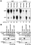
FIG. 1
FIG. 1
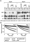
Analysis of guanine nucleotides bound to Rap2. (A) 293T cells or NIH 3T3 cells were transfected with expression vectors encoding the proteins listed at the top and labeled with 32Pi. Guanine nucleotides bound to the expressed Rap1 or Rap2 were separated by TLC. The radioactivity of GTP and GDP was quantitated, and the percentage of GTP [GTP/(GDP + GTP)] is shown at the bottom. (B) 293T cells were transfected with Flag-tagged expression vectors as indicated at the top. In one sample, cells were incubated with 100 μM Sp-cAMPs for 10 min before harvest (indicated as cAMP). Endogenous GTP-Rap2 and GTP-Rap1 were detected by the Bos method as described in the text (top). The intensity of the bands was quantitated by an image analyzer, and the fold increase is shown. The lower panels show immunoblotting (IB) of total cell lysates with anti-Rap2, anti-Rap1, or anti-Flag antibody.
FIG. 2
FIG. 2
GEF-dependent guanine nucleotide exchange reaction of Rap2. (A) 293T cells expressing GST-Rap2 or GST-Rap1 with GEFs indicated at the top were labeled with 32Pi, and guanine nucleotides bound to GST-Rap2 or GST-Rap1 were analyzed as described. In four samples, cells were incubated with 100 μM Sp-cAMPS for 10 min before harvest (indicated as cAMP). (B) Rap1 or Rap2 bound to mGDP was incubated at 20°C with or without GEFs. Epac was stimulated with 100 μM Sp-cAMPS (indicated as cAMP). The decrease in fluorescence emission at 450 nm was monitored as a function of time.
FIG. 3
FIG. 3
Guanine nucleotide exchange and GTP hydrolysis of Rap1 and Rap2 in vivo. 293T cells were labeled with 32Pi for 30 min and chased with phosphate-containing medium. Cells were harvested at the indicated time points, and the cytosolic fraction was used to load recombinant H-Ras protein with 32P-labeled guanine nucleotides in vitro. The cytosolic guanine nucleotides bound to H-Ras in vitro were separated by TLC and quantitated. In a parallel experiment, 293T cells expressing GST-Rap1 or GST-Rap2 were lysed, and the labeled guanine nucleotides bound to GST-Rap1 or GST-Rap2 were separated by TLC and quantitated. (A) The sum of radioactivity of GTP and GDP at each time point was plotted at a ratio to the radioactivity at 30 min. (B) The percentage of GTP on GST-Rap2 or GST-Rap1 at each time point was plotted. Bars indicate standard deviations.
FIG. 4
FIG. 4
Activation of Rap2 GTPase by GAPs. (A) 293T cells expressing either GST-Rap2 or GST-Rap1 and GEFs and GAPs indicated at the top were labeled with 32Pi, and guanine nucleotides bound to GST-Rap1 or GST-Rap2 were separated by TLC. (B) Rap1 and Rap2 were loaded with [γ-32P]GTP and incubated with the indicated amounts of rap1GAPII for 10 min (left panel). Similarly, the 32P-labeled Rap1 and Rap2 were incubated with (solid symbols) or without (open symbols) 1 μg of rap1GAPII for the indicated periods (right panel). The radioactivity remaining on Rap1 or Rap2 was quantitated and plotted. (C) [γ-32P]GTP-loaded Rap1 and Rap2 were incubated with buffer alone, purified rap1GAPII, or cell lysates of Jurkat cells, HT1080 cells, or 293T cells for 20 min. The radioactivity remaining on Rap1 or Rap2 was quantitated and plotted. Bars indicate standard errors.
FIG. 5
FIG. 5
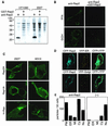
Subcellular localization of Rap2. (A) Total cell lysates of HT1080 cells and 293T cells were separated by SDS-PAGE and transferred to polyvinylidene difluoride membranes. Filters were incubated with anti-Rap2 antibody, anti-Rap2 antibody preincubated with GST-Rap2, or buffer alone. Filters were further incubated with a peroxidase-conjugated antimouse antibody, which was detected by the ECL enhanced chemiluminescence system. (B) HT1080 cells grown on fibronectin-coated coverslips were fixed with paraformaldehyde and permeabilized by Triton X-100 (PFA) or fixed with ethanol (EtOH). The cells were incubated with anti-Rap2 antibody or anti-Rap2 antibody preadsorbed by GST-Rap2. Bound antibodies are detected by the use of antimouse Alexa 488-conjugated antibody. (C) 293T and MDCK cells grown on poly-l-lysine-coated glass dishes were transfected with expression vectors of EGFP-tagged Rap2, Rap1, and H-Ras for 24 h and observed under a confocal microscope. (D) MDCK cells cultured on poly-l-lysine-coated glass dishes were transfected with expression vectors encoding ECFP-Rap2 and EYFP-ER or EYFP-Golgi for 24 h and observed with a confocal microscope. (E) HT1080 cells were collected and embedded in Lowicryl. Ultrathin sections were prepared and incubated with anti-Rap2 antibody or buffer alone (−), followed by incubation with antimouse antibody conjugated with immunogold. Cells were arbitrarily divided into plasma membrane (PM), ER, mitochondria (Mi), cytoplasm (Cy), and nucleus (Nu). Immunogold particles in these regions were counted and plotted.
FIG. 6
FIG. 6
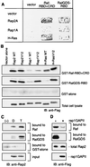
Binding of Rap2 to Raf and RalGDS. (A) Yeast strain y-190 was cotransformed with the pGAD424-derived plasmids denoted at the top and with the pGBT9-derived bait plasmids listed to the left. His activity of the transformants was assayed on histidine-deficient plates, followed by a β-galactosidase assay. (B) 293T cells expressing the Flag-tagged G proteins denoted at the top were lysed in lysis buffer and incubated with GST alone, GST-Raf-RBD+CRD, or GST-RalGDS-RBD. Proteins bound to these GST fusion proteins and the total cell lysates were separated by SDS-PAGE and probed with anti-Flag monoclonal antibody. (C) GST-Raf-RBD+CRD, GST-RalGDS-RBD, or GST alone was incubated with buffer alone (−) or recombinant Rap2 protein loaded with GDP (D) or GTP (T). Proteins bound to the beads were separated by SDS-PAGE and analyzed by immunoblotting (IB) with anti-Rap2 antibody. (D) 293T cells expressing Flag-Rap2WT alone or Flag-Rap2WT and rap1GAPII were harvested and incubated with GST-Raf-RBD+CRD or GST-RalGDS-RBD. Proteins bound to the GST fusion proteins and total cell lysates were analyzed by immunoblotting with anti-Flag antibody.
FIG. 7
FIG. 7
Inhibition of Ras-dependent transcription by Rap2. 293T cells were transfected with pFR-luc, pFA-Elk1, and pCXN2-Flag-RasV12 (H-RasV12; solid columns) or pSRα-SKR (Raf-SKR; shaded columns) in combination with wild-type (WT) and active Rap2 or Rap1 expression vectors. After 24 h, luciferase activity was measured. In this assay system, expression of luciferase is driven by the Elk1 transcription factor. Mean values obtained from three experiments are shown with standard errors.
FIG. 8
FIG. 8
Quantitative immunoblotting (IB) of Ras, Rap1, and Rap2. Thirty micrograms of proteins from 7.4 × 10⁴ 293T cells and 20 μg of proteins from 9.2 × 10⁴ HT1080 cells and recombinant Rap2, Rap1, or H-Ras, the amounts of which are indicated at the bottom of each column, were separated by SDS-PAGE and analyzed by immunoblotting with the antibodies indicated to the right. With recombinant proteins used as a standard, the amounts of Rap2, Rap1, and H-Ras in the cell lysates were determined and are shown at the bottom of each lane of cell lysates.
FIG. 9
FIG. 9
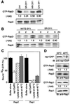
Level of GTP-Rap2 in transformed cells. (A) 3Y1 cells and 3Y1-derived transformed cells denoted at the top were lysed, and 500 μg each of the lysates was incubated with GST-RalGDS prebound to glutathione-Sepharose beads. Proteins bound to the beads and 10 μg of total cell lysates were analyzed by SDS-PAGE and immunoblotting with anti-Rap2 antibody. (B) NY72-3Y1 cells and SR-3Y1 cells expressing a temperature-sensitive mutant and wild-type v-Src, respectively, were maintained at 33°C. After a temperature shift to 40°C, cells were lysed at the times indicated and analyzed as in panel A. (C) Soluble cytosolic fraction was prepared from NY72-3Y1 cell cultures at either 40 or 33°C. [γ-32P]GTP-loaded Rap2 and Rap1 were incubated with buffer alone, purified rap1GAPII, or 30 μg of the soluble cytosolic fractions for 20 min. Radioactivity retained by Rap2 and Rap1 was quantitated and plotted. Bars indicated standard errors from three samples. (D) Cell lysates used in panel C were separated by SDS-PAGE and blotted with anti-rap1GAP (top). The levels of GTP-Rap2 and GTP-Rap1 in these cell lysates were analyzed as in panel A.
FIG. 10
FIG. 10
Inhibition of morphological transformation by Rap2. NY72-3Y1 cells were transfected with pCAGGS or pCAGGS-derived Flag-tag expression vector for wild-type Rap2 (Rap2WT), Rap2V12, Rap1WT, or Rap1V12 with an expression vector of the hygromycin resistance gene. After selection with hygromycin B at 40°C for 10 days, cells were cultured at 33°C overnight. (A) Morphology of the representative colonies at 40 and 33°C. (B) Hygromycin-resistant colonies consisting of transformed spindle cells or nontransformed flat cells were scored under the microscope. Mean values obtained from two independent experiments are shown with standard deviations. (C) Equal amounts of cell lysates were analyzed by immunoblotting (IB) with anti-Flag antibody.

Similar articles

Cited by

References

    1. Altschuler D L, Ribeiro-Neto F. Mitogenic and oncogenic properties of the small G protein Rap1b. Proc Natl Acad Sci USA. 1998;95:7475–7479. - PMC - PubMed
    1. Beranger F, Goud B, Tavitian A, de Gunzburg J. Association of the Ras-antagonistic Rap1/Krev-1 proteins with the Golgi complex. Proc Natl Acad Sci USA. 1991;88:1606–1610. - PMC - PubMed
    1. Beranger F, Tavitian A, de Gunzburg J. Post-translational processing and subcellular localization of the Ras-related Rap2 protein. Oncogene. 1991;6:1835–1842. - PubMed
    1. Bos J L. All in the family? New insights and questions regarding interconnectivity of Ras, Rap1 and Ral. EMBO J. 1998;17:6776–6782. - PMC - PubMed
    1. Bos J L. Ras-like GTPases. Biochim Biophys Acta. 1997;1333:M19–M31. - PubMed

Publication types

Substances