. 2000 Aug;20(16):6095-104.
doi: 10.1128/MCB.20.16.6095-6104.2000.

Analysis of the complex relationship between nuclear export and aryl hydrocarbon receptor-mediated gene regulation

Affiliations

Analysis of the complex relationship between nuclear export and aryl hydrocarbon receptor-mediated gene regulation

R S Pollenz et al. Mol Cell Biol. 2000 Aug.

Abstract

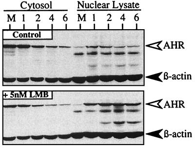
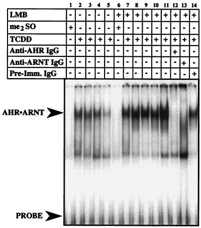
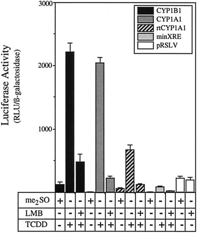
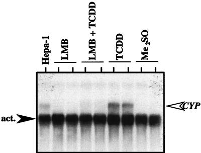
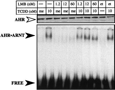
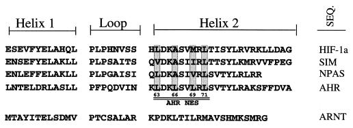
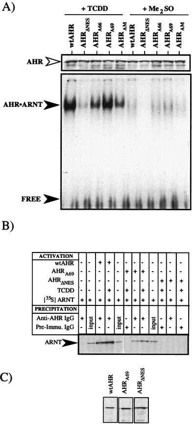
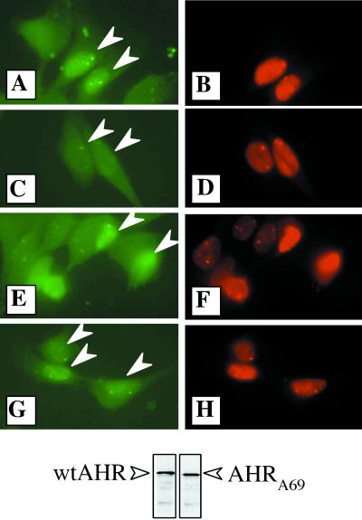
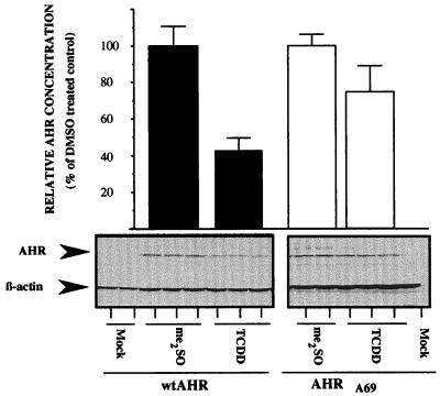
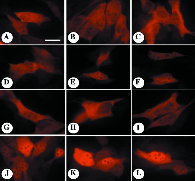
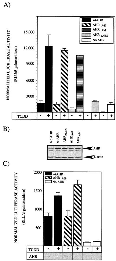
The aryl hydrocarbon receptor (AHR) contains signals for both nuclear import and nuclear export (NES). The purpose of the studies in this report was to determine the relationship between the nuclear export of the AHR and AHR-mediated gene regulation. Blockage of nuclear export in HepG2 cells with leptomycin B (LMB) resulted in increased levels of AHR-AHR nuclear translocator (ARNT) complex in the nucleus and correlative reductions in agonist-stimulated AHR degradation. However, LMB exposure inhibited agonist-mediated induction of numerous AHR-responsive reporter genes by 75 to 89% and also inhibited induction of endogenous CYP1A1. LMB did not transform the AHR to a ligand binding species or affect activation by TCDD (2, 3,7,8-tetrachlorodibenzo-p-dioxin). Mutagenesis of leucines 66 and 71 of the putative AHR NES resulted in a protein with reduced function in dimerization to ARNT and binding to DNA, while alanine substitution at leucine 69 (AHR(A69)) resulted in an AHR that bound with ARNT and associated with DNA. AHR(A69) protein injected directly into the nuclei of E36 cells remained nuclear following 6 h of agonist stimulation. In transient-transfection assays, AHR(A69) accumulated within the nucleus was not degraded efficiently following agonist exposure. Finally, AHR(A69) supported induction of AHR-responsive reporter genes in an agonist-dependent manner. These findings show that it is possible to generate an AHR protein defective in nuclear export that is functional in agonist-mediated gene induction. This implies that the negative effect of LMB on agonist-mediated gene induction is independent of the nuclear export of the AHR.


Figures

FIG. 1
FIG. 1
Accumulation of AHR in the nucleus of HepG2 cells exposed to LMB and TCDD. Duplicate plates of HepG2 cells were exposed to ethanol (0.1%) or LMB (5 nM) for 3 h followed by TCDD (2 nM) for an additional 1 to 6 h. Cell pellets were then solubilized in lysis buffer, and cytosol (10 μg) and nuclear lysates (18 μg) were resolved by SDS-PAGE. Gels were then blotted and stained with A-1A IgG (1.0 μg/ml) and β-actin IgG (1:1,000) and visualized by ECL with GAR-HRP IgG (1:10,000). 1, 2, 4, and 6 represent hours of TCDD exposure. M = cells exposed to Me2SO for 6 h.
FIG. 2
FIG. 2
Potentiation of XRE binding activity in nuclear extracts isolated from HepG2 cells treated with LMB and TCDD. Duplicate plates of HepG2 cells were exposed to ethanol (0.1%) or LMB (5 nM) for 3 h followed by TCDD (2 nM) for an additional 1 to 8 h. Nuclear extracts were prepared, and 5 μg was evaluated by EMSA. Lanes 1 and 6, 8-h exposure to Me2SO. Lanes 2, 7, and 12 to 14, 1-h TCDD exposure. Lanes 3 and 8, 2-h TCDD exposure. Lanes 4 and 9, 4-h TCDD exposure. Lane 10, 6-h TCDD exposure. Lanes 5 and 11, 8-h TCDD exposure.
FIG. 3
FIG. 3
Inhibition of TCDD-induced reporter gene activity in HepG2 cells exposed to LMB. HepG2 cells were cotransfected with reporter constructs containing the indicated promoter sequences and pSV-β-galactosidase. Twenty-four hours after transfection, triplicate plates were treated with ethanol (0.1%) or LMB (5 nM) for 3 h followed by exposure to TCDD (2 nM) or Me2SO (0.02%) for an additional 10 h. A set of cells were also transfected with a constitutive luciferase reporter (pRSLV) under the control of the Rous sarcoma virus promoter (white bars). Cells were then harvested into reporter lysis buffer and assayed for luciferase and β-galactosidase activity. Data are expressed as the means ± standard deviations for three samples of the normalized luciferase activity which represents the relative luciferase units (RLU) divided by the β-galactosidase activity. Note that LMB did not affect the activity associated with the constitutive luciferase reporter.
FIG. 4
FIG. 4
Inhibition of CYP1A1 mRNA induction in HepG2 cells exposed to LMB. LMB, HepG2 cells treated with LMB (5 nM) for 13 h. LMB + TCDD, HepG2 cells treated with LMB (5 nM) for 3 h followed by exposure to TCDD (2 nM) for an additional 10 h. TCDD, HepG2 cells treated with ethanol (0.1%) for 3 h followed by exposure to TCDD (2 nM) for an additional 10 h. Me2SO, HepG2 cells treated with ethanol (0.1%) for 3 h followed by Me2SO (0.02%) for an additional 10 h. After each incubation, total RNA was extracted from cells and evaluated for CYP1A1 and β-actin expression as described in Materials and Methods. Each lane represents an independent plate of cells. Hepa-1 = 10 μg of total RNA from murine Hepa-1 cells treated with TCDD for 24 h.
FIG. 5
FIG. 5
Analysis of LMB with AHR protein. wtAHR protein (approximately 15 ng) was expressed in vitro and mixed with an equal amount of ARNT protein. The samples were then incubated in the presence of TCDD (10 nM) or Me2SO (0.1%) in the presence or absence of varying concentrations of LMB (1.2, 12, or 60 nM) or ethanol (0.1%) for 2 h at 30°C and analyzed immediately. Equal amounts of each sample were analyzed by EMSA with 32P-labeled XRE as detailed in Materials and Methods. The specifically shifted AHR-ARNT complex and free probe are indicated by the closed arrowheads. The Western blot at the top of the EMSA represents an aliquot of the exact sample used for the EMSA that was stained for AHR (open arrowhead). Note that the concentration of AHR in each sample is similar. Numbers indicate nanomolar concentrations. me, Me2SO; et, ethanol.
FIG. 6
FIG. 6
Alignment of HLH regions of bHLH-PAS proteins. The HLH regions for the mouse HIF-1α, single-minded protein, nuclear PAS, AHR, and ARNT proteins were analyzed by Lasergene software (DNASTAR Inc., Madison, Wis.). The helix 1, loop, and helix 2 regions are shown. The putative NES within helix 2 of AHR is underlined. Conserved leucine residues with the AHR NES are indicated. SEQ., sequence.
FIG. 7
FIG. 7
Functional analysis of mutations within the NES of the AHR. The indicated AHR protein was expressed in vitro and mixed with an equal amount of cold or 35S-labeled ARNT protein (approximately 15 ng). The samples were then incubated in the presence of TCDD (10 nM) or Me2SO (0.1%) for 2 h at 30°C and analyzed immediately. (A) Equal amounts of each sample were analyzed by EMSA with 32P-labeled XRE as detailed in Materials and Methods. The specifically shifted AHR-ARNT complex and free probe are indicated by the closed arrowheads. The Western blot at the top of the EMSA represents an aliquot of the exact sample used for the EMSA that was stained for AHR (open arrowhead). Note that the concentration of AHR in each sample is similar. (B) Equal amounts of each sample were precipitated with anti-AHR IgG (A-1A) or preimmune IgG and resolved by SDS-PAGE. Specificity is demonstrated by the lack of 35S-labeled ARNT in samples precipitated with preimmune IgG. (C) The Western blot shown represents an aliquot of the exact AHR sample used for the immunoprecipitation that was stained for AHR (open arrowhead). Note that the concentration of AHR in each sample is similar.
FIG. 8
FIG. 8
Distribution of wtAHR and AHRA69 protein injected into E36 cells. wtAHR and AHRA69 proteins were expressed in vitro and mixed with 70-kDa dextran-conjugated Texas red. The samples were then injected into the nuclei of E36 cells and exposed to TCDD (2 nM) or Me2SO (0.02%) for 6 h at 30°C. Following the incubation, cells were fixed and stained with 1 μg of A-1 IgG per ml followed by goat anti-rabbit–FITC (1:500). Identical fields showing FITC fluorescence (A, C, E, and G) and Texas red fluorescence (B, D, F, and H) are shown. (A and B) Cells injected with wtAHR and exposed to Me2SO. (C and D) Cells injected with wtAHR and exposed to TCDD. (E and F) Cells injected with AHRA69 and exposed to Me2SO. (G and H) Cells injected with AHRA69 and exposed to TCDD. Arrowheads indicate the nuclei injected with AHR protein. The Western blot shown at the bottom represents an aliquot of the exact wtAHR and AHRA69 proteins used for the microinjection that were stained for AHR (open arrows).
FIG. 9
FIG. 9
Western blot analysis of recombinant AHR protein expression in E36 cells exposed to TCDD. E36 cells were transfected with expression vectors for wtAHR or AHRA69. Triplicate plates were exposed to TCDD (2 nM) or Me2SO (0.02%) for 16 h, and 18 μg of total cell lysate was resolved by SDS-PAGE. Blots were stained with A-1A IgG (1.0 μg/ml) and β-actin IgG (1:1,000) and visualized by ECL with GAR-HRP IgG (1:10,000). Bands were quantified and normalized as detailed previously (14, 17, 19, 23, 25). Data are expressed as the percentages of wtAHR or AHRA69 protein compared to that for Me2SO (DMSO)-treated controls. Bars represent the averages ± standard errors of three independent samples.
FIG. 10
FIG. 10
Subcellular localization of recombinant AHR protein expression in E36 cells exposed to TCDD. All slips were incubated with A-1 IgG (1.0 μg/ml) and visualized with goat anti-rabbit–Texas red IgG (1:750). (A to C) E36 cells expressing wtAHR and stained for AHR following a 24-h exposure to Me2SO (0.02%). (D to F) E36 cells expressing wtAHR and stained for AHR following a 24-h exposure to TCDD (2 nM). (G to I) E36 cells expressing AHRA69 and stained for AHR following a 24-h exposure to Me2SO (0.02%). (J to L) E36 cells expressing AHRA69 and stained for AHR following a 24-h exposure to TCDD (2 nM). All panels were photographed and printed for identical times. Note the greatly reduced AHR reactivity in untransfected cells. Bar = 10 μm.
FIG. 11
FIG. 11
Induction of TCDD-induced reporter gene activity in cells expressing AHRA69. Type I Hepa-1 E36 CHO cells were cotransfected with pGudLuc 1.1, pSV-β-galactosidase, and pSport expression vectors containing wtAHR, AHRA69, AHRAM, or AHRΔNES. Control plates of cells were transfected with pGudLuc 1.1, pSV-β-galactosidase, and pSport (no AHR). Eight hours after transfection, triplicate plates of cells were treated with TCDD (2 nM) or Me2SO (0.02%) for 16 h. (A) Type I cells were harvested into reporter lysis buffer and assayed for luciferase and β-galactosidase activity. Data are expressed as the means ± standard deviations for three samples of normalized luciferase activity which represents the relative luciferase units (RLU) divided by the β-galactosidase activity. (B) Western blot analysis of total cell lysates from plates of type I cells transfected with the identical DNA solutions used for panel A. Blots were stained with A-1A IgG and β-actin IgG as detailed in the text. (C) E36 cells were harvested into reporter lysis buffer and assayed for luciferase and β-galactosidase activity. Data are expressed as the means ± standard deviations for three samples of normalized luciferase activity which represents the relative luciferase units (RLU) divided by the β-galactosidase activity. Shown are results of Western blot analysis of total cell lysates from representative plates of E36 cells transfected with the identical DNA solutions used to generate the luciferase data. Blots were stained with A-1A IgG as detailed in the text.

Similar articles

Cited by

References

    1. Basci S G, Hankinson O. Functional characterization of DNA binding domains of the heterodimeric AHR complex imputing canonical bHLH protein-DNA interactions. J Biol Chem. 1996;271:8843–8850. - PubMed
    1. Carvan M J, Ponomareva L V, Solis W A, Matlib R S, Puga A, Nebert D W. Trout CYP1A3 gene: recognition of fish DNA motifs by mouse regulatory proteins. Mar Biotechnol. 1999;1:155–166. - PubMed
    1. Chang C-Y, Puga A. Constitutive activation of the aromatic hydrocarbon receptor. Mol Cell Biol. 1998;18:525–535. - PMC - PubMed
    1. Davarinos N A, Pollenz R S. Aryl hydrocarbon receptor imported into the nucleus following ligand binding is rapidly degraded via the cytoplasmic proteasome following nuclear export. J Biol Chem. 1999;274:28707–28715. - PubMed
    1. Dolwick K M, Swanson H I, Bradfield C A. In vitro analysis of Ah receptor domains involved in ligand-activated DNA recognition. Proc Natl Acad Sci USA. 1993;90:8566–8570. - PMC - PubMed

Publication types

MeSH terms

LinkOut - more resources