. 2000 Aug;20(16):6114-26.
doi: 10.1128/MCB.20.16.6114-6126.2000.

Sik (BRK) phosphorylates Sam68 in the nucleus and negatively regulates its RNA binding ability

Affiliations

Sik (BRK) phosphorylates Sam68 in the nucleus and negatively regulates its RNA binding ability

J J Derry et al. Mol Cell Biol. 2000 Aug.

Abstract

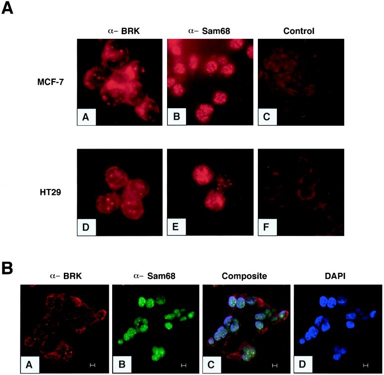
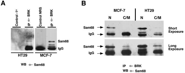
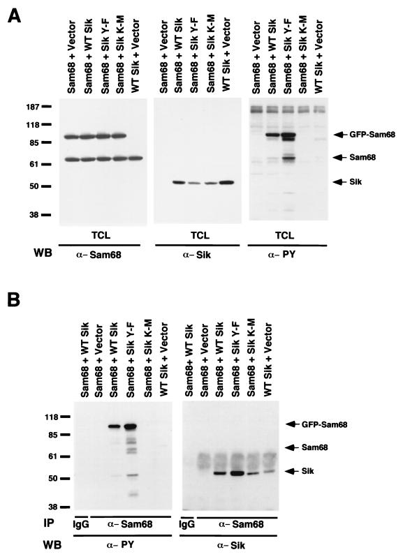
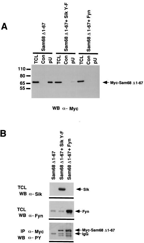
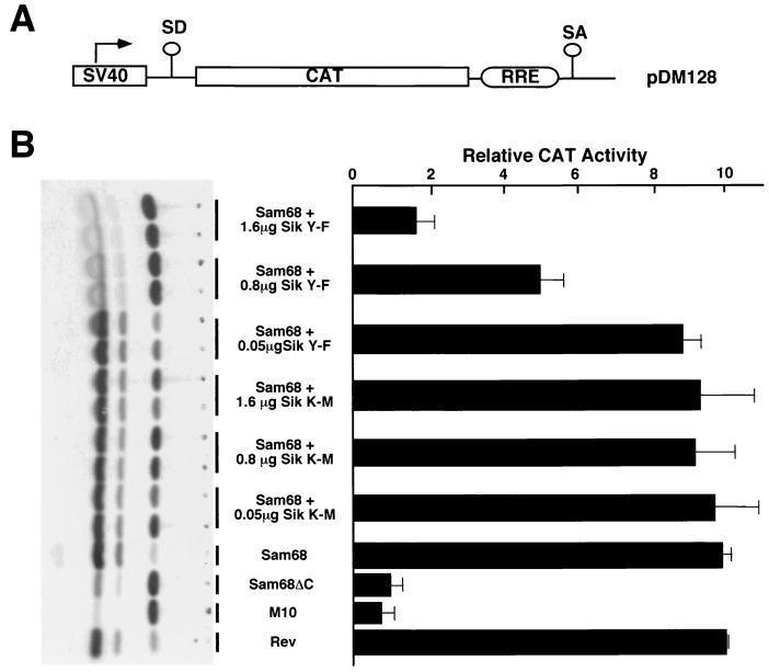
Sik (mouse Src-related intestinal kinase) and its orthologue BRK (human breast tumor kinase) are intracellular tyrosine kinases that are distantly related to the Src family and have a similar structure, but they lack the myristoylation signal. Here we demonstrate that Sik and BRK associate with the RNA binding protein Sam68 (Src associated during mitosis, 68 kDa). We found that Sik interacts with Sam68 through its SH3 and SH2 domains and that the proline-rich P3 region of Sam68 is required for Sik and BRK SH3 binding. In the transformed HT29 adenocarcinoma cell cell line, endogenous BRK and Sam68 colocalize in Sam68-SLM nuclear bodies (SNBs), while transfected Sik and Sam68 are localized diffusely in the nucleoplasm of nontransformed NMuMG mammary epithelial cells. Transfected Sik phosphorylates Sam68 in SNBs in HT29 cells and in the nucleoplasm of NMuMG cells. In functional studies, expression of Sik abolished the ability of Sam68 to bind RNA and act as a cellular Rev homologue. While Sam68 is a substrate for Src family kinases during mitosis, Sik/BRK is the first identified tyrosine kinase that can phosphorylate Sam68 and regulate its activity within the nucleus, where it resides during most of the cell cycle.


Figures

FIG. 1
FIG. 1
BRK and Sam68 localize in nuclear structures in breast and colon tumor cell lines. (A) BRK and Sam68 are found in nuclear structures in human tumor cell lines. Immunofluorescence was used to examine the localization of endogenous BRK and Sam68 in the MCF-7 (A, B) and HT29 (C, D) tumor cell lines. Cells were fixed and stained with antibodies against BRK (A, D) or Sam68 (B, E), BRK antibody preincubated with immunogenic peptide (C), or control rabbit sera (F). (B) BRK localizes to SNBs in HT29 cells. Cells were fixed and stained with antibodies against BRK followed by staining with antibodies against Sam68 and were analyzed by confocal microscopy. BRK was visualized with rhodamine (A), and Sam68 staining was visualized with FITC (B). A composite (C) shows colocalization of BRK and Sam68 as yellow spots in the nuclei (C). Nuclei were stained with DAPI (D). Bars, 5 μm.
FIG. 2
FIG. 2
BRK and Sam68 associate within the nuclei of HT29 and MCF-7 cells. (A) Sam68 coimmunoprecipitates with BRK from lysates of the HT29 colon and MCF-7 breast carcinoma cell lines. Cells were lysed, and 1 mg of total-cell lysate was incubated with anti-BRK antibodies, normal rabbit serum (NRS), or Sepharose beads alone as a control for nonspecific binding to beads. The immunoprecipitate was resolved by SDS-PAGE followed by immunoblotting with anti-Sam68 AD1 polyclonal antibodies. (B) Nuclear (N) and cytosolic and membrane (C/M) fractions from MCF-7 and HT29 cells were immunoprecipitated with anti-BRK antibodies, followed by immunoblotting with anti-Sam68 antibodies. A short exposure (10 s) shows interaction in the nuclear fraction only. A longer exposure (45 s) shows much weaker interaction in the cytosolic and membrane fraction.
FIG. 3
FIG. 3
Sik phosphorylates Sam68 in vivo. (A) Sam68 is tyrosine phosphorylated in NMuMG cells expressing wild-type Sik and Sik Y-F. NMuMG cells were cotransfected with GFP-Sam68 and Vector alone, wild-type (WT) Sik, Sik Y-F, or kinase-defective Sik K-M. Total-cell lysates were divided equally and immunoblotted with antibodies against Sam68, Sik, and phosphotyrosine. Tyrosine-phosphorylated Sam68 was detected only in lysates containing wild-type Sik or Sik Y-F. Although lower levels of Sik Y-F were expressed than wild-type Sik, the highest levels of tyrosine-phosphorylated Sam68 were detected in cells cotransfected with Sik Y-F, suggesting that tyrosine 447 of Sik negatively regulates its activity. (B) Sik associates with Sam68. Immunoprecipitations were performed with Sam68 antibody or IgG as a control and with lysates from the transfected cells in panel A. Immunoblotting was performed with antiphosphotyrosine or anti-Sik antibodies. Sik was observed to coprecipitate with Sam68 from lysates of cells transfected with Sik expression constructs. Sik antibody binding was detected using HRP-conjugated protein A. Tyrosine-phosphorylated Sik could also be detected in the Sam68 immunoprecipitates (left panel).
FIG. 4
FIG. 4
Sik Y-F has increased kinase activity and specifically phosphorylates Sam68. (A) The carboxy-terminal tyrosine of Sik functions as a negative regulatory site. HeLa cells were cotransfected with Myc-tagged Sam68 and wild-type (WT) Sik, Sik Y-F, or Sik K-M (kinase defective). Cells were lysed, the proteins were separated by SDS-PAGE, and immunoblotting with anti-Sik, anti-Myc, and antiphosphotyrosine antibodies was performed. Increased phosphorylation of Myc-Sam68 was visible in total-cell lysates from Sik Y-F-transfected cells (right panel). (B) HeLa cells were transfected with Sik Y-F or were cotransfected with Sik Y-F and Myc-Sam68, Myc-hnRNPK, or truncated Myc-Sam68 (68-347). The cells were lysed and immunoprecipitated with IgG (control) or anti-Myc antibodies. The bound proteins were analyzed by immunoblotting with antiphosphotyrosine antibodies (left panel). The same membrane was subsequently immunoblotted with anti-Myc antibodies (middle panel). Total-cell extracts (TCL) were immunoblotted with anti-Sik antibodies (right panel). The band at ∼55 kDa in lanes 1 to 16 represents the heavy chain of the immunoprecipitating antibodies. (C) Sik phosphorylates the carboxy terminus of Sam68. In vitro kinase assays were performed using full-length GST-Sik, [γ-32P]ATP, and 2 μg of the following substrates: GST-Grb2-N-SH3 (negative control), GST alone, GST-Sam68 (331–443), or GST-Sam68 (354–393). The proteins were separated by SDS-PAGE and stained with Coomassie blue (left). The gel was dried, and the phosphorylated proteins were visualized by autoradiography (right).
FIG. 5
FIG. 5
Wild-type Sik and Sik Y-F phosphorylate nuclear proteins that colocalize with GFP-Sam68 within the nucleus. (A) Localization of GFP-tagged Sam68, phosphotyrosine, and wild-type (WT) Sik in transfected NMuMG cells. Antiphosphotyrosine antibody was visualized with rhodamine (red), while anti-Sik antibody binding was visualized with avidin-Alexa 350 (blue). Wild-type Sik is present in the nucleus and at the membrane (panel C). (B) NMuMG cells were transfected with GFP-Sam68 and wild-type Sik (A to D), GFP-Sam68 and Sik Y-F (E to H), GFP-Sam68 and kinase-defective Sik K-M (I to L), or the GFP expression vector pEGFP-C1 and pcDNA3 (M to P). Cells were fixed 24 h after transfection, and tyrosine-phosphorylated proteins were localized using anti-phosphotyrosine antibodies (B, F, J, N). DAPI was used to stain the nuclei (D, H, L, P). In NMuMG cells, Sam68 displays diffuse, nuclear localization visible by green fluorescence (A, E, I). Cells cotransfected with GFP-Sam68 and wild-type Sik or Sik Y-F also stain strongly with the anti-phosphotyrosine antibody visualized using rhodamine (B, F), while no phosphotyrosine was detected in cells expressing kinase-defective Sik K-M (J). Panels C, G, K, and O are composites demonstrating colocalization that appears yellow. GFP alone is expressed throughout the cell (M) and is negative for anti-phosphotyrosine staining (N). (C) Increased phosphotyrosine in SNBs in HT29 cells following introduction of the activated Sik Y-F construct into HT29 cells. HT29 cells were transfected with GFP-Sam68 (A, C) and Sik Y-F, and tyrosine-phosphorylated proteins were localized using antiphosphotyrosine antibodies (B, C). Colocalization of Sam68 and the increased phosphotyrosine signal in SNBs are shown in panel C. No phosphotyrosine signal was detected in control cells transfected with kinase-defective Sik K-M (not shown). DAPI was used to stain the nuclei (D). Bars represent 5 μm.
FIG. 6
FIG. 6
The Sik SH2 and SH3 domains bind Sam68. (A) The ability of Sam68 to bind the SH2 and SH3 domains of Sik was tested. NMuMG cells were transfected with GFP-Sam68 and the expression vector pcDNA3 (Vector) or the wild-type Sik, activated Sik Y-F, or kinase-defective Sik K-M expression constructs. Cell lysates were divided equally and incubated with GST, GST-Sik SH2+SH3, GST-Sik SH2, and GST-SH3 covalently coupled to beads. Bound proteins as well as an aliquot of total-cell lysate from GFP-Sam68-transfected cells were separated by SDS-PAGE and immunoblotted with Sam68 AD1 polyclonal antibody. Positions of GFP-Sam68 and the endogenous Sam68 protein are indicated with arrows. GFP-Sam68 and endogenous Sam68 protein bound to the GST-Sik, SH2+SH3, and GST-Sik SH3 fusion proteins in all of the cell lysates. GFP-Sam68 binding to the GST-Sik SH2 domain was detected only in cells transfected with wild-type Sik or Sik Y-F, suggesting that phosphorylation by Sik is required for Sik SH2 binding. (B) Sam68 proline motif 3 (P3) associates with the SH3 domain of Sik. HeLa cell lysates were incubated with GST, GST-Sam68 P0, GST-Sam68 P1P2, GST-Sam68 P3, or GST Sam68 P4 covalently coupled to beads. The beads were washed, and the bound BRK was observed by immunoblotting. An aliquot of the HeLa cell lysate was used to represent total-cell lysate. BRK binding was only detected with the GST-Sam68 P3 fusion protein. (C) Beads containing covalently coupled GST-Sik SH3 protein were preincubated for 15 min at room temperature with the indicated concentration of Sam68 proline-rich peptide. Subsequently, HeLa cell lysates were added to each tube for 30 min at 4°C. The beads were washed extensively, and the bound Sam68 was quantitated by immunoblotting. Binding of Sam68 with the GST-Sik SH3 fusion protein was efficiently competed with the P3 peptide.
FIG. 7
FIG. 7
Sik negatively regulates the ability of Sam68 to bind RNA. (A) HeLa cell lysates from cells either transfected with Myc-Sam68Δ1–67 alone or cotransfected with Myc-Sam68Δ1–67 and Sik Y-F or Fyn were divided equally and precipitated with agarose (Con) or poly(U)-agarose (pU), followed by anti-Myc immunoblotting. An aliquot of total-cell lysate (TCL) was also included to monitor expression of Myc-Sam68Δ1–67. The ability of Myc-Sam68Δ1–67 to bind poly(U) agarose was inhibited in cells transfected with either Sik Y-F or Fyn. (B) Sik and Fyn are efficiently expressed and phosphorylate Myc-Sam68Δ1–67 in transfected cells. Cell lysates from transfected cells were immunoblotted with anti-Sik and anti-Fyn antibodies to demonstrate that these kinases were efficiently expressed. Immunoprecipitation of Myc-Sam68Δ1–67 followed by immunoblotting with antiphosphotyrosine confirmed that Myc-Sam68Δ1–67 was tyrosine phosphorylated by both Sik and Fyn.
FIG. 8
FIG. 8
Sik negatively regulates the ability of Sam68 to function as a cellular Rev homologue. (A) A schematic diagram of the Rev responsive element reporter CAT construct is shown. The splice acceptor and donor sites are indicated as SA and SD. CAT indicates the chloramphenicol acetyltransferase cDNA, and RRE is the HIV Rev responsive element. (B) COS7 cells were transfected with the RRE-CAT reporter plasmid in the presence of the indicated expression vectors and pCH110. CAT activity was normalized to β-galactosidase activity. Each bar represents CAT activity from at least eight samples from at least three separate experiments and the standard deviation indicated. The autoradiogram shown on the left represents a typical experiment: the two rows of dots on the left represent monoacetylated [14C]chloramphenicol and the row on the right represents unacetylated [14C]chloramphenicol.

References

    1. Baehrecke E H. who encodes a KH RNA binding protein that functions in muscle development. Development. 1997;124:1323–1332. - PubMed
    1. Barker K T, Jackson L E, Crompton M R. BRK tyrosine kinase expression in a high proportion of human breast carcinomas. Oncogene. 1997;15:799–805. - PubMed
    1. Barlat I, Maurier F, Duchesne M, Guitard E, Tocque B, Schweighoffer F. A role for Sam68 in cell cycle progression antagonized by a spliced variant within the KH domain. J Biol Chem. 1997;272:3129–3132. - PubMed
    1. Chen T, Boisvert F M, Bazett-Jones D P, Richard S. A role for the GSG domain in localizing Sam68 to novel nuclear structures in cancer cell lines. Mol Biol Cell. 1999;10:3015–3033. - PMC - PubMed
    1. Chen T, Damaj B B, Herrera C, Lasko P, Richard S. Self-association of the single-KH-domain family members Sam68, GRP33, GLD-1, and Qk1: role of the KH domain. Mol Cell Biol. 1997;17:5707–5718. - PMC - PubMed

Publication types