. 2000 Aug;20(16):6159-69.
doi: 10.1128/MCB.20.16.6159-6169.2000.

BCR-ABL prevents c-jun-mediated and proteasome-dependent FUS (TLS) proteolysis through a protein kinase CbetaII-dependent pathway

Affiliations

BCR-ABL prevents c-jun-mediated and proteasome-dependent FUS (TLS) proteolysis through a protein kinase CbetaII-dependent pathway

D Perrotti et al. Mol Cell Biol. 2000 Aug.

Abstract

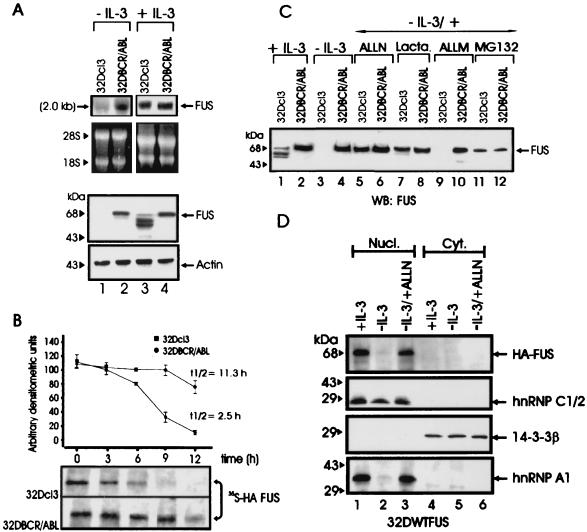
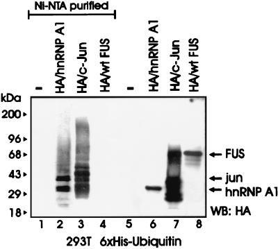
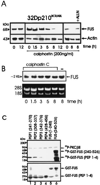
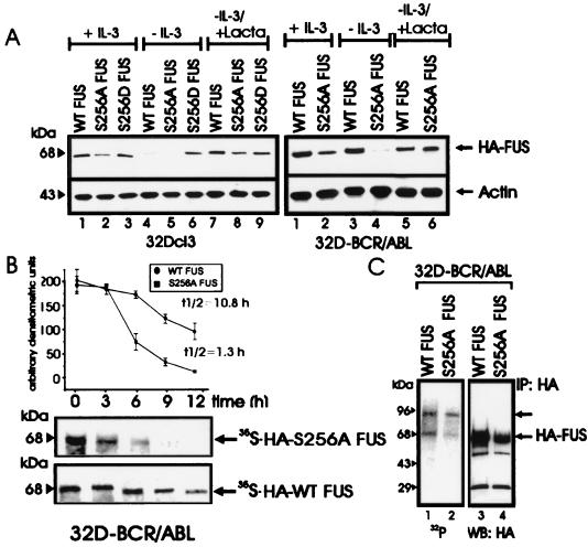
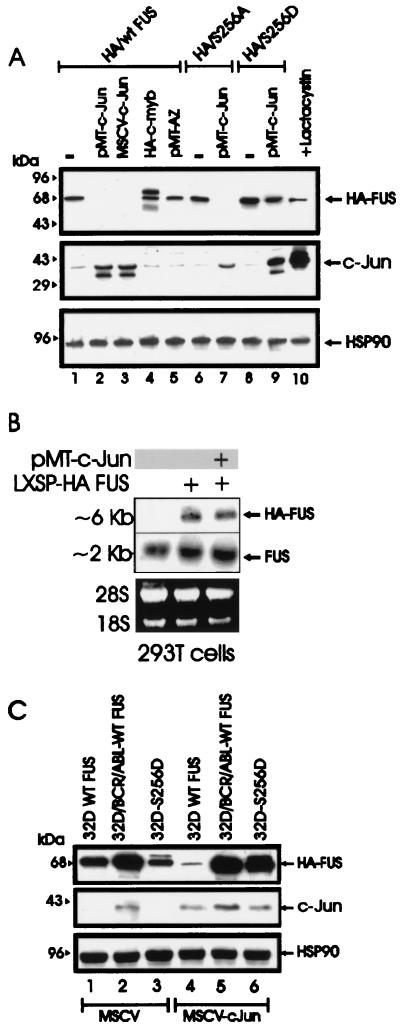
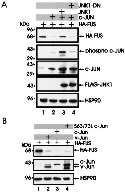
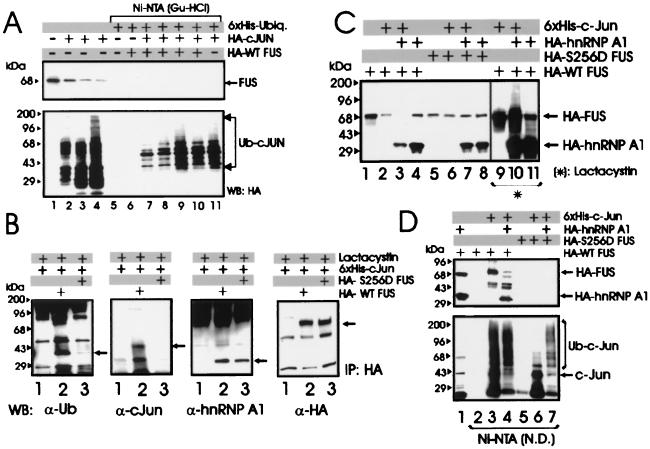
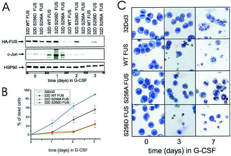
The DNA binding activity of FUS (also known as TLS), a nuclear pro-oncogene involved in multiple translocations, is regulated by BCR-ABL in a protein kinase CbetaII (PKCbetaII)-dependent manner. We show here that in normal myeloid progenitor cells FUS, although not visibly ubiquitinated, undergoes proteasome-dependent degradation, whereas in BCR-ABL-expressing cells, degradation is suppressed by PKCbetaII phosphorylation. Replacement of serine 256 with the phosphomimetic aspartic acid prevents proteasome-dependent proteolysis of FUS, while the serine-256-to-alanine FUS mutant is unstable and susceptible to degradation. Ectopic expression of the phosphomimetic S256D FUS mutant in granulocyte colony-stimulating factor-treated 32Dcl3 cells induces massive apoptosis and inhibits the differentiation of the cells escaping cell death, while the degradation-prone S256A mutant has no effect on either survival or differentiation. FUS proteolysis is induced by c-Jun, is suppressed by BCR-ABL or Jun kinase 1, and does not depend on c-Jun transactivation potential, ubiquitination, or its interaction with Jun kinase 1. In addition, c-Jun-induced FUS proteasome-dependent degradation is enhanced by heterogeneous nuclear ribonucleoprotein (hnRNP) A1 and depends on the formation of a FUS-Jun-hnRNP A1-containing complex and on lack of PKCbetaII phosphorylation at serine 256 but not on FUS ubiquitination. Thus, novel mechanisms appear to be involved in the degradation of FUS in normal myeloid cells; moreover, the ability of the BCR-ABL oncoprotein to suppress FUS degradation by the induction of posttranslational modifications might contribute to the phenotype of BCR-ABL-expressing hematopoietic cells.


Figures

FIG. 1
FIG. 1
FUS expression, stability, and proteasome-dependent degradation. (A) Northern (top panel) and Western blot (bottom panel) analysis of FUS expression in parental and BCR-ABL-expressing 32Dcl3 cells in the presence of IL-3 (lanes 3 and 4) or after IL-3-deprivation for 8 h (lanes 1 and 2). rRNA and actin levels were used as controls for RNA and protein loading, respectively. (B) Stability of FUS in IL-3-cultured parental and BCR-ABL-expressing 32Dcl3 cells ectopically expressing the HA-tagged WT FUS. The turnover of FUS was monitored by a pulse-chase assay and quantitated by densitometry. Each point on the graph represents the mean and standard deviation of the relative amount of FUS during the chase period; t1/2 values were calculated using the formula reported in Materials and Methods. The graph is representative of three independent experiments with similar results. (C) Effect of proteasome (lactacystin [Lacta.] and MG132), calpain and proteasome (ALLN), and calpain (ALLM) inhibitors on endogenous FUS expression in IL-3-deprived (8 h), parental, and BCR-ABL-expressing cells. FUS was detected using antiserum raised against the N-terminal region (amino acids 1 to 240) of FUS. (D) Effect of ALLN on nuclear and cytoplasmic levels of HA-tagged FUS. Western blots show expression of HA-tagged FUS, hnRNP C1/2, 14-3-3β, and hnRNP A1 in nuclear and cytoplasmic fractions of 32Dcl3 cells (cultured in IL-3, IL-3 starved [for 8 h], or IL-3 starved in the presence of ALLN). Expression of hnRNP C1/2 was used as nuclear marker, while that of 14-3-3β was used as cytoplasmic marker. The anti-hnRNP C1/2 (4F4) and the anti-hnRNP A1 (9H10) monoclonal antibodies were a kind gift of G. Dreyfuss (Howard Hughes Medical Institute, University of Pennsylvania School of Medicine, Philadelphia, Pa.), while HA-tagged FUS and 14-3-3β were detected using monoclonal anti-HA and anti 14-3-3β (Santa Cruz Biotechnology, Santa Cruz, Calif.) antibodies. Data are representative of three different experiments with similar results.
FIG. 2
FIG. 2
FUS proteolysis does not require its ubiquitination. An in vivo ubiquitination assay of c-Jun, hnRNP A1, and FUS was performed. Shown is a Western blot with an anti-HA antibody on nickel chromatography-purified (Ni-NTA resin under denaturing conditions) His6-ubiquitinated proteins (lanes 1 to 4) or on total-cell lysates (lanes 5 to 8) from 293T cells transfected with His6-tagged ubiquitin (lanes 1 to 8) along with HA-hnRNP A1 (lanes 2 and 6), HA-c-Jun (lanes 3 and 7), or HA-FUS (lanes 4 and 8). Data are representative of three independent experiments with similar results.
FIG. 3
FIG. 3
PKC-dependent FUS expression and identification of FUS PKCβII phosphorylation sites. (A) Western blot of FUS expression in BCR-ABL-expressing 32Dcl3 cells untreated or treated for the indicated times with calphostin C, alone or in the presence of the proteasome inhibitor ALLN. Actin expression was used as a control. (B) Northern blot of FUS expression in calphostin C-treated (1.5 to 8 h) BCR-ABL-expressing 32Dcl3 cells. (C) In vitro kinase assay (top panel) with recombinant PKCβII as the active kinase and GST-FUS fusion proteins as the substrate. The N-terminal (amino acids 1 to 240) (lane 5) and the C-terminal (amino acids 240 to 526) (lane 6) regions of FUS and four different FUS peptides (lanes 1 to 4) containing the putative PKC phosphorylation sites fused to GST are visible after Coomassie staining of the SDS-PAGE-fractionated kinase reaction products (bottom panel). Data are representative of three different experiments with similar results.
FIG. 4
FIG. 4
Role of serine 256 in expression, stability, proteasome-mediated degradation, and in vivo phosphorylation of FUS. (A) HA-FUS levels in parental (left panel) and BCR-ABL-expressing (right panel) 32Dcl3 cells stably expressing WT FUS or the S256A or S256D mutant. Cells were maintained in the presence of IL-3 or were IL-3 deprived (for 8 h) in the presence or absence of the proteasome inhibitor lactacystin (Lacta). Actin levels were used as a control. (B) Stability of newly synthesized WT FUS and S256A FUS mutant in IL-3- and serum-deprived (8 h) 32DBCR-ABL cells. Each point of the graph represents the mean and standard deviation of the relative amounts of WT FUS and S256A FUS during the chase period. Values on the graph are representatives of three independent experiments. (C) FUS phosphorylation in in vivo 32P-labeled WT FUS- and S256A-expressing 32DBCR-ABL cells (lanes 1 and 2), and amount of immunoprecipitated (IP) WT and S256A FUS (lanes 3 and 4). Data are representative of three experiments with similar results.
FIG. 5
FIG. 5
c-Jun requirement for FUS proteasome-dependent degradation. (A) HA-FUS expression (top panel) in transiently transfected 293T cells (lane 1), cotransfected with two different c-Jun expression plasmids (pMT-c-Jun [pMT35] and MSCV-c-Jun) (lanes 2 and 3, respectively), with pMT HA-c-Myb (lane 4), or with a CMV-based vector containing the full-length antizyme (AZ) cDNA (lane 5). Expression of S256A FUS and S256D FUS mutants upon transient transfection in 293T cells (lanes 6 and 8) or cotransfection with pMT-c-Jun (lanes 7 and 9) is also shown. 293T cells were also cotransfected with WT HA FUS and pMT-c-Jun and treated for 8 h with the proteasome inhibitor lactacystin before being subjected to lysis (lane 10). c-Jun (middle panel) and HSP90 (bottom panel) expression were monitored as controls. (B) Ectopic (top panel) and endogenous (middle panel) FUS mRNA expression in parental 293T cells (lane 1) or in cells transfected with the LXSP HA-FUS plasmid alone (lane 2) or cotransfected with pMT-c-Jun (lane 3). Ethidium bromide staining of rRNA is shown as a control for equal loading (bottom panel). (C) Effect of transient expression of c-Jun (middle panel) on HA-FUS levels (top panel) in retrovirus-infected parental or BCR-ABL-expressing 32Dcl3 cells constitutively expressing HA-tagged WT FUS or S256D FUS. HSP90 levels (bottom panel) were monitored as a control of equal loading.
FIG. 6
FIG. 6
Effect of JNK1, v-Jun, and S63/73L c-Jun mutant on FUS expression. (A) HA-FUS expression (top row) in lysates of 293T cells transfected with WT HA-FUS alone (lanes 1 to 4) or with c-Jun (lanes 2 to 4), FLAG-tagged WT JNK1 (lane 3), or a FLAG-tagged dominant-negative JNK1 (lane 4). Phospho-c-Jun levels (second row) were detected using an anti-phospho-Jun antibody (Santa Cruz Biotechnology, Inc.). Total c-Jun levels (third row) were monitored using a mix (1:1) of polyclonal anti-c-Jun antibodies (Santa Cruz Biotechnology and Oncogene Sciences). Levels of exogenous JNK1 (fourth row) were detected using an anti-FLAG antibody (Sigma). (B) HA-FUS expression (top row) in lysates of 293T cells transfected with WT HA-FUS alone (lanes 1 to 4) or with v-Jun (lane 2), c-Jun (lane 3), or the transactivation-deficient S63/73L c-Jun mutant (lane 4). c-Jun and v-Jun levels (second row) were detected using the polyclonal anti-c-Jun antibodies mix described for panel A. HSP90 levels were monitored as control for equal loading.
FIG. 7
FIG. 7
Role of ubiquitination and of hnRNP A1 expression in c-Jun-induced FUS degradation. (A) Endogenous FUS levels in 293T cells that were not transfected (lane 1) or transfected with HA-c-Jun and harvested 16, 24, and 36 h after transfection (lanes 2 to 4), and an in vivo ubiquitination assay in 293T cells cotransfected with His6-tagged ubiquitin, HA-c-Jun, and HA-WT FUS expression plasmids and harvested 16, 24, 36, and 48 h after transfection (lanes 7 to 10). Western blot with anti-FUS (upper panel) or anti-HA (lower panel) antibody on total-cell lysate (lanes 1 to 4) or Ni-NTA-purified proteins (lanes 5 to 11) from nontransfected cells (lane 1), cells transfected with HA-c-Jun only (lanes 2 to 4), cells transfected with His6-tagged ubiquitin (lanes 5 to 11), plus HA-WT FUS (lane 6), cotransfected with HA-WT FUS and HA-c-Jun (lanes 7–10), or plus HA-c-Jun only (lane 11). (B) Identification of WT and S256D FUS-associated proteins in lactacystin-treated 293T cells. Shown are Western blots with anti-ubiquitin (first panel), anti-c-Jun (second panel), anti-hnRNP A1 (third panel), and anti-HA (fourth panel) antibody on HA-immunoprecipitates (IP) from lysates of 293T cells transfected with His6-tagged c-Jun (lanes 1 to 3) alone (lane 1) or with HA-tagged WT FUS (lane 2) or S256D FUS (lane 3) and treated for 8 h with 10 μM lactacystin before being subjected to lysis. (C) Effect of hnRNP A1 on c-Jun-induced degradation of FUS. Shown are Western blots with an anti-HA antibody on total-cell lysates from 293T cells transfected (1:1:1 molar ratio) with HA-tagged WT FUS (lanes 1 to 4 and 9 to 11) or S256D FUS mutant (lanes 5 to 8), plus His6-tagged c-Jun (lanes 2, 3, 6, 7, 9, and 10) or HA-tagged hnRNP A1 (lanes 3, 4, 7, 8, 10, and 11) left untreated (lanes 2 to 8) or treated (lanes 9 to 11) with the proteasome inhibitor lactacystin. c-Jun and HSP90 levels were measured as internal controls of transfection efficiency and equal loading (data not shown). (D) Ni-NTA pull-down assay performed with the same lysate (1.5 mg) used in the experiment in Fig. 6C. Shown are Western blots with an anti-HA (upper panel) or anti-c-Jun (lower panel) antibody on total-cell lysates (lane 1) or on nondenatured (N.D.) Ni-NTA-purified fractions (lanes 2 to 7) from 293T cells transfected with HA-tagged WT FUS (lanes 1 to 4) or S256D FUS (lanes 5 to 7) along with His6-tagged c-Jun (lanes 3, 4, 6, and 7) or HA-hnRNP A1 (lanes 1, 4, and 7). Data are representative of three experiments with similar results.
FIG. 8
FIG. 8
Effect of Ser 256 FUS mutant expression on G-CSF-induced differentiation of 32Dcl3 cells. (A) Kinetics of FUS, c-Jun, and HSP90 expression (Western blotting) in a representative (of three for each transfectant) clone of WT FUS, S256D FUS, or S256A FUS-expressing 32Dcl3 cells cultured in the presence of G-CSF for 0, 1, 2, or 3 days. (B) Effect of G-CSF on the viability of parental and derivative cell lines ectopically expressing WT FUS, S256A FUS, or S256D FUS. Each point represents the average of three independent experiments and standard deviation. The percentage cell death was determined by trypan blue exclusion. (C) G-CSF-induced differentiation of parental and representative (of three for each transfectant) 32Dcl3-derived cell lines. Representative micrographs of May-Grunwald-Giemsa-stained cytospins are shown.

Similar articles

Cited by

References

    1. Bercovich Z, Rosenberg-Hasson Y, Ciechanover A, Kahana C. Degradation of ornithine decarboxylase in reticulocyte lysate is ATP-dependent but ubiquitin-independent. J Biol Chem. 1989;264:15949–15952. - PubMed
    1. Bouvet P, Diaz J J, Kindbeiter K, Madjar J J, Amalric F. Nucleolin interacts with several ribosomal proteins through its RGG domain. J Biol Chem. 1998;273:19025–19029. - PubMed
    1. Bureau J P, Henry L, Baz A, Scherrer K, Chateau M T. Prosome (proteasome) changes during differentiation are related to the type of inducer. Mol Biol Rep. 1997;24:57–62. - PubMed
    1. Campanero M R, Flemington E K. Regulation of E2F through ubiquitin-proteasome-dependent degradation: stabilization by the pRB tumor suppressor protein. Proc Natl Acad Sci USA. 1997;94:2221–2226. - PMC - PubMed
    1. Cartegni L, Maconi M, Morandi E, Cobianchi F, Riva S, Biamonti G. hnRNP A1 selectively interacts through its Gly-rich domain with different RNA-binding proteins. J Mol Biol. 1996;259:337–348. - PubMed

Publication types

MeSH terms

Substances