Comparative Study
. 2000 Aug;66(8):3234-40.
doi: 10.1128/AEM.66.8.3234-3240.2000.

Comparative experiments to examine the effects of heating on vegetative cells and spores of Clostridium perfringens isolates carrying plasmid genes versus chromosomal enterotoxin genes

Affiliations
Comparative Study

Comparative experiments to examine the effects of heating on vegetative cells and spores of Clostridium perfringens isolates carrying plasmid genes versus chromosomal enterotoxin genes

M R Sarker et al. Appl Environ Microbiol. 2000 Aug.

Retraction in

Abstract

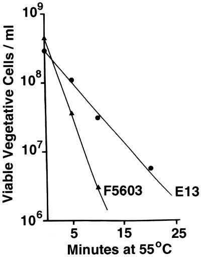
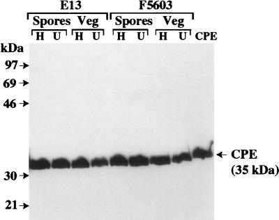
Clostridium perfringens enterotoxin (CPE) is an important virulence factor for both C. perfringens type A food poisoning and several non-food-borne human gastrointestinal diseases. Recent studies have indicated that C. perfringens isolates associated with food poisoning carry a chromosomal cpe gene, while non-food-borne human gastrointestinal disease isolates carry a plasmid cpe gene. However, no explanation has been provided for the strong associations between certain cpe genotypes and particular CPE-associated diseases. Since C. perfringens food poisoning usually involves cooked meat products, we hypothesized that chromosomal cpe isolates are so strongly associated with food poisoning because (i) they are more heat resistant than plasmid cpe isolates, (ii) heating induces loss of the cpe plasmid, or (iii) heating induces migration of the plasmid cpe gene to the chromosome. When we tested these hypotheses, vegetative cells of chromosomal cpe isolates were found to exhibit, on average approximately twofold-higher decimal reduction values (D values) at 55 degrees C than vegetative cells of plasmid cpe isolates exhibited. Furthermore, the spores of chromosomal cpe isolates had, on average, approximately 60-fold-higher D values at 100 degrees C than the spores of plasmid cpe isolates had. Southern hybridization and CPE Western blot analyses demonstrated that all survivors of heating retained their cpe gene in its original plasmid or chromosomal location and could still express CPE. These results suggest that chromosomal cpe isolates are strongly associated with food poisoning, at least in part, because their cells and spores possess a high degree of heat resistance, which should enhance their survival in incompletely cooked or inadequately warmed foods.


Figures

FIG. 1
FIG. 1
Thermal death curves for vegetative cells of strain E13, which carries a chromosomal cpe gene, and strain F5603, which carries a plasmid cpe gene. Vegetative (FTG medium) cultures of E13 and F5603 were heated at 55°C for specified times, and the number of viable bacteria per milliliter of each heated culture was then determined (see Materials and Methods). The results are the results of representative experiments; these results were highly reproducible (data not shown).
FIG. 2
FIG. 2
Thermal death curves for sporulating cultures of strain E13, which carries a chromosomal cpe gene, and strain F5603, which carries a plasmid cpe gene. Heat-shocked DS medium cultures of E13 and F5603 were heated at 100°C for specified times, and the number of viable spores per milliliter of each culture was determined (see Materials and Methods). The results are the results of representative experiments; these results were highly reproducible (data not shown).
FIG. 3
FIG. 3
RFLP analysis of NruI-digested total DNA isolated from strain E13, which carries a chromosomal cpe gene, and strain F5603, which carries a plasmid cpe gene. Southern blots were probed with a 639-bp DIG-labeled cpe-specific fragment. The representative results shown for heated (lanes H) and unheated (lanes U) FTG medium (Veg) or DS medium (Spores) cultures of E13 are results obtained for survivors of heating for 20 min at 55°C and heating for 60 min at 100°C, respectively. The representative results shown for heated (lanes H) and unheated (lanes U) FTG medium (Veg) or DS medium (Spores) cultures of F5603 are results obtained for survivors of heating for 10 min at 55°C and heating for 1 min at 100°C, respectively. The molecular sizes (in kilobase pairs [Kb]) of the DNA markers are indicated in the center.
FIG. 4
FIG. 4
PFGE-Southern hybridization analysis of undigested genomic DNA from strain E13, which carries a chromosomal cpe gene, and strain F5603, which carries a plasmid cpe gene. The blot was probed with a 639-bp DIG-labeled cpe-specific fragment. The representative results shown for heated (lanes H) and unheated (lanes U) FTG medium (Veg) or DS medium (Spores) cultures of E13 are results obtained for survivors of heating for 20 min at 55°C and heating for 60 min at 100°C, respectively. The representative results shown for heated (lanes H) and unheated (lanes U) FTG medium (Veg) or DS medium (Spores) cultures of F5603 are results obtained for survivors of heating for 10 min at 55°C and heating for 1 min at 100°C, respectively. The molecular sizes (in kilobase pairs [kb]) of the DNA markers are indicated between the two blots. The cpe probe reactive material appearing at the top of all gel lanes was found previously with both chromosomal and plasmid cpe isolates subjected to PFGE-Southern hybridization analyses (–11); this material apparently was cpe-containing DNA that was trapped in gel wells.
FIG. 5
FIG. 5
CPE Western immunoblot analysis of sporulating cultures of unheated and heated C. perfringens isolates. The representative results shown are the results obtained for DS medium lysates of strain E13 (which carries a chromosomal cpe gene) and strain F5603 (which carries a plasmid cpe gene), which were analyzed for CPE expression by an immunoblot analysis performed with CPE antiserum. The representative results shown for heated (lanes H) and unheated (lanes U) FTG medium (Veg) and DS medium (Spores) cultures of E13 are results obtained for survivors of heating for 20 min at 55°C and heating for 60 min at 100°C, respectively. The representative results shown for heated (lanes H) and unheated (lanes U) FTG medium (Veg) or DS medium (Spores) cultures of F5603 are results obtained for survivors of heating for 10 min at 55°C and heating for 1 min at 100°C, respectively. The molecular sizes of the protein markers (in kilodaltons) are indicated on the left.

References

    1. Ando Y, Tsuzuki T, Sunagawa H, Oka S. Heat resistance, spore germination, and enterotoxigenicity of Clostridium perfringens. Microbiol Immunol. 1985;29:317–326. - PubMed
    1. Bean N H, Goulding J S, Lao C, Angulo F J. Surveillance for foodborne-disease outbreaks—United States, 1988–1982. Morb Mortal Wkly Rep. 1996;45:1–54. - PubMed
    1. Bean N H, Griffen P M. Foodborne disease outbreaks in the United States, 1973–1987; pathogens, vehicles and trends. J Food Prot. 1990;53:804–817. - PubMed
    1. Billington S J, Wieckowski E U, Sarker M R, Bueschel D, Songer J G, McClane B A. Clostridium perfringens type E animal enteritis isolates with highly conserved, silent enterotoxin sequences. Infect Immun. 1998;66:4531–4536. - PMC - PubMed
    1. Brynestad S, Granum P E. Evidence that Tn5565, which includes the enterotoxin gene in Clostridium perfringens, can have a circular form which may be a transposition intermediate. FEMS Microbiol Lett. 1999;170:281–286. - PubMed

Publication types

MeSH terms

LinkOut - more resources