

Immunomagnetic purification of Colletotrichum lindemuthianum appressoria
- PMID: 10919807
- PMCID: PMC92171
- DOI: 10.1128/AEM.66.8.3464-3467.2000
Immunomagnetic purification of Colletotrichum lindemuthianum appressoria
Abstract
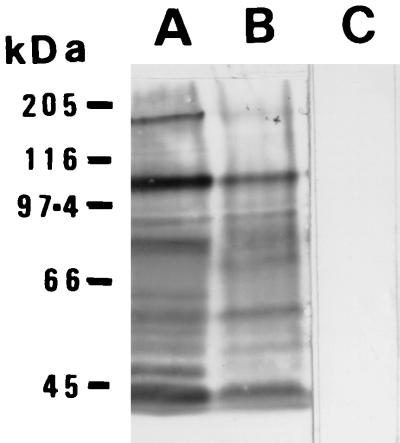
We developed a method to purify appressoria of the bean anthracnose fungus Colletotrichum lindemuthianum for biochemical analysis of the cell surface and to compare appressoria with other fungal structures. We used immunomagnetic separation after incubation of infected bean leaf homogenates with a monoclonal antibody that binds strongly to the appressoria. Preparations with a purity of >90% could be obtained. Examination of the purified appressoria by transmission electron microscopy showed that most had lost their cytoplasm. However, the plasma membrane was retained, suggesting that there is some form of attachment of this membrane to the cell wall. The purified appressoria can be used for studies of their cell surface, and we have shown that there are clear differences in the glycoprotein constituents of cell walls of appressoria compared with mycelium.
Figures


References
-
- Cantrill L C, Deverall B J. Isolation of haustoria from wheat leaves infected by the leaf rust fungus. Physiol Mol Plant Pathol. 1993;42:337–343.
-
- Clark J S C, Spencer-Phillips P T N. Isolation of endophytic mycelia by enzymic maceration of Peronospora-infected leaves. Mycol Res. 1990;94:283–287.
-
- Gil F, Gay J L. Ultrastructural and physiological properties of the host interfacial components of haustoria of Erysiphe pisi in vivo and in vitro. Physiol Plant Pathol. 1977;10:1–12.
-
- Hahn M, Mendgen K. Isolation by ConA binding of haustoria from different rust fungi and comparison of their surface qualities. Protoplasma. 1992;170:95–103.
-
- Hansel T T, Pound J D, Pilling D, Kitas G D, Salmon M, Gentle T A, Lee S S, Thompson R A. Purification of human blood eosinophils by negative selection using immunomagnetic beads. J Immunol Methods. 1989;122:97–103. - PubMed
Publication types
MeSH terms
Substances
LinkOut - more resources
Full Text Sources
