

Analysis of gyrB and toxR gene sequences of Vibrio hollisae and development of gyrB- and toxR-targeted PCR methods for isolation of V. hollisae from the environment and its identification
- PMID: 10919814
- PMCID: PMC92178
- DOI: 10.1128/AEM.66.8.3506-3514.2000
Analysis of gyrB and toxR gene sequences of Vibrio hollisae and development of gyrB- and toxR-targeted PCR methods for isolation of V. hollisae from the environment and its identification
Abstract
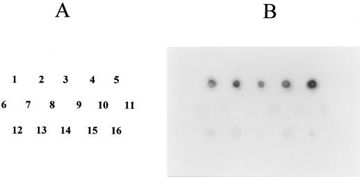
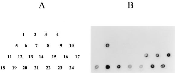
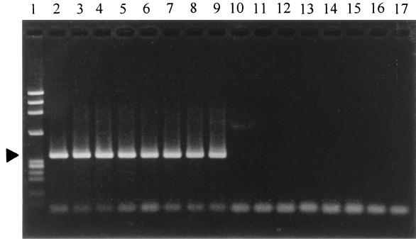
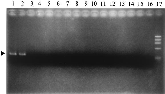
Isolation of Vibrio hollisae strains, particularly from the environment, is rare. This may be due, in part, to the difficulty encountered when using conventional biochemical tests to identify the microorganism. In this study, we evaluated whether two particular genes may be useful for the identification of V. hollisae. The two genes are presumed to be conserved among the bacterial species (gyrB) or among the species of the genus Vibrio (toxR). A portion of the gyrB sequence of V. hollisae was cloned by PCR using a set of degenerate primers. The sequence showed 80% identity with the corresponding Vibrio parahaemolyticus gyrB sequence. The toxR gene of V. hollisae was cloned utilizing a htpG gene probe derived from the V. parahaemolyticus htpG gene, which is known to be linked to the toxR gene in V. hollisae. The coding sequence of the cloned V. hollisae toxR gene had 59% identity with the V. parahaemolyticus toxR coding sequence. The results of DNA colony hybridization tests using the DNA probes derived from the two genes of V. hollisae indicated that these gene sequences could be utilized for differentiation of V. hollisae from other Vibrio species and from microorganisms found in marine fish. PCR methods targeting the two gene sequences were established. Both PCR methods were shown to specifically detect the respective target sequences of V. hollisae but not other organisms. A strain of V. hollisae added at a concentration of 1 to 10(2) CFU/ml to alkaline peptone water containing a seafood sample could be detected by a 4-h enrichment incubation in alkaline peptone water at 37 degrees C followed by quick DNA extraction with an extraction kit and 35-cycle PCR specific for the V. hollisae toxR gene. We conclude that screening of seafood samples by this 35-cycle, V. hollisae toxR-specific PCR, followed by isolation on a differential medium and identification by the above htpG- and toxR-targeted PCR methods, can be useful for isolation from the environment and identification of V. hollisae.
Figures
References
-
- Abbott S L, Janda J M. Severe gastroenteritis associated with Vibrio hollisae infection: report of two cases and review. Clin Infect Dis. 1994;18:310–312. - PubMed
-
- Casadaban M J, Cohen S N. Analysis of gene control signals by DNA fusion and cloning in Escherichia coli. J Mol Biol. 1980;138:179–207. - PubMed
-
- Feinberg A P, Vogelstein B. A technique for radiolabelling DNA restriction endonuclease fragments to high specific activity. Anal Biochem. 1983;132:6–13. - PubMed
-
- Gras-Rouzet S, Donnio P Y, Juguet F, Plessis P, Minet J, Avril J L. First European case of gastroenteritis and bacteremia due to Vibrio hollisae. Eur J Clin Microbiol Infect Dis. 1996;15:864–866. - PubMed
Publication types
MeSH terms
Substances
Associated data
- Actions
- Actions
LinkOut - more resources
Full Text Sources
Other Literature Sources
Molecular Biology Databases
