. 2000 Aug;66(8):3586-91.
doi: 10.1128/AEM.66.8.3586-3591.2000.

Real-time measurements of the interaction between single cells of Listeria monocytogenes and nisin on a solid surface

Affiliations

Real-time measurements of the interaction between single cells of Listeria monocytogenes and nisin on a solid surface

B B Budde et al. Appl Environ Microbiol. 2000 Aug.

Abstract

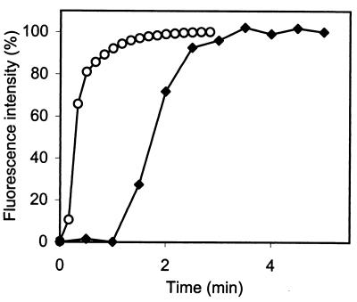
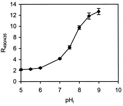
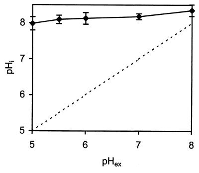
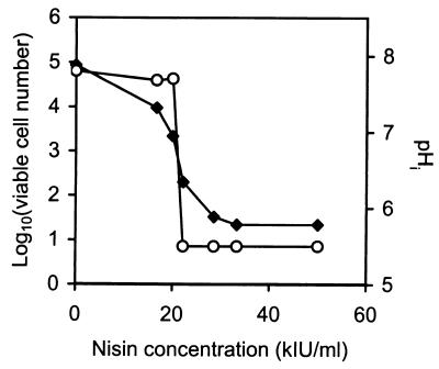
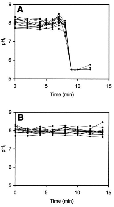
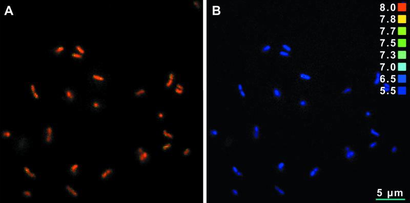
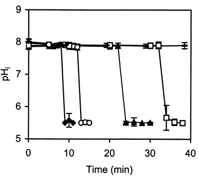
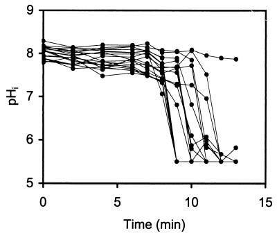
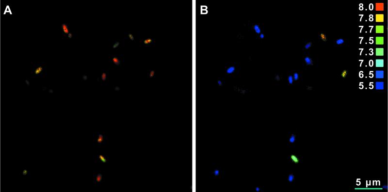
A method to obtain real-time measurements of the interactions between nisin and single cells of Listeria monocytogenes on a solid surface was developed. This method was based on fluorescence ratio-imaging microscopy and measurements of changes in the intracellular pH (pH(i)) of carboxyfluorescein succinimidyl ester-stained cells during exposure to nisin. Immobilized cells were placed in a chamber mounted on a microscope and attached to a high-precision peristaltic pump which allowed rapid changes in the nisin concentration. In the absence of nisin, the pH(i) of L. monocytogenes was almost constant (approximately pH 8.0) and independent of the external pH in the pH range from 5.0 to 9.0. In the presence of nisin, dissipation of the pH gradient (DeltapH) was observed, and this dissipation was both time and nisin concentration dependent. The dissipation of DeltapH resulted in cell death, as determined by the number of CFU. In the model system which we used the immobilized cells were significantly more resistant to nisin than the planktonic cells. The kinetics of DeltapH dissipation for single cells revealed a variable lag phase depending on the nisin concentration, which was followed by a very rapid decrease in pH(i) within 1 to 2 min. The differences in nisin sensitivity between single cells in a L. monocytogenes population were insignificant for cells grown to the stationary phase in a liquid laboratory substrate, but differences were observed for cells grown on an agar medium under similar conditions, which resulted in some cells having increased resistance to nisin.


Figures

FIG. 1
FIG. 1
Increase in fluorescence intensity (wavelength, 435 nm) from time zero of perfusion through the chamber until a constant fluorescence intensity was reached when we used 5 mM fluorescein in PBS at pH 7.4 and a flow rate of 9 μl/s without the filter membrane (○) and with the filter membrane inserted in the chamber (⧫). Measurements were recorded in the center of the chamber.
FIG. 2
FIG. 2
Relationship between R490/435 of the individual cells of L. monocytogenes 4140 and pHi. The pHi was equilibrated to pHex by incubating preparations with 63% (vol/vol) ethanol. The ratio values are averages based on 40 single cells. The error bars indicate standard deviations.
FIG. 3
FIG. 3
pHi values of energized L. monocytogenes 4140 cells at various pHex values. The buffer used was 50 mM potassium phosphate (pH 5.0 to 8.0). The dashed line is the line for the equation pHi = pHex. The pHi values are averages based on 45 single cells. The error bars indicate standard deviations.
FIG. 4
FIG. 4
Viable cell counts (⧫) and pHi values (○) for L. monocytogenes 4140 when it was perfused with different concentrations of nisin for 12 min at pH 5.5. The cell counts were determined based on detachment of the immobilized cells from the filter after whirl mixing in peptone saline water, plating of appropriate dilutions onto BHI agar plates, and subsequent incubation at 37°C. The ratio values are averages based on 13 to 15 single cells. The detection limit was 101 cells/ml.
FIG. 5
FIG. 5
pHi values of single immobilized L. monocytogenes 4140 cells from an 18-h stationary culture as a function of time following exposure to nisin (50 kIU/ml) at pH 5.5 (A) and potassium phosphate buffer containing 10 mM glucose (pH 5.5) (B). The results for 15 individual cells are shown.
FIG. 6
FIG. 6
Ratio images of immobilized L. monocytogenes 4140 cells at pHex 5.5, showing the pHi of each single cell before exposure to nisin (A) and after 12 min of exposure to nisin (50 kIU/ml) at pH 5.5 (B). A color-coded pH scale is shown on the right. Ratio images were saved as TIF files, and Adobe Photoshop 5.5 was used for contrast enhancement.
FIG. 7
FIG. 7
Influence of nisin concentration (⧫, 50 kIU/ml; ○, 25 kIU/ml; ▴, 12.5 kIU/ml; □, 8.3 kIU/ml; –, control without nisin) on the time to reach dissipation of ΔpH in L. monocytogenes 4140. The pHi values are averages based on 15 single cells. The error bars indicate standard deviations.
FIG. 8
FIG. 8
pHi values of 20 single cells of L. monocytogenes 4140 from colonies grown for 20 to 22 h on an agar plate at 37°C as a function of the time of exposure to nisin (50 kIU/ml) at pH 5.5.
FIG. 9
FIG. 9
Ratio images of immobilized L. monocytogenes 4140 cells originating from colonies grown on an agar plate. The pHi of each cell is shown at pHex 5.5 before exposure to nisin (A) and after 12 min of exposure to nisin (50 kIU/ml) at pH 5.5 (B). A color-coded pH scale is shown on the right. Ratio images were saved as TIF files, and Adobe Photoshop 5.5 was used for contrast enhancement.

References

    1. Abee T, Kröckel L, Hill C. Bacteriocins: modes of action and potentials in food preservation and control of food poisoning. Int J Food Microbiol. 1995;28:169–185. - PubMed
    1. Blom H, Katla T, Hagen B F, Axelsson L. A model assay to demonstrate how intrinsic factors affect diffusion of bacteriocins. Int J Food Microbiol. 1997;38:103–109. - PubMed
    1. Breeuwer P, Drocourt J L, Rombouts F M, Abee T. A novel method for continuous determination of the intracellular pH in bacteria with the internally conjugated fluorescent probe 5 (and 6-)-carboxyfluorescein succinimidyl ester. Appl Environ Microbiol. 1996;62:178–183. - PMC - PubMed
    1. Bruno M E, Kaiser A, Montville T J. Depletion of proton motive force by nisin in Listeria monocytogenes cells. Appl Environ Microbiol. 1992;58:2255–2259. - PMC - PubMed
    1. Correa O S, Rivas E A B A J. Cellular envelopes and tolerance to acid pH in Mesorhizobium loti. Curr Microbiol. 1999;38:329–334. - PubMed

Publication types

MeSH terms

LinkOut - more resources