. 2000 Aug;66(8):3592-602.
doi: 10.1128/AEM.66.8.3592-3602.2000.

Community structure, cellular rRNA content, and activity of sulfate-reducing bacteria in marine arctic sediments

Affiliations

Community structure, cellular rRNA content, and activity of sulfate-reducing bacteria in marine arctic sediments

K Ravenschlag et al. Appl Environ Microbiol. 2000 Aug.

Abstract

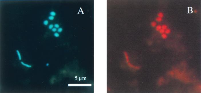
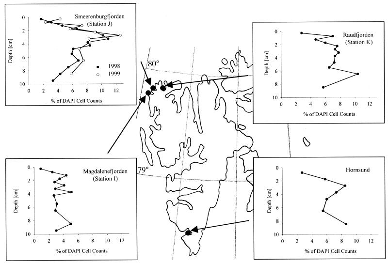
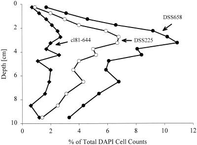
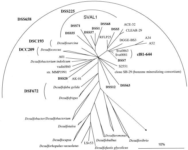
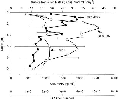
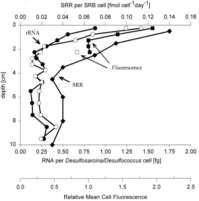
The community structure of sulfate-reducing bacteria (SRB) of a marine Arctic sediment (Smeerenburgfjorden, Svalbard) was characterized by both fluorescence in situ hybridization (FISH) and rRNA slot blot hybridization by using group- and genus-specific 16S rRNA-targeted oligonucleotide probes. The SRB community was dominated by members of the Desulfosarcina-Desulfococcus group. This group accounted for up to 73% of the SRB detected and up to 70% of the SRB rRNA detected. The predominance was shown to be a common feature for different stations along the coast of Svalbard. In a top-to-bottom approach we aimed to further resolve the composition of this large group of SRB by using probes for cultivated genera. While this approach failed, directed cloning of probe-targeted genes encoding 16S rRNA was successful and resulted in sequences which were all affiliated with the Desulfosarcina-Desulfococcus group. A group of clone sequences (group SVAL1) most closely related to Desulfosarcina variabilis (91.2% sequence similarity) was dominant and was shown to be most abundant in situ, accounting for up to 54. 8% of the total SRB detected. A comparison of the two methods used for quantification showed that FISH and rRNA slot blot hybridization gave comparable results. Furthermore, a combination of the two methods allowed us to calculate specific cellular rRNA contents with respect to localization in the sediment profile. The rRNA contents of Desulfosarcina-Desulfococcus cells were highest in the first 5 mm of the sediment (0.9 and 1.4 fg, respectively) and decreased steeply with depth, indicating that maximal metabolic activity occurred close to the surface. Based on SRB cell numbers, cellular sulfate reduction rates were calculated. The rates were highest in the surface layer (0.14 fmol cell(-1) day(-1)), decreased by a factor of 3 within the first 2 cm, and were relatively constant in deeper layers.


Figures

FIG. 1
FIG. 1
Epifluorescence micrographs of bacteria in sediment samples from Smeerenburgfjorden, Svalbard, Arctic Ocean. (A) DAPI staining. (B) FISH performed with probe DSS658 specific for the Desulfosarcina-Desulfococcus group (same microscopic field).
FIG. 2
FIG. 2
Detection and quantification of the Desulfosarcina-Desulfococcus group at various stations along the coast of Svalbard by FISH (probe DSS658).
FIG. 3
FIG. 3
Depth profile for subgroups of the Desulfosarcina-Desulfococcus group as detected by FISH (Smeerenburgfjorden sediment). Symbols: ●, probe DSS658 targeting the Desulfosarcina-Desulfococcus group; ○, probe DSS225 targeting group SVAL1; ⧫, probe cl81-644 targeting previously cloned 16S rDNA sequences from the same habitat.
FIG. 4
FIG. 4
Phylogenetic tree showing the affiliations of 16S rDNA clone sequences with selected reference sequences of members of the delta subclass of the Proteobacteria. The tree was calculated by using maximum-likelihood analysis and was corrected with filters which considered only 50% conserved regions of the 16S rRNAs of members of the delta subclass of the Proteobacteria. The DSS clones, as well as clone sequences A01, SB-29, RFLP25, ACE-32, CLEAR-29, A52, A34, and DGGE-BS3, are not full-length sequences (length, 650 to 900 bp) and therefore were added to the existing tree by using a special algorithm included in the ARB software without allowing changes in the tree topology based on almost complete sequences. Different calculations of phylogenetic trees did not result in a stable branching order for some subgroups. Consequently, the phylogenetic affiliations of these subgroups are shown as multifurcations. New cloned 16S rDNA sequences are indicated by boldface type. The group consisting of clone sequences DSS1, DSS5, DSS55, DSS71, and DSS68 was designated SVAL1. Bar = 10% estimated phylogenetic divergence.
FIG. 5
FIG. 5
Depth profiles for SRB abundance, SRB rRNA concentrations, and SRRs. Numbers of SRB cells (○) and rRNA concentrations (●) were determined by adding the values for the groups targeted by probes DSS658 (Desulfosarcina-Desulfococcus group), DSR651 (Desulforhopalus spp.), DSV698 (Desulfovibrio spp.), Sval428 (Desulfotalea spp.), DSB985 (Desulfobacter spp.), 221 (Desulfobacterium spp.), and 660 (Desulfobulbus spp.); means based on the values for two cores are shown. The SRRs (□) are mean values based on the values for three cores.
FIG. 6
FIG. 6
Depth profiles for specific rRNA contents and mean cell fluorescence for DSS658 (Desulfosarcina-Desulfococcus group)-targeted cells and SRRs per SRB cell. The average cellular rRNA contents were determined by combining FISH and rRNA hybridization data (●, core A; ○, core B). The mean fluorescence of hybridized cells was quantified by laser scanning microscopy in the following way. For each depth a mean cell fluorescence was calculated; the lowest mean cell fluorescence value was defined as 1, and the mean fluorescence values for cells in the other layers were expressed relative to this value (■, core A; □, core B). SRRs per SRB cell (⧫) were based on SRB abundance.

References

    1. Amann R I, Binder B J, Olson R J, Chisholm S W, Devereux R, Stahl D A. Combination of 16S rRNA-targeted oligonucleotide probes with flow cytometry for analyzing mixed microbial populations. Appl Environ Microbiol. 1990;56:1919–1925. - PMC - PubMed
    1. Amann R I, Ludwig W, Schleifer K-H. Phylogenetic identification and in situ detection of individual microbial cells without cultivation. Microbiol Rev. 1995;59:143–169. - PMC - PubMed
    1. Canfield D E, Jørgensen B B, Fossing H, Glud R, Gundersen J, Ramsing N B, Thamdrup B, Hansen J W, Nielsen L P, Hall P O J. Pathways of organic carbon oxidation in three continental margin sediments. Mar Geol. 1993;113:27–40. - PubMed
    1. Coleman M L, Hedrick D B, Lovley D R, White D C, Pye K. Reduction of Fe(III) in sediments by sulphate-reducing bacteria. Nature. 1993;361:436–438.
    1. Dannenberg S, Kroder M, Dilling W, Cypionka H. Oxidation of H2, organic compounds and inorganic sulfur compounds coupled to reduction of O2 or nitrate by sulfate-reducing bacteria. Arch Microbiol. 1992;158:93–99.

Publication types

MeSH terms

LinkOut - more resources