. 2000 Aug 15;97(17):9498-503.
doi: 10.1073/pnas.150006697.

A role for transcriptional repression of p21CIP1 by c-Myc in overcoming transforming growth factor beta -induced cell-cycle arrest

Affiliations

A role for transcriptional repression of p21CIP1 by c-Myc in overcoming transforming growth factor beta -induced cell-cycle arrest

G F Claassen et al. Proc Natl Acad Sci U S A. .

Abstract

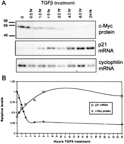
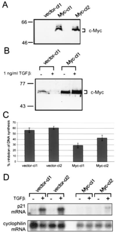
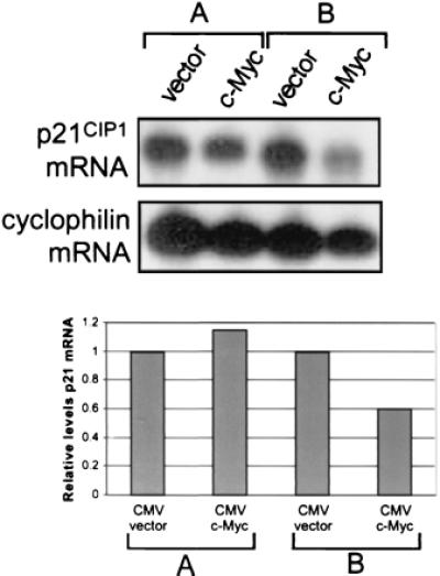
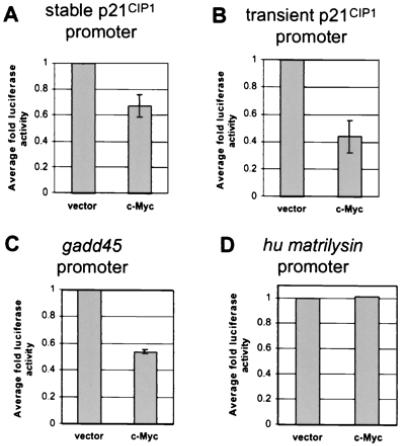
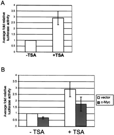
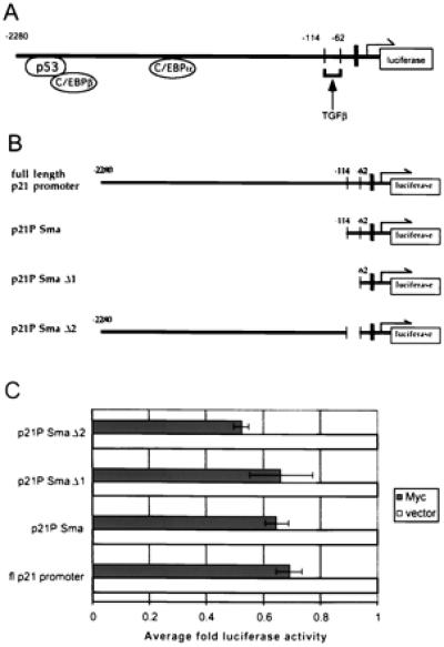
c-Myc plays a vital role in cell-cycle progression. Deregulated expression of c-Myc can overcome cell-cycle arrest in order to promote cellular proliferation. Transforming growth factor beta (TGFbeta) treatment of immortalized human keratinocyte cells inhibits cell-cycle progression and is characterized by down-regulation of c-Myc followed by up-regulation of p21(CIP1). A direct role of c-Myc in this pathway was demonstrated by the observation that ectopic expression of c-Myc overcame the cell-cycle block induced by TGFbeta treatment. The induction of p21(CIP1) transcription by TGFbeta was blocked in human keratinocyte cells stably expressing c-Myc. Furthermore, overexpression of c-Myc in NIH 3T3 cells repressed the basal levels of p21(CIP1) mRNA. Repression of p21(CIP1) transcription by c-Myc occurred at the promoter level in a region near the start site of transcriptional initiation and was independent of histone deacetylase activity. These data suggest that the down-regulation of c-Myc after TGFbeta signaling is important for subsequent regulation of p21(CIP1) and cell-cycle inhibition. Thus, repression of the cell-cycle inhibitory gene p21(CIP1) plays a role in c-Myc-dependent cell-cycle progression.


Figures

Figure 1
Figure 1
p21CIP1 up-regulation is temporally related to c-Myc down-regulation after TGFβ treatment of HaCaT cells. (A) HaCaT cells were treated with 1 ng/ml TGFβ for the time periods indicated. Samples were analyzed by Western blot with anti-Mycfl for c-Myc protein expression and by Northern blot for p21CIP1 mRNA expression. The Northern blot was subsequently probed for cyclophilin as a control for equal loading. (B) Densitometric analysis of Western and Northern blots. The highest value for each c-Myc protein and normalized p21CIP1 mRNA was set to 1.0 for graphical representation. c-Myc protein levels are indicated by diamonds, and p21CIP1 mRNA levels normalized to cyclophilin expression are represented by squares.
Figure 2
Figure 2
Expression of c-Myc protein in HaCaT lines blocks TGFβ-mediated cell-cycle arrest and subsequent up-regulation of p21CIP1 mRNA. (A) Ectopic expression of c-Myc in stable lines. Stable clonal HaCaT lines vector-cl1 (clone 1), Myc-cl1, and Myc-cl2 were analyzed for ectopic c-Myc expression by Western blot. Blots were probed with anti-av-myc 12C, which recognizes only exogenous c-Myc protein. (B) Down-regulation of c-Myc after TGFβ treatment. HaCaT clonal lines vector-cl1 and Myc-cl1 were left untreated or treated for 20 hr with 1 ng/ml TGFβ. Whole-cell lysates were analyzed by Western blot by using anti-Mycfl. (C) Inhibition of entry into S phase after TGFβ treatment. Cell-cycle inhibition by TGFβ treatment was determined by [3H]thymidine incorporation. Clonal HaCaT lines were plated in duplicate and re-fed the next day with fresh media with or without 1 ng/ml TGFβ. Cells were treated for 16 hr, then labelled with 1 mCi/ml [3H]thymidine for an additional 4 hr with or without TGFβ. [3H]thymidine incorporation was determined by scintillation counting. Inhibition of DNA synthesis is represented as the percent difference between untreated and treated cells. Error bars indicate standard deviation of the average of three independent experiments. (D) Up-regulation of p21CIP1 after TGFβ treatment is blocked by ectopic c-Myc expression. Clonal HaCaT control or c-Myc-expressing lines as indicated were left untreated or treated with 1 ng/ml TGFβ for 20 hr. PolyA+ mRNA was isolated, and 2 μg mRNA per sample was analyzed by Northern blot analysis. The blot was sequentially probed for both p21CIP1 (Top) and cyclophilin (Bottom).
Figure 3
Figure 3
p21CIP1 levels are diminished in cells overexpressing c-Myc. NIH 3T3 cells were plated at 1.5 × 10⁶ cells/100-mm dish (A) or 7 × 10⁵ cells/100-mm dish (B) and transiently transfected 24 hr later with either empty CMV vector or CMV-Myc. PolyA+ mRNA was isolated, and approximately 1 μg RNA per sample was analyzed by Northern blot analysis. The blot was sequentially probed for both p21CIP1 (Top, Upper) and cyclophilin (Top, Lower). The Northern blot was then analyzed by densitometry, and relative levels of p21CIP1 mRNA normalized by levels of cyclophilin were plotted on a bar graph, where p21CIP1 levels in vector-only-transfected cells were set to a value of one for comparison with c-Myc transfected cells (Bottom).
Figure 4
Figure 4
c-Myc-mediated transcriptional repression. (A) Repression of the p21CIP1 promoter in stable reporter lines by c-Myc. Generation of NIH 3T3 cell lines stably expressing p21CIP1-luciferase reporter constructs is described in Materials and Methods. Cells were analyzed for luciferase activity 24 hr after plating and normalized by standard protein assay. Fold luciferase activity was determined as compared to vector alone. Error bars represent standard deviation of the average of duplicate readings from four independent stable clones. (B) Repression of the p21CIP1 promoter by c-Myc. A 2.3-kb fragment of the p21CIP1 promoter linked to a luciferase reporter gene was transiently transfected into NIH 3T3 fibroblasts. Cells were cotransfected with a βgal vector for standardization and 1 μg of either empty CMV vector or c-Myc expression vector. Luciferase activity was determined 48 hr after transfection and normalized to β-gal activity. Fold luciferase activity was determined as compared to vector alone. Error bars represent standard deviation of the average of three independent experiments. (C) Repression of the gadd45 promoter by c-Myc. Reporter assays were performed as described for B, where 1 μg of either empty CMV or c-Myc expression vector was cotransfected into cells with a gadd45 luciferase reporter construct. Error bars represent standard deviation of the average of three independent experiments. (D) c-Myc expression does not affect transcription of the human matrilysin promoter. NIH 3T3 fibroblasts were transiently transfected with a reporter vector containing the human matrilysin promoter. Cells were then cotransfected with 1 μg of either empty CMV vector or c-Myc expression vector. Fold luciferase activity was determined as described for B.
Figure 5
Figure 5
TSA treatment does not affect p21CIP1 repression by c-Myc. (A) Effect of TSA on basal p21CIP1 promoter activity. NIH 3T3 p21CIP1 reporter lines infected with vector only were left untreated (−TSA) or treated with 500 ng/ml TSA overnight (+TSA). Cells were analyzed for luciferase activity 24 hr after plating and normalized by standard protein assay. Fold luciferase activity was determined as compared to untreated cells for each line. Error bars represent standard deviation of the average of duplicate readings from four independent stable clones. (B) Effect of TSA on repression of the p21CIP1 promoter by c-Myc. NIH 3T3 p21CIP1 reporter lines infected with either empty retroviral vector (white bars) or c-Myc expression vector (dark bars) were left untreated (−TSA) or treated with 500 ng/ml TSA overnight (+TSA). Cells were analyzed for luciferase activity after treatment and normalized by standard protein assay. Fold luciferase activity was determined as compared to untreated vector only lines. Error bars represent standard deviation of the average of duplicate readings for four independent clones.
Figure 6
Figure 6
Sequences immediately upstream of the transcriptional initiation site are sufficient for repression of p21CIP1 by c-Myc. (A) Diagram of the p21CIP1 promoter illustrating regions bound by some of the major regulatory proteins, as well as the TGFβ-responsive element. The TATA box, located at −45 bp relative to start of transcription, is indicated by the black bar. The arrow indicates the site of transcriptional initiation. (B) Diagrams of 5′ deletion constructs of p21CIP1 promoter. Numbers in top three diagrams indicate 5′ end of the deletion construct relative to the transcription start site. The last diagram depicts deletion p21 pSmaΔ2, in which only the region from −114 to −62, including the TGFβ-responsive element, is deleted. (C) Luciferase activity for each promoter deletion construct depicted in B was determined, and fold luciferase activity in cells cotransfected with c-Myc expression vector (dark solid bars) was determined as compared to cells cotransfected with empty CMV vector (white solid bars). Error bars represent the standard deviation of the average of three independent experiments.

References

    1. Mateyak M K, Obaya A J, Adachi S, Sedivy J M. Cell Growth Diff. 1997;8:1039–1048. - PubMed
    1. Karn J, Watson J V, Lowe A D, Green S M, Vedeckis W. Oncogene. 1989;4:773–787. - PubMed
    1. Eilers M, Schirm S, Bishop J M. EMBO J. 1991;10:133–141. - PMC - PubMed
    1. Cole M D, McMahon S B. Oncogene. 1999;18:2916–2924. - PubMed
    1. Claassen G F, Hann S R. Oncogene. 1999;18:2925–2933. - PubMed

Publication types

MeSH terms

LinkOut - more resources