. 2000 Aug 15;97(17):9585-90.
doi: 10.1073/pnas.160274797.

The human VASA gene is specifically expressed in the germ cell lineage

Affiliations

The human VASA gene is specifically expressed in the germ cell lineage

D H Castrillon et al. Proc Natl Acad Sci U S A. .

Abstract

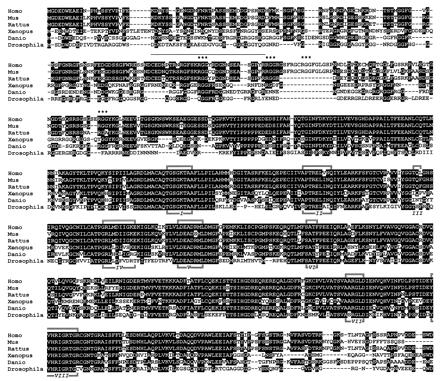
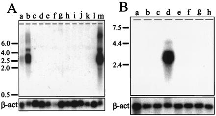
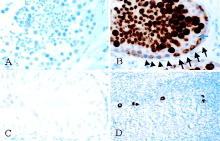
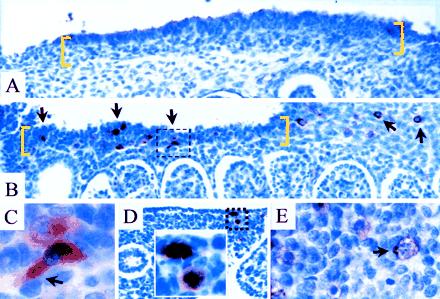
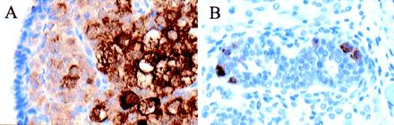
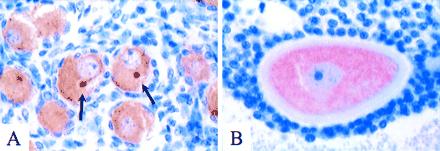
To understand the origins and function of the human germ cell lineage and to identify germ cell-specific markers we have isolated a human ortholog of the Drosophila gene vasa. The gene was mapped to human chromosome 5q (near the centromere) by radiation hybrid mapping. We show by Northern analysis of fetal and adult tissues that expression of the human VASA gene is restricted to the ovary and testis and is undetectable in somatic tissues. We generated polyclonal antibodies that bind to the VASA protein in formalin-fixed, paraffin-embedded tissue and characterized VASA protein expression in human germ cells at various stages of development. The VASA protein is cytoplasmic and expressed in migratory primordial germ cells in the region of the gonadal ridge. VASA protein is present in fetal and adult gonadal germ cells in both males and females and is most abundant in spermatocytes and mature oocytes. The gene we have isolated is thus a highly specific marker of germ cells and should be useful for studies of human germ cell determination and function.


Figures

Figure 1
Figure 1
Alignment of VASA protein sequences from human and major experimental organisms, including Mus musculus (Genbank accession no. 2500525), Rattus norvegicus (Genbank accession no. 2500526), Xenopus laevis (Genbank accession no. 2896107), Danio reio (Genbank accession no. 2558535), and Drosophila melanogaster (Genbank accession no. 1352826). Residues conserved in ≥ 50% of the sequences are highlighted in black. The eight conserved homology boxes present in all DEAD box proteins are indicated by I–VIII. * indicate the four copies of the RGG motif in the human protein; all VASA proteins contain multiple copies of this motif in their N termini. A bar overlies the region corresponding to the synthetic peptide used for antibody generation.
Figure 2
Figure 2
(A) Northern analysis of VASA in fetal tissues (lanes a, testis; b, ovary; c, brain; d, thymus; e, lung; f, liver; g, spleen; h, pancreas; i, small intestine, colon, and stomach; j, kidney; k, adrenal, and l, placenta) and adult testis (lane m). The positions of size standards are indicated on the left. Approximately 5 μg of total RNA was loaded per lane. The blot was reprobed with a human β-actin probe to confirm RNA loading. (B) Northern analysis of VASA in adult tissues: lanes a, spleen; b, thymus; c, prostate; d, testis; e, ovary; f, small intestine; g, colon; and h, peripheral blood leukocytes. Each lane was loaded with 2.5 μg of poly(A)+ RNA from pooled specimens. The ovarian sample was prepared from ovaries of pre- and postmenopausal patients.
Figure 3
Figure 3
Expression of VASA in adult gonads. (A) Testis, preimmune control. No immunoreactivity was observed. (B) Testis, VASA immunostaining. Strong signal (brown stain) was observed within the seminiferous tubules. The spermatogonia, directly in contact with the basement membrane, exhibited either weak (arrowheads) or intermediate staining (arrows). The Sertoli cells (blue arrow) formed a distinct layer directly above the spermatogonia and showed lack of immunoreactivity. Spermatocytes (large cells) and spermatids (small cells) above the Sertoli layer exhibited strong immunoreactivity. Scattered spermatozoa, identifiable by their small, darkly staining nuclei, were negative for VASA. (C) Testicular biopsy from infertile patient with azoospermia and complete absence of germ cells (Sertoli cell only syndrome). No VASA immunoreactivity was observed. (D) Ovary of premenopausal female. The scattered oocytes in the ovarian cortex were intensely VASA-positive.
Figure 4
Figure 4
Study of VASA-positive PGCs in the region of the gonadal ridge (sagittal sections). (A) Six weeks (estimated gestational age) before arrival of PGCs at gonadal ridge (bracketed in yellow). No VASA-positive cells were identified. (BF) Seven weeks during arrival of PGCs at the gonadal ridge. (B) Strongly VASA-positive cells were present in the gonadal ridge (multiple arrows) and also in the mesenchyme at the coelomic angle, apparently migrating toward the ridge (upper right hand corner). A box indicates the area magnified in C. (C) High magnification of a PGC with distinct pseudopodium (arrow). A second VASA-positive PGC, slightly out of focus, is above. (D) Primitive gut. VASA-positive cells, one of which was undergoing mitosis in the gut mesenchyme. (E) C-kit receptor immunoreactivity. Membrane-associated signal was observed in presumptive PGCs; area corresponds to the upper right hand corner in B. A distinct pseudopodium points in the direction of the gonadal ridge (arrow).
Figure 5
Figure 5
VASA expression in fetal gonads. (A) 17-week (estimated gestational age) ovary. VASA immunostaining was observed in undifferentiated cortical germ cells and maturing oocytes. (B) 17-week testis. Only scattered VASA-positive germ cells were present in the seminiferous tubules at this developmental stage.
Figure 6
Figure 6
VASA protein localization in fetal and adult ovaries. (A) 35-week (estimated gestational age) ovary with numerous primordial follicles. The oocytes contained VASA-positive cytoplasm. In many oocytes (two in the center of this field), a distinct compact perinuclear structure exhibited accentuated VASA immunostaining (arrows). This structure was always round, of uniform size, and centrally located relative to the nucleus. (B) Maturing antral follicle in adult ovary, consisting of large oocyte with prominent zona pellucida and surrounding granulosa cells. The ooplasm was diffusely VASA-positive, with no distinct subcellular localization.

References

    1. Wylie C. Cell. 1999;96:165–174. - PubMed
    1. Saffman E E, Lasko P. Cell Mol Life Sci. 1999;55:1141–1163. - PMC - PubMed
    1. Lasko P F, Ashburner M. Nature (London) 1988;335:611–617. - PubMed
    1. Hay B, Jan L Y, Jan Y N. Cell. 1988;55:577–587. - PubMed
    1. Styhler S, Nakamura A, Swan A, Suter B, Lasko P. Development (Cambridge, UK) 1998;125:1569–1578. - PubMed

Publication types

MeSH terms

Associated data