. 2000 Aug 1;19(15):3849-56.
doi: 10.1093/emboj/19.15.3849.

Crystal structures of the metal-dependent 2-dehydro-3-deoxy-galactarate aldolase suggest a novel reaction mechanism

Affiliations

Crystal structures of the metal-dependent 2-dehydro-3-deoxy-galactarate aldolase suggest a novel reaction mechanism

T Izard et al. EMBO J. .

Abstract

Carbon-carbon bond formation is an essential reaction in organic chemistry and the use of aldolase enzymes for the stereochemical control of such reactions is an attractive alternative to conventional chemical methods. Here we describe the crystal structures of a novel class II enzyme, 2-dehydro-3-deoxy-galactarate (DDG) aldolase from Escherichia coli, in the presence and absence of substrate. The crystal structure was determined by locating only four Se sites to obtain phases for 506 protein residues. The protomer displays a modified (alpha/beta)(8) barrel fold, in which the eighth alpha-helix points away from the beta-barrel instead of packing against it. Analysis of the DDG aldolase crystal structures suggests a novel aldolase mechanism in which a phosphate anion accepts the proton from the methyl group of pyruvate.


Figures

None
Fig. 1. The reactions catalyzed by DDG aldolase (EC 4.1.2.20). Components are depicted in the Fisher projection. The equilibrium constant lies far in the direction of cleavage (Fish and Blumenthal, 1966).
None
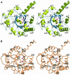
Fig. 2. (A) Stereo cartoon drawing (Bacon and Anderson, 1988; Kraulis, 1991; Merritt and Murphy, 1994) of the DDG aldolase protomer; α-helices are depicted as yellow helical ribbons and β-strands as blue arrows. The phosphate anions in the active site and the catalytic magnesium are shown. Oxygen atoms are colored in red, nitrogen in blue, sulfur in green, carbon in yellow, phosphate in black and magnesium in light blue. This color coding is used throughout all figures. β-strands β4 and β6 are immediately followed by more than one helix of which helix 4′ is a short 310-helix. Prolines 7 and 107, located on the loop preceding the N-terminal α-helix and on α-helix α4, respectively, are in their cis conformations. In addition to the assigned elements of secondary structure, there is a section of sequence that presents an extended conformation. This section involves residues 122–125 residing between helices α4 and 4′. (B) Stereo Cα trace of the DDG aldolase protomer shown in the same orientation as in (A). Every tenth Cα atom is labeled.
None
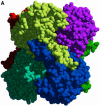
Fig. 3. (A) Space filling representation of the hexameric DDG aldolase looking down the non-crystallographic dyad. Each of the six subunits is colored differently. (B) Cartoon drawing of DDG aldolase oligomer shown in the same orientation as in (A) illustrating the active site pocket location between two 3-fold-related protomers. For clarity, only four of the six protomers within the hexamer are shown. The two phosphates located in the active site pocket and the catalytic magnesium are shown in space filling representation. The active site pocket is mainly lined by residues belonging to one protomer. The 3-fold-related subunit makes contacts with the ‘second’ phosphate.
None
Fig. 4. Stereo views of ligands binding to DDG aldolase. The bonds of the ligands are shown in pink while the bonds of the enzyme are shown in white. For clarity, water molecules (drawn as red spheres) are not labeled. The magnesium site is shown and possible ligands coordinating the Mg2+ are indicated. (A)–(D) are in the same orientation. (A) Residues in contact with the two phosphate anions bound to the active site pocket as seen in the substrate-free DDG aldolase structure. Final σA-weighted FoFc omit electron density map for ligands bound to the enzyme. The contour level of the electron density map is 3σ and the resolution is 1.8 Å. Four solvent molecules and the catalytic magnesium interact with the ‘first’ phosphate and five water molecules are hydrogen bonded to the ‘second’ phosphate. Ser124′ and Val125′ belong to a 3-fold-related protomer. (B) Residues in contact with pyruvate. Final σA-weighted FoFc omit electron density map for pyruvate bound to the enzyme. The contour level of the electron density map is 3σ and the resolution is 2.6 Å. The ligand’s carbonyl and carboxyl reside 2.5 and 2.4 Å, respectively, away from the magnesium. All contacts are made by one subunit within the hexamer. (C) Superposition of the substrate-free structure (white) onto the aldolase–pyruvate complexed structure (gray) to illustrate the possible role of the ‘second’ phosphate in the reaction mechanism (gray dotted line). The anion’s oxygen is 3.4 Å away from the methyl carbon atom. The current distance of 3.9 Å between Arg75 and the ‘second’ phosphate is easily decreased to hydrogen bonding distance by a slight side chain movement without steric hindrance and/or by moving the ‘second’ phosphate deeper into the active site. (D) Modeling of the condensed substrate, DDG, into the active site based upon the aldolase–pyruvate structure. The carboxylate at C6 of DDG fills the cavity occupied by the ‘second’ phosphate. A possible role during catalysis for the solvent molecule bridging the ligand’s O4 and His50 is indicated (gray dotted line).

Similar articles

Cited by

References

    1. Bacon D.J. and Anderson,W.F. (1988) A fast algorithm for rendering space-filling molecule pictures. J. Mol. Graph., 6, 219–220.
    1. Barbas C.F. III et al. (1997) Immune versus natural selection: antibody aldolases with enzymic rates but broader scope. Science, 278, 2085–2092. - PubMed
    1. Bennett M.J., Schlunegger,M.P. and Eisenberg,D. (1995) 3D domain swapping: a mechanism for oligomer assembly. Protein Sci., 4, 2455–2468. - PMC - PubMed
    1. Blackwell N.C. (2000) Mechanistic and structural investigation of DDG aldolase. PhD thesis, University of Leicester, Leicester, UK.
    1. Blackwell N.C., Cullis,P.M., Cooper,R.A. and Izard,T. (1999) Rhombohedral crystals of 2-dehydro-3-deoxy galactarate aldolase from Escherichia coli. Acta Crystallogr. D, 55, 1368–1369. - PubMed

Publication types

LinkOut - more resources