Comparative Study
. 2000 Aug 1;19(15):3857-69.
doi: 10.1093/emboj/19.15.3857.

Crystal structure of a repair enzyme of oxidatively damaged DNA, MutM (Fpg), from an extreme thermophile, Thermus thermophilus HB8

Affiliations
Comparative Study

Crystal structure of a repair enzyme of oxidatively damaged DNA, MutM (Fpg), from an extreme thermophile, Thermus thermophilus HB8

M Sugahara et al. EMBO J. .

Abstract

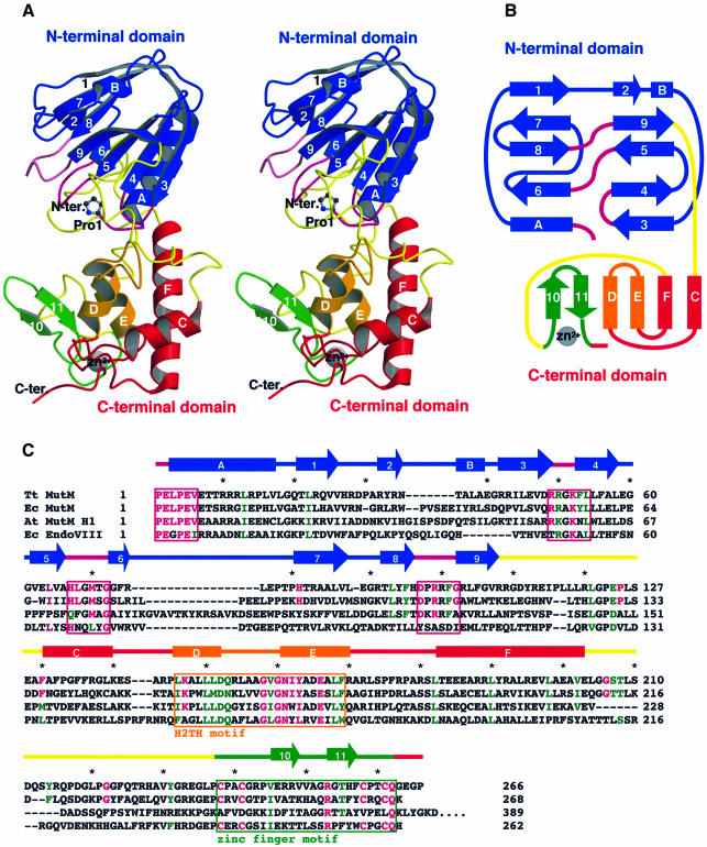
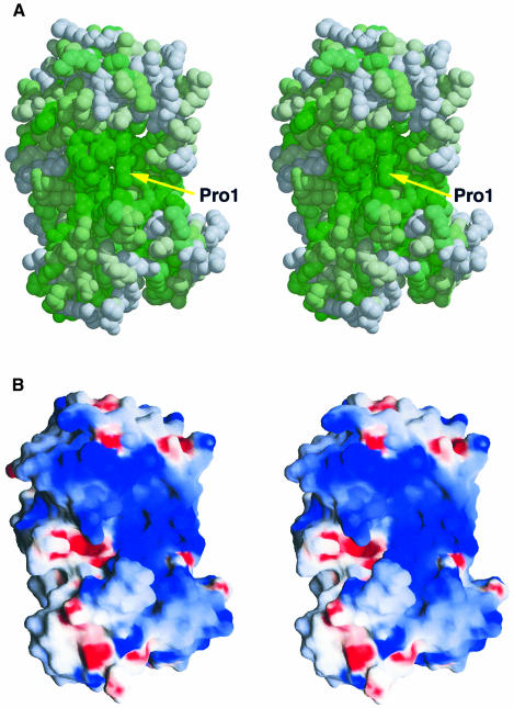
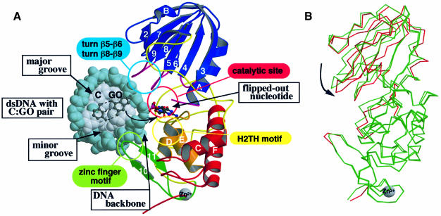
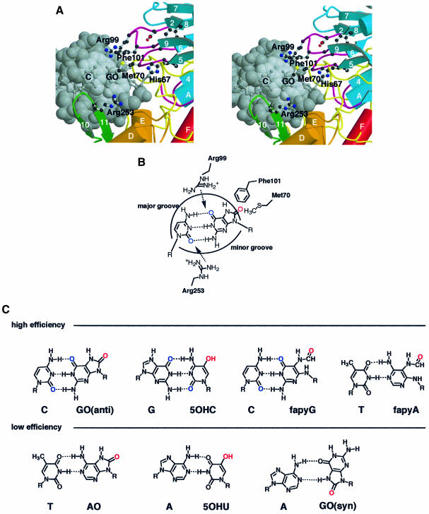
The MutM [formamidopyrimidine DNA glycosylase (Fpg)] protein is a trifunctional DNA base excision repair enzyme that removes a wide range of oxidatively damaged bases (N-glycosylase activity) and cleaves both the 3'- and 5'-phosphodiester bonds of the resulting apurinic/apyrimidinic site (AP lyase activity). The crystal structure of MutM from an extreme thermophile, Thermus thermophilus HB8, was determined at 1.9 A resolution with multiwavelength anomalous diffraction phasing using the intrinsic Zn(2+) ion of the zinc finger. MutM is composed of two distinct and novel domains connected by a flexible hinge. There is a large, electrostatically positive cleft lined by highly conserved residues between the domains. On the basis of the three-dimensional structure and taking account of previous biochemical experiments, we propose a DNA-binding mode and reaction mechanism for MutM. The locations of the putative catalytic residues and the two DNA-binding motifs (the zinc finger and the helix-two-turns-helix motifs) suggest that the oxidized base is flipped out from double-stranded DNA in the binding mode and excised by a catalytic mechanism similar to that of bifunctional base excision repair enzymes.


Figures

None
Fig. 1. Overall structure, topology and amino acid sequence of MutM. (A) Stereo ribbon diagram and (B) topology of MutM. The MutM molecule consists of an N-terminal domain (residues 1–109, blue), a C-terminal domain [residues 131–203 (red and orange) and 238–266 (green)] and two long loops [110–130 and 204–237 (yellow)]. The N-terminal domain consists of a two-layered β-sandwich with two α-helices. The C-terminal domain consists of four α-helix bundles (red and orange) and a β-hairpin loop of the zinc finger motif (green). There are two conformers in an asymmetrical unit (see Figure 3B). The conformations of the two long loops (in yellow) in the interdomain cleft differ between the two conformers. These two long loops could work as a hinge in domain movement. Pro1 (shown by a ball-and-stick model), which works as a nucleophile in the glycosylase/AP lyase reaction, is situated at the bottom of the cleft. The highly conserved regions (purple) in the N-terminal domain, the H2TH motif (orange) and the zinc finger motif (green) are located around the cleft (see Figure 2A). (C) Aligned sequences of bacterial T.thermophilus MutM (Tt MutM) (Mikawa et al., 1998), E.coli MutM (Ec MutM) (Boiteux et al., 1987), A.thaliana MutM homologue 1 (At MutM H1) (Murphy and Gao, 1998; Ohtsubo et al., 1998) and E.coli endonuclease VIII (Ec EndoVIII) (Jiang et al., 1997). The α-helices (bars) and β-strands (arrows) are shown above the aligned sequences. The amino acid numbering refers to the T.thermophilus HB8 protein. Coloured letters (homologous residues in magenta, similar residues in green) represent the residues conserved among 16 MutM proteins (see also Figure 2A). The asterisks indicate every tenth amino acid residue of the T.thermophilus sequence. The highly conserved regions in the N-terminal domain (purple), the H2TH motif (orange) and the zinc finger motif (green) are boxed.
None
Fig. 2. Conserved residues and electrostatic potential surface of MutM. (A) Stereo CPK model of MutM looking towards the interdomain large cleft. The surface is shown by a colour gradient according to residue conservation, with absolutely conserved residues in dark green. Homology was based on multiple alignment of the MutMs from T.thermophilus HB8 (Mikawa et al., 1998), E.coli (Boiteux et al., 1987), Neisseria meningitidis (Swartley and Stephens, 1995), Bacillus firmus (Ivey, 1990), Synechococcus elongatus naegeli (Floss et al., 1997), Bacillus subtilis (Lapidus et al., 1997), Salmonella typhimurium (Suzuki et al., 1997), Lactococcus lactis subsp. cremoris (Duwat et al., 1995), Haemophilus influenzae (sp|P44948), Synechocystis PCC6803 (sp|P74290), Mycobacterium leprae (emb|CAA19197.1), Mycobacterium tuberculosis (sp|Q10959), Streptococcus mutans (sp|P55045), Streptomyces coelicolor A3(2) (emb|CAB63194.1), Mycoplasma pneumoniae (sp|P75402) and Mycoplasma genitalium (sp|P55825). Homologous sequences were identified with PSI-BLAST (Altschul et al., 1997) and sequences were aligned with CLUSTAL W (Thompson et al., 1994). (B) The solvent-accessible surface is coloured according to electrostatic potential, with positive in blue and negative in red. The deep cleft between the two domains is electrostatically positive. Damaged DNA may bind to this large cleft.
None
Fig. 3. The model of the MutM–DNA complex and its hinge movement. (A) The model of the complex between the flipped out DNA and MutM in the closed form was obtained by molecular dynamics calculation. The viewing direction is similar to that in Figure 1A. The kinked DNA is drawn as a CPK model with backbones coloured in blue and with bases in grey. C and GO bases and their sugar residues before and after flipping out are shown by ball-and-stick models. All four conserved regions [(i) turns β5–β6 and β8–β9 in the sky-blue circle, (ii) the catalytic site in the pink circle, (iii) the H2TH motif in the yellow circle and (iv) the zinc finger motif in the light-green circle] are in the large cleft of the MutM molecule. The N-terminal domain can access the major groove of DNA and the zinc finger motif of the C-terminal domain can access the minor groove. The H2TH motif of the C-terminal domain is situated near the active site and may interact with the DNA backbone with the damaged base. (B) The superimposed structures of the two molecules in an asymmetrical unit. The superimposition was carried out to minimize the r.m.s.d. of the main chain in the C-terminal domain only. The main chain that deviates by >1 Å from the other molecule is only coloured red in the closed form monomer.
None
Fig. 4. Two DNA-binding motifs in the MutM structure. (A) Superimposition of the MutM H2TH motif (purple) upon the HhH motif of E.coli EndoIII (green; PDB code 2ABK) (Thayer et al., 1995) and the H3TH motif of Pyrococcus furiosus Flap endonuclease-1 (FEN-1) (cyan; PDB code 1B43) (Hosfield et al., 1998). In the H2TH motif of MutM, there are two turns between α-helix D (αD) and α-helix E (αE). The amino acid sequences are aligned based on structural superimposition. The amino acid residues conserved among MutM, EndoIII and FEN-1 are coloured red. The HhH motif in EndoIII and the H3TH motif in FEN-1 are thought to interact with the dsDNA backbone (Doherty et al., 1996; Artymiuk et al., 1997). Therefore, the H2TH motif of MutM is presumed to play a similar role to the HhH and H3TH motifs and may contribute to non-sequence-specific recognition. (B) The ε-amino group of Lys147 forms hydrogen bonds (dashed lines) to the side chain of conserved residues and the carbonyl oxygen atoms of the main chain (ball-and-stick) at the interface of the H2TH motif (orange) and in the zinc finger motif (green). The decrease in catalytic efficiency caused by replacement of Lys147 with alanine (Rabow and Kow, 1997) can be attributed to the disruption of these hydrogen bonds and loss of folding integrity in this region of DNA interaction.
None
Fig. 5. The structural basis for lesion recognition in MutM. (A) Model of recognition of GO:C-paired DNA (grey) by conserved amino acid residues (ball-and-stick) of MutM. (B) Schematic representation of the proposed lesion-recognition mechanism for the GO:C pair in dsDNA. Arg99 and Arg253 can interact with the keto groups of the GO and C base pair (blue) in both sides of dsDNA to disrupt the pairing hydrogen bonds in the model. The C8 oxo group of the GO base (red) in the DNA major groove could be pulled off by Met70 or Phe101 in the N-terminal domain. Phe101 could be inserted into the vacant site after nucleotide flipping to compensate for base stacking in the dsDNA. (C) Pairing of the oxidized bases in high- and low-efficiency MutM substrates is shown in the upper and lower rows, respectively (Hatahet et al., 1994; Tchou et al., 1994).
None
Fig. 6. Architecture of the MutM active site. (A) The nucleophile Pro1 of MutM is surrounded by the invariant charged residues Glu2, Glu5 and Lys52, accompanied by several bound water molecules. The hydrophilic active site is very positive electrostatically (Figure 2B), so Lys52 is expected to be a proton donor for glycosidase activity in MutM. (B) Model of docking of the flipped out GO nucleotide to MutM active site in Figure 3A. The 8-oxo group of the GO base is located near the ε-amino group of Lys52, which as a proton donor may help the protonation of the oxo group and lead to depurination. (C) The putative reaction intermediate adduct with Pro1 after β-elimination, which is configurationally definitive due to its conjugated double bonding (Bhagwat and Gerlt, 1996), can fit into the active site and the hydroxyl group at C4′ of the opened deoxyribose reaches the carboxylic acid of Glu2, which is a good candidate for a proton acceptor.
None
Fig. 7. Schematic representation of the reaction mechanism of MutM N-glycosylase/AP lyase. The reaction scheme is proposed on the basis of the active site architecture according to the mechanism proposed by Castaing et al. (1999). We propose that the invariant amino acid residues (Glu2, Glu5 and Lys52) in the vicinity of the primary catalytic residue Pro1, whose N-terminal amine forms a Schiff base with the C1′ of damaged deoxyribose (Zharkov et al., 1997), act as the additional catalytic residues for the enzyme action and, together with their bound water molecules, form a hydrogen bond network in the active site. In this highly electrostatically positive environment, Lys52 may act as a proton donor for the depurination of the damaged base (Figure 6B). After C2′ of deoxyribose has formed a Schiff base with Pro1, Glu5 could withdraw the proton of C2′ via a bound water, leading to β-elimination. The resulting adduct intermediate (Figure 6C) would deprotonate at C4′ of the opened deoxyribose, leading to δ-elimination. Finally, regain of the proton by Lys52 would release the other product, 4-oxo-2-pentenal, to form the gapped dsDNA product (Bhagwat and Gerlt, 1996). The residues believed to contribute to each reaction step were deduced from the crystal structure and are shown in red.

Similar articles

Cited by

References

    1. Altschul S.F., Madden,T.L., Schaffer,A.A., Zhang,J., Zhang,Z., Miller,W. and Lipman,D.J. (1997) Gapped BLAST and PSI-BLAST: a new generation of protein database search programs. Nucleic Acids Res., 25, 3389–3402. - PMC - PubMed
    1. Ames B.N., Gold,L.S. and Willett,W.C. (1995) The causes and prevention of cancer. Proc. Natl Acad. Sci. USA, 92, 5258–5265. - PMC - PubMed
    1. Artymiuk P.J., Ceska,T.A., Suck,D. and Sayers,J.R. (1997) Prokaryotic 5′–3′ exonucleases share a common core structure with γδ-resolvase. Nucleic Acids Res., 25, 4224–4229. - PMC - PubMed
    1. Bhagwat M. and Gerlt,J.A. (1996) 3′- and 5′-strand cleavage reactions catalyzed by the Fpg protein from Escherichia coli occur via successive β- and δ-elimination mechanisms, respectively. Biochemistry, 35, 659–665. - PubMed
    1. Boiteux S., O’Connor,T.R. and Laval,J. (1987) Formamidopyrimidine-DNA glycosylase of Escherichia coli: cloning and sequencing of the fpg structural gene and overproduction of the protein. EMBO J., 6, 3177–3183. - PMC - PubMed

Publication types

MeSH terms

Substances

Associated data