. 2000 Aug 1;19(15):3945-55.
doi: 10.1093/emboj/19.15.3945.

APC(ste9/srw1) promotes degradation of mitotic cyclins in G(1) and is inhibited by cdc2 phosphorylation

Affiliations

APC(ste9/srw1) promotes degradation of mitotic cyclins in G(1) and is inhibited by cdc2 phosphorylation

M A Blanco et al. EMBO J. .

Abstract

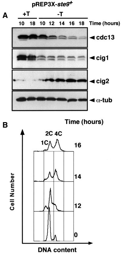
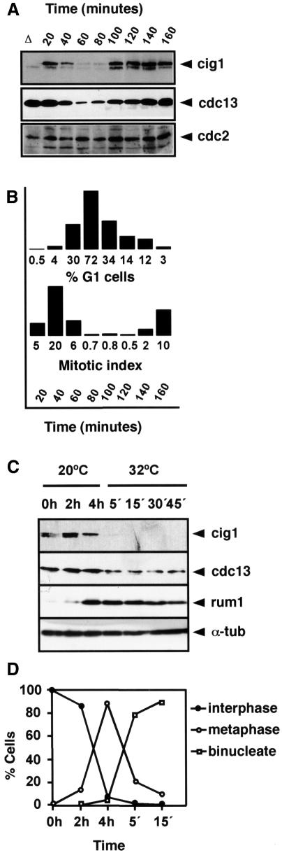
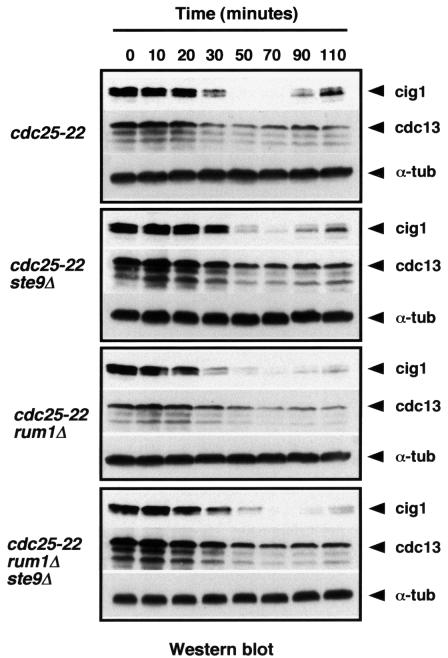
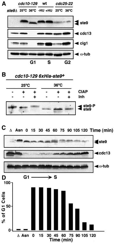
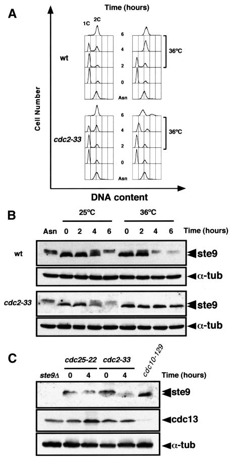
Fission yeast ste9/srw1 is a WD-repeat protein highly homologous to budding yeast Hct1/Cdh1 and Drosophila Fizzy-related that are involved in activating APC/C (anaphase-promoting complex/cyclosome). We show that APC(ste9/srw1) specifically promotes the degradation of mitotic cyclins cdc13 and cig1 but not the S-phase cyclin cig2. APC(ste9/srw1) is not necessary for the proteolysis of cdc13 and cig1 that occurs at the metaphase-anaphase transition but it is absolutely required for their degradation in G(1). Therefore, we propose that the main role of APC(ste9/srw1) is to promote degradation of mitotic cyclins when cells need to delay or arrest the cell cycle in G(1). We also show that ste9/srw1 is negatively regulated by cdc2-dependent protein phosphorylation. In G(1), when cdc2-cyclin kinase activity is low, unphosphorylated ste9/srw1 interacts with APC/C. In the rest of the cell cycle, phosphorylation of ste9/srw1 by cdc2-cyclin complexes both triggers proteolysis of ste9/srw1 and causes its dissociation from the APC/C. This mechanism provides a molecular switch to prevent inactivation of cdc2 in G(2) and early mitosis and to allow its inactivation in G(1).


Figures

None
Fig. 1. Overproduction of ste9+ promotes degradation of cdc13 and cig1 cyclins but not of cig2. An S.pombe leu1-32 strain was transformed with the plasmid pREP3X-ste9+. Transformants were selected on plates containing minimal medium with thiamine. Cells were grown in the presence (+T, repressed conditions) or absence of thiamine (–T, derepressed conditions) and samples were taken at the indicated times. (A) Extracts were prepared from these samples and the amounts of cdc13, cig1, cig2 and α-tubulin were determined. (B) FACS analysis of the cells.
None
Fig. 2. Cig1 oscillates during the mitotic cell cycle. (A) Cig1 protein levels were measured in a synchronous culture of the temperature-sensitive wee1-50 strain. A homogeneous population of cells in early G2 was selected by centrifugal elutriation of the wee1-50 strain at 25°C. This culture was incubated for 20 min at 25°C and then shifted up to 36°C. Samples were taken every 20 min to determine cig1, cdc13 and cdc2 protein levels. Both cig1 and cdc13 protein levels decreased in anaphase and increased at the end of G1. A cig1 deletion (Δ) cell extract was used as a negative control. (B) Percentage of G1 cells and mitotic index of the synchronous culture. (C) Cig1 is degraded during mitosis. Early G2 cells of nda3-KM311 were isolated by centrifugal elutriation at 32°C and blocked in metaphase for 4 h at 20°C. The culture was then released at 32°C. Samples were taken at the indicated times to determine cig1, cdc13, rum1 and α-tubulin protein levels. (D) Percentage of cells in interphase, metaphase and anaphase determined by DAPI staining.
None
Fig. 3. Degradation of cdc13 and cig1 in mitosis does not require ste9 or rum1. Wild-type, ste9Δ, rum1Δ and ste9Δ rum1Δ cells were synchronized in G2 using the cdc25-22 mutant. After 4 h at 36°C, the cultures were release to 25°C and samples were taken to measure cig1, cdc13 and α-tubulin levels.
None
Fig. 4. ste9 is phosphorylated in S-phase and G2 but not in G1. (A) ste9, cdc13 and cig1 protein levels in cells arrested in G1 with the cdc10-129 mutant, in S-phase with hydroxyurea and in G2 with the cdc25-22 mutant. As a negative control, we used an extract prepared from the ste9Δ mutant and α-tubulin as loading control. (B) A His6-ste9 allele introduced by gene replacement into the ste9 locus was purified on an Ni2+-NTA column from cdc10-129 cells growing at 25°C or after 4 h at 36°C. The purified His6-ste9 was treated with calf intestine alkaline phosphatase (CIAP) in the absence (–) or presence (+) of phosphatase inhibitors (Inh). (C) ste9 phosphorylation occurs at the G1–S transition. A cdc10-129 culture grown at 25°C to mid-exponential phase in minimal medium was shifted to 36°C for 4 h and then released at 25°C. Samples for western blot and flow cytometry were taken before the shift to 36°C (Asn), 4 h after the shift to 36°C (t = 0 min) and every 15 min after the release to 25°C. The levels of ste9, cdc13 and α-tubulin were determined. ste9 was unphosphorylated at the block point (t = 0 min) and became phosphorylated at the onset of S-phase (t = 60 min). Cdc13 protein levels were low in G1 cells while ste9 was unphosphorylated and started to accumulate when ste9 became phosphorylated and the cells initiated S-phase. Δ is a negative control from ste9Δ and Asn is an extract from the asynchronous culture of cdc10-129 at 25°C. (D) Percentage of cells in G1 during the course of the experiment.
None
Fig. 5. ste9 phosphorylation depends on cdc2 function. Wild-type and cdc2-33 cells were nitrogen starved for 12 h at 25°C. NH4Cl was added to the culture and half of the cells were incubated at 25°C and the rest at 36°C. Samples were taken for flow cytometry (A) and for western blots (B) at 0, 2, 4 and 6 h after the addition of NH4Cl to determine ste9 and α-tubulin protein levels. Asn corresponds to a sample taken from the asynchronous culture before nitrogen starvation. (C) ste9 mobility in cdc25-22 and cdc2-33 extracts prepared from cells growing exponentially at 25°C (t = 0) and then shifted to 36°C for 4 h (t = 4). The levels of cdc13, cdc2 and α-tubulin in these extracts are also shown. As a control, we used an extract from cdc10-129 cells incubated at 36°C for 4 h.
None
Fig. 6. Expression of ste9 phosphorylation mutants induces diploidization. Three mutants, ste9-4A, ste9-10A and ste9-13A, containing four, 10 and the 13 putative cdk phosphorylation sites were mutated to alanine by site-directed in vitro mutagenesis. These mutant alleles were introduced into the fission yeast genome by gene replacement. (A) Schematic representation of the ste9 protein with the seven WD repeats and the position of the 13 putative cdk phosphorylation sites. (B) FACS profile of cells replaced with the different ste9 mutant alleles. (C) Electrophoretic mobility of the different ste9 alleles. The asterisk corresponds to a non-specific band recognized by the anti-ste9 antibody that it is also detected in ste9Δ. (D) Half-lives of ste9, ste9-4A, ste9-10A and ste9-13A in exponentially growing cultures after the addition of 100 µg/ml of cycloheximide. The asterisk corresponds to a non-specific band recognized by the anti-ste9 antibody that it is also detected in ste9Δ and serves as loading control.
None
Fig. 7. ste9 associates with APC/C only in G1. (A) Extracts from cdc10-129, cdc10-129 cut9⋅HA ste9Δ and cdc10-129 cut9⋅HA mutants grown at 25 or 36°C were immunoprecipitated with anti-HA antibodies (IP α-HA) and then western blotted with anti-HA or anti-ste9 antibodies. Total cell extracts were separated on an SDS–polyacrylamide gel, transferred to a nitrocellulose membrane and probed with anti-HA, anti-ste9, anti-cdc13, anti-cig1 and anti-α-tubulin antibodies. (B) ste9 phosphorylation mutants associate with APC/C in G2. Extracts from cdc25-22, cdc25-22 cut9⋅HA and cdc10-129 cut9⋅HA mutants grown at 36°C for 4 h were immunoprecipitated with anti-HA antibodies (IP α-HA) and then western blotted with anti-HA and anti-ste9 antibodies. Total cell extracts were separated on an SDS–polyacrylamide gel, transferred to a nitrocellulose membrane and probed with anti-HA, anti-ste9 and anti-α-tubulin antibodies.

Similar articles

Cited by

References

    1. Amon A., Irniger,S. and Nasmyth,K. (1994) Closing the cell cycle circle in yeast: G2 cyclin proteolysis initiated at mitosis persits until the activation of G1 cyclins in the next cycle. Cell, 77, 1037–1050. - PubMed
    1. Baümer M., Braus,G.H. and Irniger,S. (2000) Two different modes of cyclin Clb2 proteolysis during mitosis in Saccharomyces cerevisae. FEBS Lett., 468, 142–148. - PubMed
    1. Benito J., Martín-Castellanos,C. and Moreno,S. (1998) Regulation of the G1 phase of the cell cycle by periodic stabilization and degradation of the p25rum1 Cdk inhibitor. EMBO J., 17, 482–497. - PMC - PubMed
    1. Berry L.D., Feoktistova,A., Wright,M.D. and Gould,K.L. (1999) The Schizosaccharomyces pombe dim1+ gene interacts with the anaphase-promoting complex or cyclosome (APC/C) component lid1+ and is required for APC/C function. Mol. Cell Biol., 19, 2535–2546. - PMC - PubMed
    1. Boeke J.D., LaCroute,F. and Fink,G.R. (1984) A positive selection for mutants lacking orotidine-5′-phosphate decarboxylase activity in yeast: 5-fluoro-orotic acid resistance. Mol. Gen. Genet., 197, 345–346. - PubMed

Publication types

MeSH terms