. 2000 Aug 1;19(15):3968-77.
doi: 10.1093/emboj/19.15.3968.

Fission yeast Fizzy-related protein srw1p is a G(1)-specific promoter of mitotic cyclin B degradation

Affiliations

Fission yeast Fizzy-related protein srw1p is a G(1)-specific promoter of mitotic cyclin B degradation

S Yamaguchi et al. EMBO J. .

Abstract

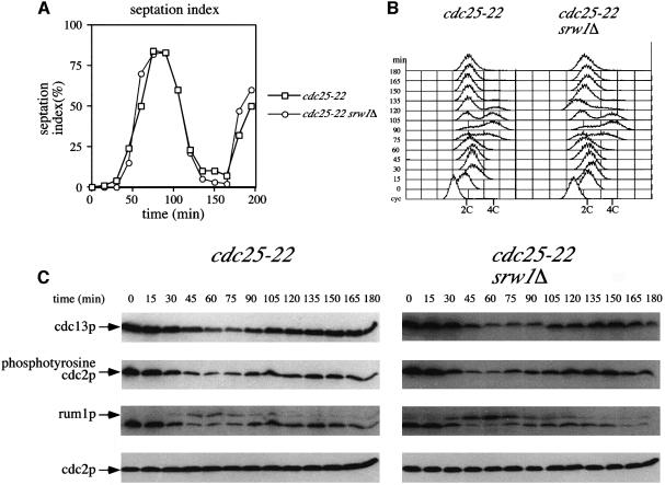
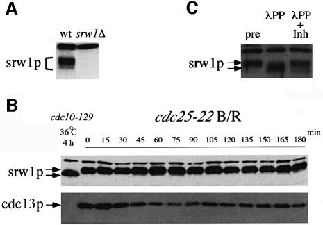
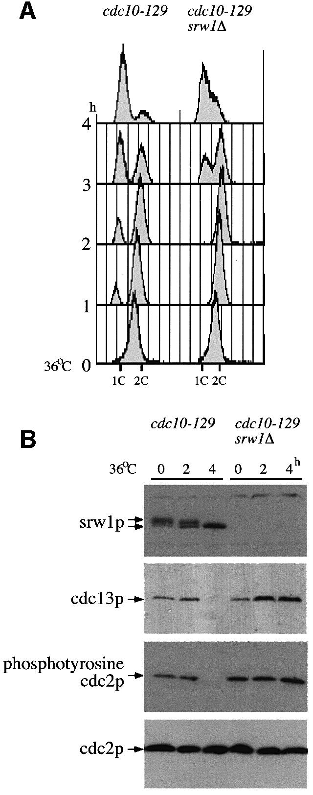
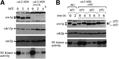
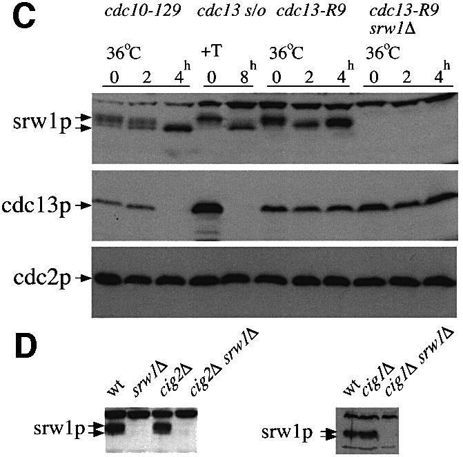
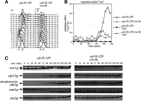
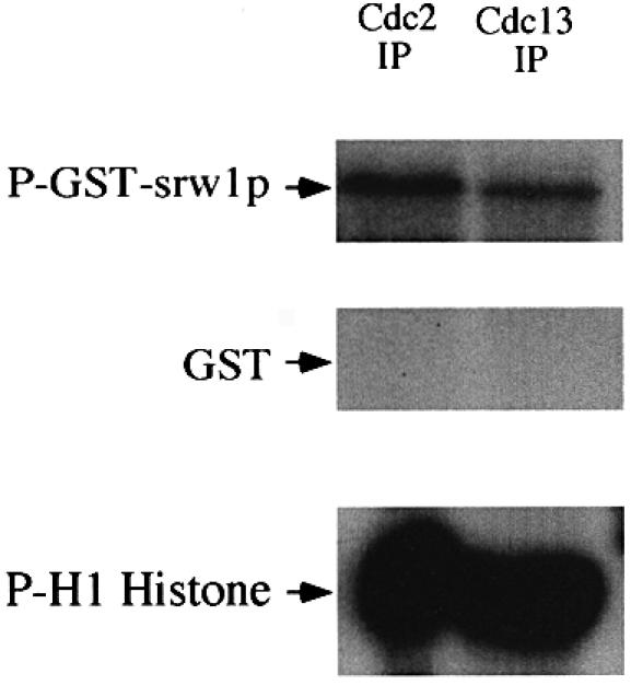
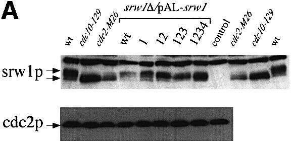
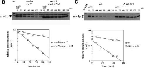
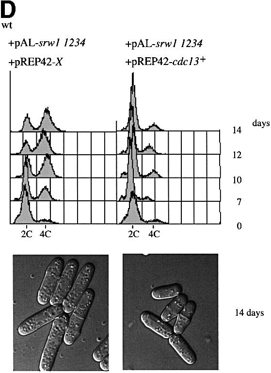
Downregulation of cyclin-dependent kinase (Cdk)-mitotic cyclin complexes is important during cell cycle progression and in G(1) arrested cells undergoing differentiation. srw1p, a member of the Fizzy-related protein family in fission yeast, is required for the degradation of cdc13p mitotic cyclin B during G(1) arrest. Here we show that srw1p is not required for the degradation of cdc13p during mitotic exit demonstrating that there are two systems operative at different stages of the cell cycle for cdc13p degradation, and that srw1p is phosphorylated by Cdk-cdc13p only becoming dephosphorylated during G(1) arrest. We propose that this phosphorylation targets srw1p for proteolysis and inhibits its activity to promote cdc13p turnover.


Figures

None
Fig. 1. srw1p is not required for cdc13p degradation at mitotic exit. (A) Septation index and (B) flow cytometric analysis of G2 block and release experiments. cdc25-22 and cdc25-22 srw1Δ strains were grown to exponential phase at 25°C (cyc), blocked for 4 h at 36°C, released at 25°C and incubated for the time indicated. (C) Western blots of the experiments described above using anti-cdc13p, anti-phosphotyrosine cdc2p, anti-rum1p and anti-cdc2p antibodies. For anti-rum1p antibody blotting, the same blots were used after the anti-phosphotyrosine cdc2p antibody.
None
Fig. 2. srw1p is phosphorylated throughout the cell cycle. (A) Extracts were prepared from exponentially growing wild-type (wt) cells and srw1Δ cells, western blotted and probed with anti-srw1p antibody. The upper band is a non-specific cross-reacting protein. (Bcdc25-22 strain was blocked and released (B/R) as described in Figure 1. Samples were western blotted and probed with anti-srw1p and anti-cdc13p antibodies. An extract from G1 arrested cdc10-129 cells was loaded as a control. (C) Extracts were prepared from exponentially growing wild-type cells (pre) and incubated with λ phosphatase in the absence (λPP) or presence (λPP + Inh) of phosphatase inhibitors.
None
Fig. 3. srw1p becomes dephosphorylated during G1 arrest, which coincides with the degradation of cdc13p. (A) Flow cytometry of G1 block experiments. cdc10-129 and cdc10-129 srw1Δ strains were shifted to 36°C and incubated for the indicated time in hours. (B) Western blots of the experiments above using anti-srw1p, anti-cdc13p, anti-phosphotyrosine cdc2p and anti-cdc2p antibodies.
None
Fig. 4. srw1p phosphorylation is cdc2p dependent. (Acdc2-M26 and cdc2-M26 srw1Δ strains were shifted to 36°C and incubated for the indicated time in hours. Extracts were western blotted and probed with anti-srw1p, anti-cdc13p and anti-cdc2p antibodies. Extracts were immunoprecipitated with anti-cdc2p antibody, and H1 kinase activity of each immunoprecipitate was measured. (Bcdc2-M26 strain was blocked with 11 mM HU for the indicated time in hours. Further 11 mM HU was added after 4 h to prevent cells from leaking from the block. Cells were incubated for 4 h at 25°C and then incubated for another 2 h at either 36 or 25°C. Extracts from cdc2-M26 cells blocked in the absence of HU were also loaded. The arrows indicate the migration of srw1p in experiments at 36 and 25°C, respectively. Extracts were immunoprecipitated with anti-cdc2p antibody, and H1 kinase activity of each immunoprecipitate was measured. (Ccdc13 was shut off by adding thiamine (+T) to the cdc13Δ REP45-cdc13+ strain (cdc13 s/o) and cells were incubated at 30°C for the indicated time in hours. cdc13-R9 and cdc13-R9 srw1Δ strains were shifted to 36°C and incubated for the indicated time in hours. Extracts from cdc10-129 cells were loaded as controls. Extracts were western blotted and probed with anti-srw1p, anti-cdc13p and anti-cdc2p antibodies. (D) Extracts were taken from exponentially growing wild-type (wt), srw1Δ, cig2Δ, cig2Δ srw1Δ, cig1Δ and cig1Δ srw1Δ cells, western blotted and probed with anti-srw1p antibody.
None
Fig. 5. srw1p becomes phosphorylated just before S-phase starts, which coincides with the accumulation of cdc13p. (A) Flow cytometry and (B) septation index and percentage of ‘cut’ cells in G1 block and release experiments. cdc10-129 and cdc10-129 srw1Δ strains were blocked for 4 h at 36°C and released into 25°C and incubated for the times indicated. (C) Western blots of the experiments described above using anti-srw1p, anti-cdc13p, anti-phosphotyrosine cdc2p, anti-rum1p and anti-cdc2p antibodies. For anti-rum1p antibody blotting, the same blots were used after the anti-phosphotyrosine cdc2p antibody.
None
Fig. 6. srw1p is phosphorylated by cdc2p in vitro. cdc2p and cdc13p-associated cdc2p were immunoprecipitated from wild-type extracts and assayed for protein kinase activity using GST–srw1p or histone H1 as substrates. GST was used as a control.
None
Fig. 7. srw1p stability is regulated by phosphorylation on Cdk consensus sites. (A) Samples were taken from exponentially growing srw1Δ cells carrying pAL-srw1 with no (wt), single (1), double (12), triple (123) or quadruple (1234) mutations in Cdk consensus sites or empty vector (pAL-X). Extracts from wild-type cells, cdc10-129 blocked cells and cdc2-M26 blocked cells were loaded as controls. Extracts were western blotted and probed with anti-srw1p and anti-cdc2p antibodies. (Bsrw1 was shut off by adding thiamine (+T) to srw1Δ cells expressing wild-type srw1 (srw1Δ nmt81-srw1+) or quadruple mutant srw1 (srw1Δ nmt81-srw1 1234) and cells were incubated at 30°C for the times indicated. Extracts from wild-type cells were loaded to show the endogenous level of srw1p. Extracts were western blotted and probed with anti-srw1p antibody, the srw1p protein was quantified with NIH image analyser. (C) Wild-type cells and cdc10-129 cells were incubated for 4 h at 36°C, cycloheximide (CYH) was added, and cells were incubated at 36°C for the times indicated. (D) Flow cytometry and morphology of wild-type cells co-expressing quadruple mutant srw1 (pAL-srw1 1234) and either pREP42-X or pREP42-cdc13+. Cells were incubated for the number of days indicated at 30°C in minimal medium in the absence of thiamine. (Ecdc13 expression was turned off by adding thiamine to cdc13Δ REP41-cdc13+ and cdc13Δ REP41-cdc13+ srw1:: srw1 1234 strains and cells were further incubated at 32°C for the time indicated in minutes. Extracts were western blotted and probed with anti-cdc13p, anti-srw1p and anti-cdc2p antibodies.

Similar articles

Cited by

References

    1. Amon A., Irniger,S. and Nasmyth,K. (1994) Closing the cell cycle circle in yeast: G2 cyclin proteolysis initiated at mitosis persists until the activation of G1 cyclins in the next cycle. Cell, 77, 1037–1050. - PubMed
    1. Ayscough K. and Warren,G. (1994) Inhibition of protein synthesis disrupts the Golgi apparatus in the fission yeast, Schizosaccharomyces pombe. Yeast, 10, 1–11. - PubMed
    1. Bähler J., Wu,J.Q., Longtine,M.S., Shah,N.G., McKenzie,A.,III, Steever,A.B., Wach,A., Philippsen,P. and Pringle,J.R. (1998) Heterologous modules for efficient and versatile PCR-based gene targeting in Schizosaccharomyces pombe. Yeast, 14, 943–951. - PubMed
    1. Baum B., Wuarin,J. and Nurse P. (1997) Control of S-phase periodic transcription in the fission yeast mitotic cycle. EMBO J., 16, 4676–4688. - PMC - PubMed
    1. Baum B., Nishitani,H., Yanow,S. and Nurse,P. (1998) Cdc18 transcription and proteolysis couple S phase to passage through mitosis. EMBO J., 17, 5689–5698. - PMC - PubMed

Publication types

MeSH terms