. 2000 Aug 1;19(15):3978-89.
doi: 10.1093/emboj/19.15.3978.

Mammalian LIN-7 PDZ proteins associate with beta-catenin at the cell-cell junctions of epithelia and neurons

Affiliations

Mammalian LIN-7 PDZ proteins associate with beta-catenin at the cell-cell junctions of epithelia and neurons

C Perego et al. EMBO J. .

Abstract

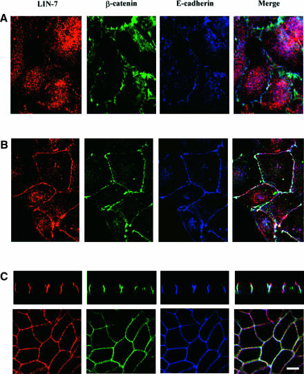
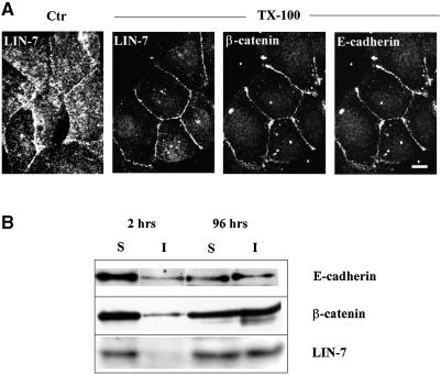
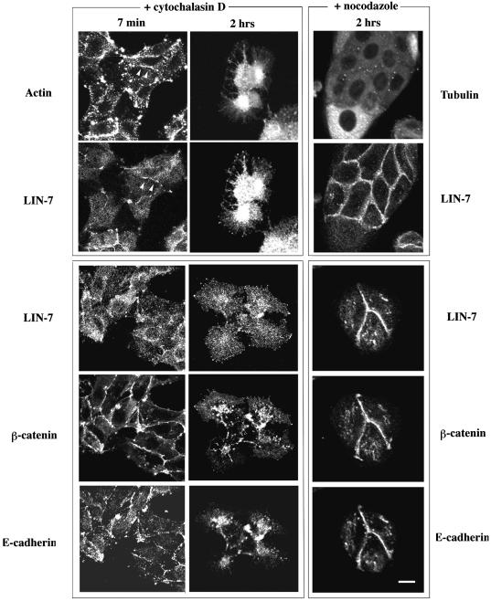
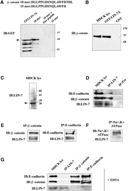
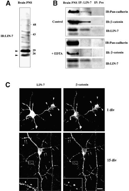
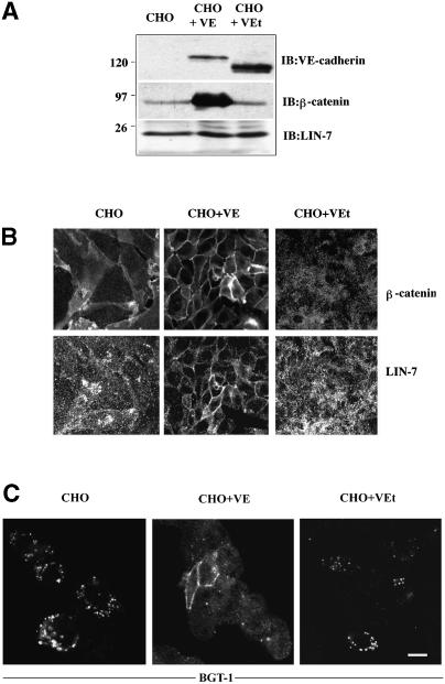
The heterotrimeric PDZ complex containing LIN-2, LIN-7 and LIN-10 is known to be involved in the organization of epithelial and neuronal junctions in Caenorhabditis elegans and mammals. We report here that mammalian LIN-7 PDZ proteins form a complex with cadherin and beta-catenin in epithelia and neurons. The association of LIN-7 with cadherin and beta-catenin is Ca(2+) dependent and is mediated by the direct binding of LIN-7 to the C-terminal PDZ target sequence of beta-catenin, as demonstrated by means of co-immunoprecipitation experiments and in vitro binding assays with the recombinant glutathione S-transferase:LIN-7A. The presence of beta-catenin at the junction is required in order to relocate LIN-7 from the cytosol to cadherin-mediated adhesions, thus indicating that LIN-7 junctional recruitment is beta-catenin dependent and that one functional role of the binding is to localize LIN-7. Moreover, when LIN-7 is present at the beta-catenin-containing junctions, it determines the accumulation of binding partners, thus suggesting the mechanism by which beta-catenin mediates the organization of the junctional domain.


Figures

None
Fig. 1. The junctional localization of LIN-7 parallels the maturation of the junctions and the acquisition of cell polarity. MDCK cells were plated at high density onto poly-l-lysine-coated glass coverslips, cultured for different times and then fixed in 4% paraformaldehyde. LIN-7 distribution was compared with that of E-cadherin and β-catenin by means of confocal analyses of the triple immunofluorescence stainings. Rabbit anti-LIN-7 (red), mouse anti-β-catenin (green) and rat anti-E-cadherin (blue) antibodies were used. In the merged images, the white color indicates the co-localization of the three proteins, yellow the co-localization of LIN-7 with β-catenin, magenta the co-localization of LIN-7 with E-cadherin, and light blue the co-localization of β-catenin with E-cadherin. (A) In cells cultured for 2 h, E-cadherin appears as irregular and often punctate staining at the cell–cell boundary where it co-localizes with β-catenin (light blue in the merged image); predominantly, diffuse cytoplasmic staining for LIN-7 can be seen. (B) In cells cultured for 10–10–18 h, LIN-7 largely co-localizes with E-cadherin and β-catenin at the cell–cell contact regions. (C) In fully polarized MDCK cells, LIN-7 co-localizes with E-cadherin and β-catenin along the lateral plasma membrane domain, but LIN-7 labeling can also be seen in a more apical region (red in the merged image of the vertical section). Bar, 15 µm.
None
Fig. 2. LIN-7 is recruited progressively in the Triton X-100-insoluble fraction together with β-catenin and E-cadherin. (A) Immunofluorescence confocal analysis of LIN-7, β-catenin and E-cadherin staining after extraction with Triton X-100. Cells seeded in culture for 10–18 h were extracted (+ TX-100) or not (Ctr) with 0.5% Triton X-100 for 20 min before fixation with 4% paraformaldehyde. The distribution of LIN-7 in the control and treated cells is shown. On the Triton X-100-treated coverslips, the localization of LIN-7 is compared with that of β-catenin and E-cadherin. A horizontal section of triple immunofluorescence staining is shown. The detergent treatment largely extracts the cytosolic LIN-7, but the junctional staining is preserved. Bar, 15 µm. (B) Western blot analysis of the amount of LIN-7, β-catenin and E-cadherin in the Triton X-100-soluble (S) and -insoluble (I) fractions. High-density cultures of MDCK cells were extracted in Triton X-100 2 or 96 h after seeding in culture. Equivalent volumes of the Triton X-100-soluble or -insoluble fractions were separated by 10% SDS–PAGE and immunostained using the indicated antibodies (IB:). A concomitant association of these proteins with cytoskeletal elements during the junctional maturation is suggested by their similar reaction to Triton X-100 extraction.
None
Fig. 3. The junctional localization of LIN-7 depends on a stably assembled actin cytoskeleton but not on the microtubule cytoskeleton. The localization of LIN-7, E-cadherin and β-catenin after disassembly of the actin (+ cytochalasin D) or the microtubule (+ nocodazole) cytoskeleton is shown. Low-density MDCK cells were treated with 2 µM cytochalasin D for 7 min or 2 h, or with 33 µM nocodazole for 2 h. The coverslips were cut into two parts: half was double stained with the LIN-7 antibody and phalloidin–FITC (actin) or with anti-tubulin antibodies (tubulin) in order to assess cytoskeleton depolymerization (upper panels); the other half was triple stained with LIN-7, β-catenin and E-cadherin antibodies (lower panels). Confocal analysis of the horizontal sections is shown. Partial depolymerization of the actin cytoskeleton (7 min) mainly affected the junctional localization of LIN-7, whereas the E-cadherin and β-catenin remained predominantly at the cell–cell contacts. The arrowheads indicate a contact region positive for LIN-7 staining in which the cytoskeleton is still assembled. Longer treatment (2 h) completely depolymerizes the actin cytoskeleton causing the cytosolic redistribution of the three proteins. Nocodazole treatment does not affect the junctional localization of the three proteins. Bar, 20 µm.
None
Fig. 4. LIN-7 forms a Ca2+-dependent complex with E-cadherin and β-catenin via its interaction with the β-catenin PDZ target motif. (A) Recombinant GST:LIN-7A was retained by the 18mer peptide containing the β-catenin PDZ target sequence, but not by the 15mer peptide. The sequences of the β-catenin 18mer and 15mer peptides immobilized on CNBr-activated Sepharose 4B are shown. A lysate from bacteria expressing the GST:LIN-7A fusion protein was incubated with the indicated immobilized peptides. Bound GST:LIN-7A was detected using a monoclonal antibody directed against GST (IB:GST). Purified GST:LIN-7A fusion protein was loaded in the gel as control (GST:LIN-7A). In addition to the band at the position expected for the fusion protein (arrowhead), lower molecular weight bands, presumably due to the proteolysis of the products, are visible. Molecular weight standards expressed in kilodaltons are indicated on the right. (B) β-catenin is retained specifically by the GST:LIN-7A but not by GST. MDCK cell lysates were incubated with immobilized GST or GST:LIN-7A fusion protein. The bound material was resolved by 10% SDS–PAGE and immunostained with monoclonal antibodies directed against β-catenin (IB:β-catenin). Ten percent of the total MDCK cell lysate (MDCK lys) used in the experiment was probed with the same antibody. Molecular weight standards expressed in kilodaltons are indicated on the right. (C) The LIN-7 antibody recognizes a band of the expected molecular weight in immunoblotting of MDCK cell lysate (arrowhead). A 20 µg aliquot of cell lysate was probed with the LIN-7 antibody (IB:LIN-7) raised against synthetic peptides designed in the LIN-7 PDZ domain (see Materials and methods). Molecular weight standards expressed in kilodaltons are indicated on the right. (D) Co-immunoprecipitation of E-cadherin and β-catenin with the LIN-7 antibody. (E) Co-immunoprecipitation of LIN-7 with the β-catenin or E-cadherin antibodies. (F) LIN-7 is not co-immunoprecipitated by the Na+,K+ ATPase antibody. (G) LIN-7 is not found in the complex with β-catenin and E-cadherin after pre-treatment with 5 mM EDTA. (D–G) The MDCK cell cultures were extracted in lysis buffer and immunoprecipitated with the indicated antibodies (IP:). The immunoprecipitates were loaded onto an 11% SDS–polyacrylamide gel followed by immunoblot analysis with the indicated antibodies (IB:). As controls, immunoprecipitation experiments were performed using a LIN-7 pre-immune serum (Pre) or the monoclonal antibody raised against Na+,K+ ATPase. When indicated, 10% of the cell lysates (MDCK lys) used in the immunoprecipitation experiments were probed with the indicated antibodies.
None
Fig. 5. LIN-7 associates with the cadherin–catenin adhesion system in neurons. (A) The LIN-7 antibody recognizes two main bands of the expected molecular weights in immunoblots of PNS brain fractions (arrowheads). A 20 µg aliquot of PNS was probed with the LIN-7 antibody (IB:LIN-7). Molecular weight standards expressed in kilodaltons are indicated on the right. (B) LIN-7 forms a Ca2+-dependent immunocomplex with β-catenin and cadherin in brain. PNS brain fractions were extracted with lysis buffer (Control) or lysis buffer containing 5 mM EDTA (+ EDTA). The brain extracts were immunoprecipitated with LIN-7 (IP:LIN-7) or pre-immune serum (IP:Pre). The immunoprecipitates were separated by SDS–PAGE, and immunoprobed with the LIN-7, the monoclonal β-catenin and the Pan-cadherin antibodies (IB:). Ten percent of the PNS extract (Brain PNS) was immunostained with the same antibodies. (C) LIN-7 co-localizes with β-catenin in cultured rat hippocampal neurons. One-day-old (1 div) or 15-day-old (15 div) hippocampal neurons were fixed and double stained with the LIN-7 and β-catenin antibodies. In the undifferentiated neurons (1 div), the arrows indicate the dendritic growth cones, and the arrowheads the filopodia emanating from the growing axon and the axonal growth cone. In the differentiated neurons (15 div), the arrows indicate sites reminiscent of synaptic labeling and positive for β-catenin and LIN-7. The boxed region is shown at higher magnification (3.5×) in the inset. Bar, 20 µm.
None
Fig. 6. The junctional localization of β-catenin mediates the redistribution of LIN-7. (A) Western blot analyses of LIN-7, β-catenin and VE-cadherin expression in untransfected CHO cells (CHO), and CHO cells transfected with wild-type (CHO + VE) or truncated VE-cadherin (CHO + VEt). A 20 µg aliquot of the total proteins from each cell line was loaded onto an 11% SDS–polyacrylamide gel and immunoprobed with the indicated antibodies (IB:). Bands corresponding to the wild-type (∼130 kDa) and truncated (∼100 kDa) VE-cadherin were revealed by specific staining. The CHO cells transfected with wild-type VE-cadherin express larger amounts of β-catenin. An equal level of LIN-7 expression was detected in the three cell lines. Molecular weight standards expressed in kilodaltons are indicated on the left. (B) Immunolocalization of β-catenin and LIN-7 in untransfected and transfected CHO cells. The cells were fixed in 4% paraformaldehyde and double stained with the β-catenin and LIN-7 antibodies. The presence of β-catenin at the junction determines the redistribution of LIN-7 from the cytosol to the junctions. (C) Immunolocalization of the epithelial GABA transporter BGT-1 in untransfected and transfected CHO cells. The cells were transiently transfected with the BGT-1 cDNA and, 48 h later, were fixed in 4% paraformaldehyde and processed for immunofluorescence using an antibody raised against the BGT-1 transporter (BGT-KLH antibody; Perego et al., 1997). Only CHO cells localizing LIN-7 at the junctions retain and accumulate the BGT-1 transporter at the cell surface. Bar: (B) 20 µm; (C) 15 µm.

Similar articles

Cited by

References

    1. Adams C.L., Nelson,W.J. and Smith,S.J. (1996) Quantitative analysis of cadherin–catenin–actin reorganization during development of cell–cell adhesion. J. Cell Biol., 135, 1899–1911. - PMC - PubMed
    1. Angres B., Barth,A. and Nelson,W.J. (1996) Mechanism for transition from initial to stable cell–cell adhesion: kinetic analysis of E-cadherin-mediated adhesion using a quantitative adhesion assay. J. Cell Biol., 134, 549–557. - PMC - PubMed
    1. Borg J.P., Straight,S.W., Kaech,S.M., de Taddeo-Borg,M., Kroon,D.E., Karnak,D., Turner,R.S., Kim,S.K. and Margolis,B. (1998) Identification of an evolutionarily conserved heterotrimeric protein complex involved in protein targeting. J. Biol. Chem., 273, 31633–31636. - PubMed
    1. Butz S., Okamoto,M. and Sudhof,T.C. (1998) A tripartite protein complex with the potential to couple synaptic vesicle exocytosis to cell adhesion in brain. Cell, 94, 773–782. - PubMed
    1. Cameron P.L., Sudhof,T.C., Jahn,R. and De Camilli,P. (1991) Colocalization of synaptophysin with transferrin receptors: implications for synaptic vesicle biogenesis. J. Cell Biol., 115, 151–164. - PMC - PubMed