. 2000 Aug 1;19(15):3990-4003.
doi: 10.1093/emboj/19.15.3990.

Skin and hair follicle integrity is crucially dependent on beta 1 integrin expression on keratinocytes

Affiliations

Skin and hair follicle integrity is crucially dependent on beta 1 integrin expression on keratinocytes

C Brakebusch et al. EMBO J. .

Abstract

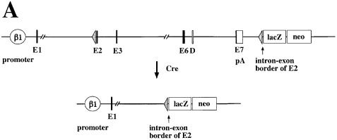
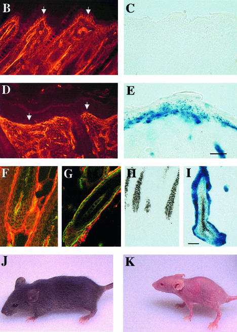
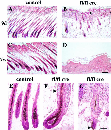
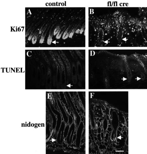
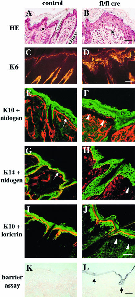
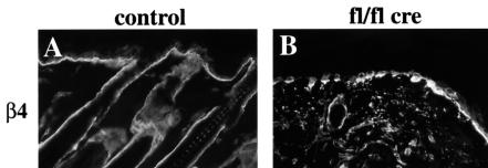
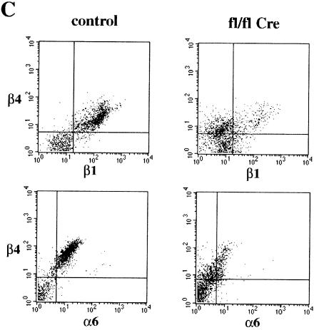
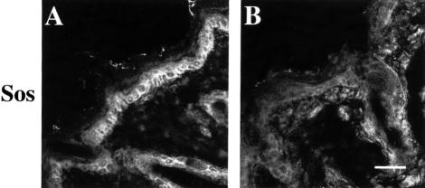
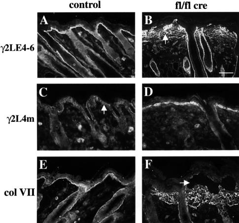
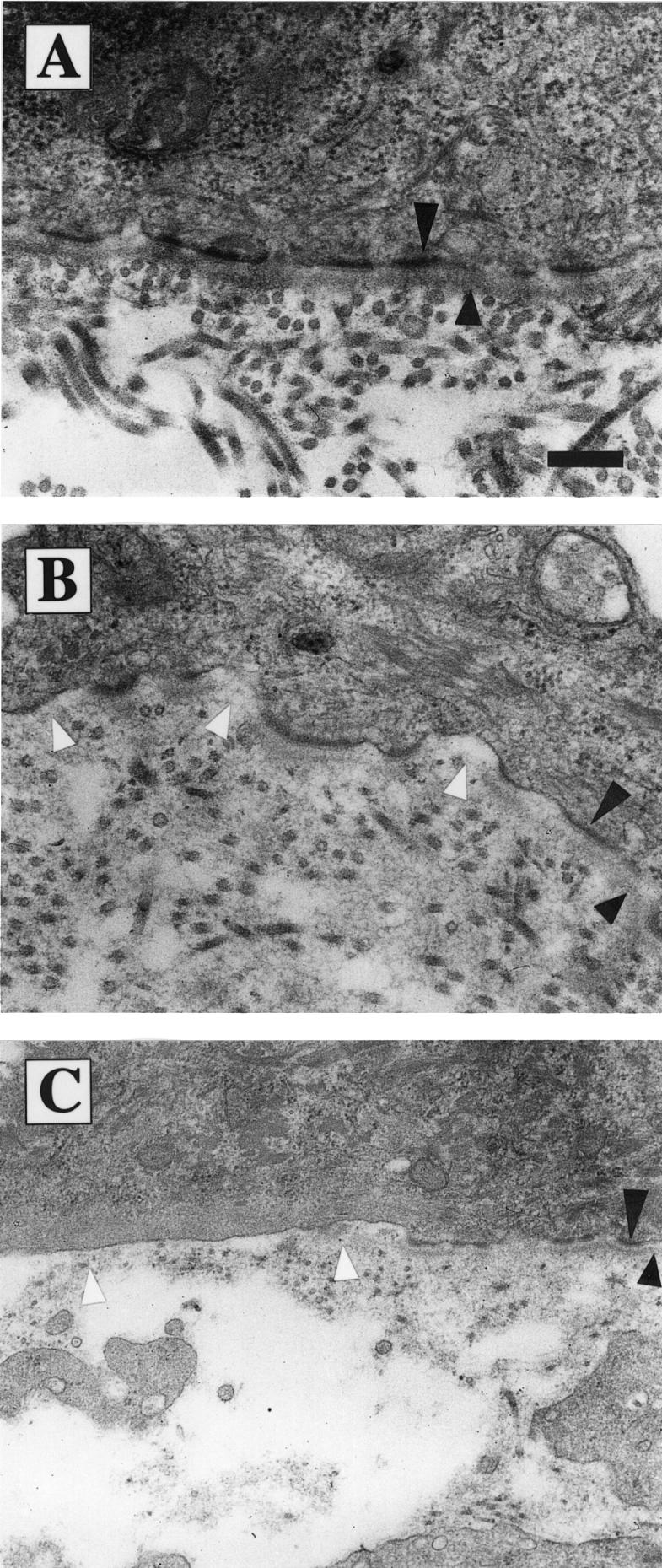
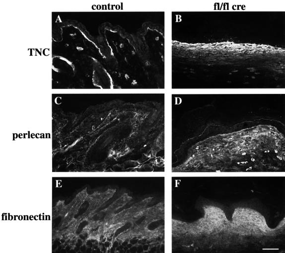
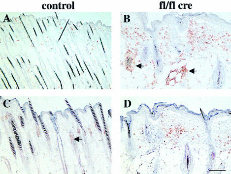
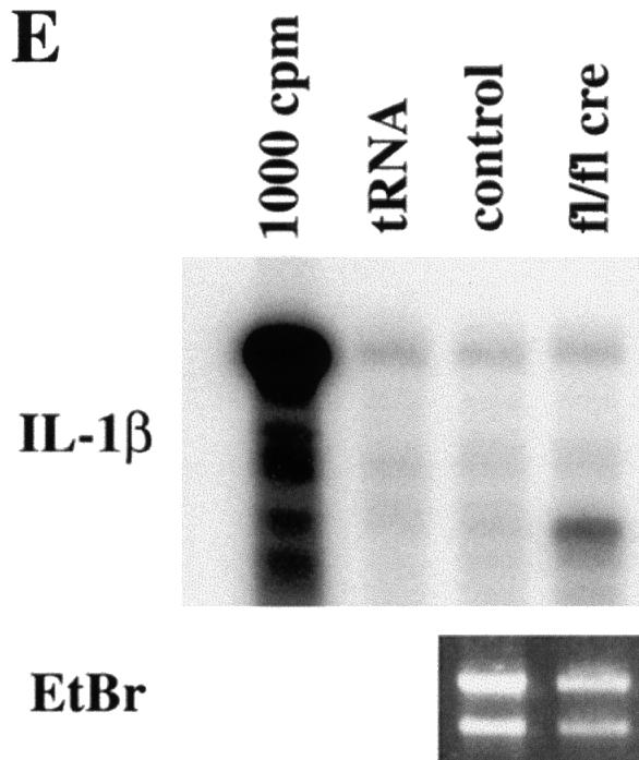
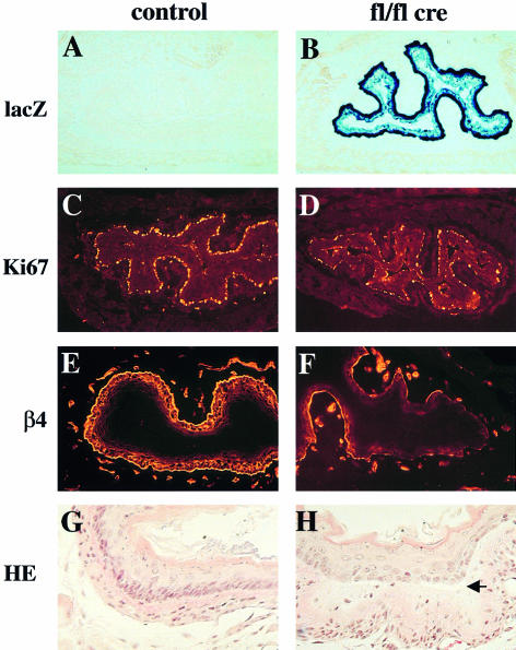
beta 1 integrins are ubiquitously expressed receptors that mediate cell-cell and cell-extracellular matrix interactions. To analyze the function of beta1 integrin in skin we generated mice with a keratinocyte-restricted deletion of the beta 1 integrin gene using the cre-loxP system. Mutant mice developed severe hair loss due to a reduced proliferation of hair matrix cells and severe hair follicle abnormalities. Eventually, the malformed hair follicles were removed by infiltrating macrophages. The epidermis of the back skin became hyperthickened, the basal keratinocytes showed reduced expression of alpha 6 beta 4 integrin, and the number of hemidesmosomes decreased. Basement membrane components were atypically deposited and, at least in the case of laminin-5, improperly processed, leading to disruption of the basement membrane and blister formation at the dermal-epidermal junction. In contrast, the integrity of the basement membrane surrounding the beta 1-deficient hair follicle was not affected. Finally, the dermis became fibrotic. These results demonstrate an important role of beta 1 integrins in hair follicle morphogenesis, in the processing of basement membrane components, in the maintenance of some, but not all basement membranes, in keratinocyte differentiation and proliferation, and in the formation and/or maintenance of hemidesmosomes.


Figures

None
Fig. 1. Generation of mice with a keratinocyte-restricted ablation of the β1 integrin gene. (A) Schematic presentation of the floxed β1 integrin gene and the cre-mediated deletion of the gene. After deletion of the β1 integrin gene, the β1 integrin promoter will transcribe the lacZ cDNA. (E, exons in arbitrary numbering; D, splice variant; pA, polyadenylation sequence of β1 integrin gene). Epidermis of back skin of 9-day-old control (B and C) and mutant mice (D and E) stained for β1 integrin (B and D) and lacZ activity (C and E) (bar, 50 µm). Note the loss of β1 integrin expression in the mutant basal keratinocytes (arrows in B and D). Bulb region of hair follicles of back skin of 9-day-old control (F and H) and mutant mice (G and I) double-stained for β1 (red) and α6 integrin (green) (F and G) and for lacZ activity (H and I) (bar, 50 µm). Note the loss of α6 and β1 integrin in the hair matrix and the thin unstained region visible between the α6 integrin expressing ORS cells and β1 integrin expressing cells surrounding the hair follicle (G). Control (J) and mutant (K) mice at 4 weeks of age. Mutant mice were approximately half the weight of control littermates, lost nearly all hair, and had frequent wounds, especially in mechanically stressed regions of the skin.
None
Fig. 2. Aberrant hair follicle morphogenesis and loss of subcutaneous fat layer in keratinocyte-restricted β1-deficient mice. Hematoxylin–eosin stained sections of back skin of 9-day (A and B) and 7-week-old (C and D) control (A and C) and mutant mice (B and D). At 9 days of age the mutant hair follicles have grown less deeply into the subcutis than normal hair follicles (A, B). Seven-week-old mutant mice had lost all hair follicles and had no subcutaneous fat layer (C, D; bar, 200 µm). Hematoxylin–eosin staining of hair follicles derived from 9-day-old control (E) and mutant mice (F, G). Hair follicles in mutant mice showed gross abnormalities, ranging from multilayered ORS (F, arrow) to severely malformed follicles with peripheral hair deposition (G, arrow; bar, 100 µm).
None
Fig. 3. Reduced proliferation of hair matrix cells lacking β1 integrin. Back skin sections of 16-day-old control (A, C and E) and mutant mice (B, D and F) were stained for Ki67 (A and B), TUNEL (C and D) and nidogen (E and F). Hair matrix cells in the hair bulb (arrows) of the mutant mice showed no Ki67-positive proliferating cells (B), in contrast to normal mice (A). Ki67-positive cells between the mutant hair follicles were proliferating dermal fibroblasts. No increased apoptosis was observed in the matrix cells of mutants (D). Nidogen deposition around the β1-deficient hair follicles was thickened but continuous (F; bar, 100 µm).
None
Fig. 4. Abnormalities in keratinocyte morphology and differentiation. Back skin sections of 9-day-old control (A, C, E, G and I) and mutant mice (B, D, F, H and J) were stained with hematoxylin–eosin (A and B; bar, 30 µm) and with antibodies for keratin-6 (K6; C and D; bar, 50 µm), keratin-10 (K10; green; E, F, I and J), keratin-14 (K14; green; G and H), nidogen (red; E–H) and loricrin (red; I and J). Basal keratinocytes from normal mice (A, arrow) attach to a nidogen-containing basement membrane (E and G, arrows). Mutant mice showed basal keratinocytes with an aberrant morphology, a thickened epidermis and blistering between epidermis and dermis (arrow and arrowheads in B, F, H and J). Keratin-6 is upregulated in mutant epidermis (D). Basal cells did not express keratin-10 either in normal or mutant skin (E, F, I and J). Suprabasal cells were positive for keratin-10 and keratin-14 (E–J). Most of the keratin-10 positive cells in the mutant skin showed no expression of loricrin (J), indicating a delayed terminal differentiation (E–J; bar, 40 µm). The barrier function of the skin was tested using a hematoxylin dye penetration assay (for details see Materials and methods). Sections of back skin of 4-week-old control (K) and mutant mice (L) were counterstained with eosin. Hematoxylin penetrated the superficial layers of the stratum corneum (violet), but neither the lower layers (L, arrows) nor any underlying tissue in the mutant mice, indicating an intact barrier function of the skin (bar, 50 µm).
None
Fig. 5. Reduced expression of α6β4 integrin in mutant epidermis. Back skin sections of 16-day-old control (A) and mutant (B) mice were stained for β4 integrin. In mutants, skin expression of β4 integrin by the basal keratinocytes was reduced and discontinuous (B) (bar, 50 µm). (C) FACS analysis of keratinocyte preparations from back skin of 3-week-old normal and mutant mice double stained for β4 and β1 integrin, and for β4 and α6 integrin, respectively. Basal keratinocytes expressing high amounts of β1, α6 and β4 integrin were found in normal, but hardly in mutant skin. Instead, mutant skin preparations contained a population of β1-null cells expressing low amounts of β4 and α6 integrin.
None
Fig. 6. Reduced Sos expression of basal keratinocytes in mutant skin. Back skin sections of 9-day-old control (A) and mutant (B) mice were stained for Sos. Sos expression was significantly reduced in basal keratinocytes of mutant skin compared with normal (bar, 50 µm).
None
Fig. 7. Aberrant processing and deposition of laminin-5 and collagen VII in mutant skin. Back skin sections of 16-day-old control (A, C and E) and mutant mice (B, D and F) were stained for the γ2 chain of laminin-5 using antibodies recognizing both the unprocessed and the processed form (γ2LE4-6; A and B), and antibodies specific for a fragment present only in the unprocessed form (γ2L4m; C and D). γ2LE4-6 showed diffuse staining in the dermis of mutant skin (B, arrow). Significantly increased amounts of unprocessed laminin γ2 were detected with the γ2L4m specific antibody around the basal keratinocytes of mutant mice, while dermally deposited laminin-5 was processed normally (D). Note the virtual absence of unprocessed laminin γ2 in the basement membrane of normal epidermis (C) and the normal γ2LE4-6 staining around the hair follicle in mutant skin (B; bar, 50 µm). Staining for the anchoring fibril component collagen VII (E, F) revealed a diffuse, dermal staining in mutant mice (F). In blisters (F, arrow), small amounts of collagen VII could be detected at the epidermal side.
None
Fig. 8. Distortion of the basement membrane at the dermal–epidermal junction. Electron microscopy of back skin sections of 4-week-old control (A) and mutant (B and C) mice. The lamina densa is marked by short and hemidesmosomes by long arrowheads. Loss of β1 integrin expression on keratinocytes resulted in a discontinuous lamina densa [gaps marked by white arrowheads in (B)] and in a reduction of hemidesmosomes [region without hemidesmosomes and basement membrane marked by white arrowheads in (C)]. (A and B) Bar, 0.25 µm; (C) bar, 0.42 µm.
None
Fig. 9. Increased deposition of extracellular matrix components in mutant skin. Back skin sections of 6-week-old control (A, C and E) and mutant mice (B, D and F) were stained for tenascinC (TNC) (A, B), perlecan (C, D), and fibronectin (E, F). All three proteins showed increased staining in the mutant dermis (bar, 100 µm).
None
Fig. 10. Infiltration of inflammatory cells in the mutant skin and RNase protection assay. Back skin sections of 16-day-old control (A and C) and mutant (B and D) mice were stained for F4/80 (macrophage/monocyte specific antigen; A, B) and Gr-1 (granulocyte specific antigen; C, D). In mutant skin, an infiltration of macrophages and granulocytes was observed. Some deformed hair follicles were surrounded by macrophages (arrows in B). (Staining of the sebaceous glands in (C) (arrow) and (D) is background staining; bar, 100 µm.) (E) RNA isolated from skin of 4-week-old mutant and normal mice was analyzed by RNase protection (for details see Materials and methods). Mutant skin (fl/fl cre) showed a strong upregulation of IL-1β mRNA compared with littermate controls (control) (1000 c.p.m.: radiolabeled RNA probe; tRNA: negative control). Gel separated RNA samples were stained with ethidium bromide (EtBr) to control RNA amounts used in the assay.
None
Fig. 11. Defects in the esophagus of mutant mice. Esophagus sections of 2- (AF) and 6-week-old (G and H) control (A, C, E and G) and mutant mice (B, D, F and H) were stained for lacZ activity (A and B), Ki67 (C and D), β4 integrin (E and F), and with hematoxylin–eosin (G and H). Basal cells in the mutant esophagus showed strong lacZ activity (B), reduced Ki67-positive proliferating cells (D) and a reduced staining for β4 integrin (F). In 6-week-old mice, mutant basal cells (H) displayed a different morphology than in normal mice (G). In addition, blisters were observed (H, arrow).

Similar articles

Cited by

References

    1. Aumailley M. and Rousselle,P. (1999) Laminins of the dermo-epidermal junction. Matrix Biol., 18, 19–28. - PubMed
    1. Bagutti C., Wobus,A.M., Fässler,R. and Watt,F.M. (1996) Differentiation of embryonal stem cells into keratinocytes: comparison of wild-type and β1 integrin-deficient cells. Dev. Biol., 179, 184–196. - PubMed
    1. Bloch W. et al. (1997) β1 integrin is essential for teratoma growth and angiogenesis. J. Cell Biol., 139, 265–278. - PMC - PubMed
    1. Bottazzi M.E. and Assoian,R.K. (1997) The extracellular matrix and mitogenic growth factors control G1 phase cyclins and cyclin-dependent kinase inhibitors. Trends Cell Biol., 7, 348–352. - PubMed
    1. Brakebusch C., Hirsch,E., Potocnik,A. and Fässler,R. (1997) Genetic analysis of β1 integrin function: confirmed, new and revised roles for a crucial family of cell adhesion molecules. J. Cell Sci., 110, 2895–2904. - PubMed

Publication types

Substances