. 2000 Aug 1;19(15):4056-63.
doi: 10.1093/emboj/19.15.4056.

The RET proto-oncogene induces apoptosis: a novel mechanism for Hirschsprung disease

Affiliations

The RET proto-oncogene induces apoptosis: a novel mechanism for Hirschsprung disease

M C Bordeaux et al. EMBO J. .

Abstract

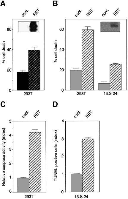
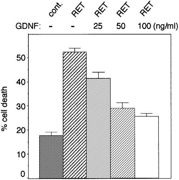
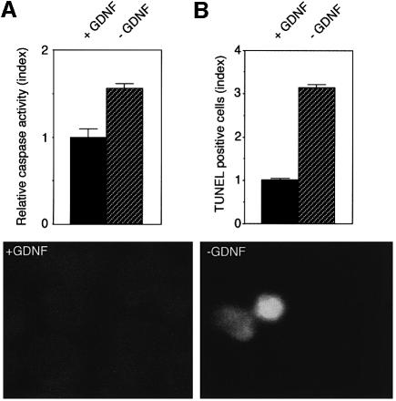
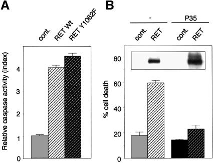
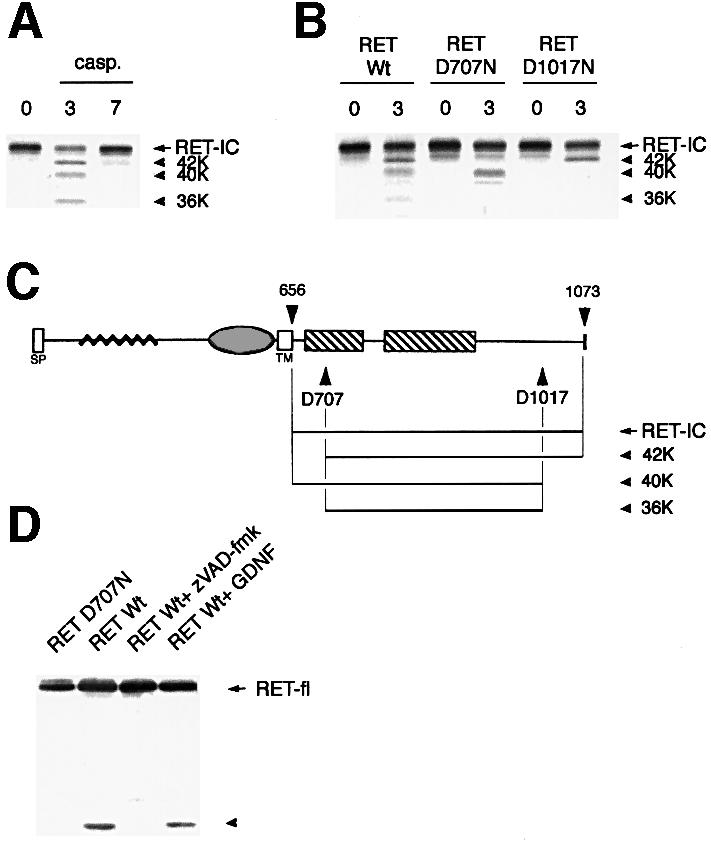
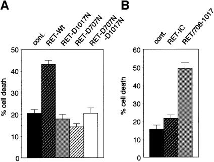
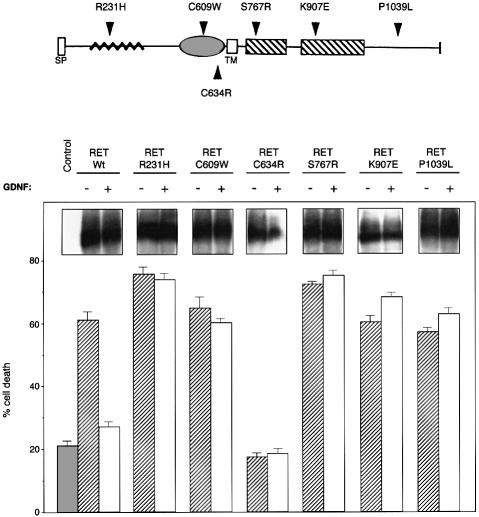
The RET (rearranged during transfection) proto-oncogene encodes a tyrosine kinase receptor involved in both multiple endocrine neoplasia type 2 (MEN 2), an inherited cancer syndrome, and Hirschsprung disease (HSCR), a developmental defect of enteric neurons. We report here that the expression of RET receptor induces apoptosis. This pro-apoptotic effect of RET is inhibited in the presence of its ligand glial cell line-derived neurotrophic factor (GDNF). Furthermore, we present evidence that RET induces apoptosis via its own cleavage by caspases, a phenomenon allowing the liberation/exposure of a pro-apoptotic domain of RET. In addition, we report that Hirschsprung-associated RET mutations impair GDNF control of RET pro-apoptotic activity. These results indicate that HSCR may result from apoptosis of RET-expressing enteric neuroblasts.


Figures

None
Fig. 1. RET induces apoptosis in the absence of GDNF. 293T or 13.S.24 cells were transiently transfected with either the RET expression plasmid pJ7Ω-ret9 (RET) or pCMV control plasmid (cont.). (A) 293T cell death induced by RET expression monitored by trypan blue staining. The percentage of cell death is presented as the percentage of trypan blue-positive cells in the different transfected cell populations. (B) 293T or 13.S.24 cells were co-transfected with the GFRα-1 expression plasmid (GFRα-1) and either pJ7Ω-ret9 (RET) or pCMV control plasmid (cont.). Cell death was estimated as in (A). Insets (A) and (B): 293T or 13.S.24 cells transfected with either a mock plasmid (cont.) or the RET-expressing plasmid (RET) were analyzed for RET expression by western blot assay using anti-RET antibody. While RET is not endogenously expressed in 293T or 13.S.24 cells, transfection assays allow the detection of RET in both cell lines. (C and D) RET induces caspase activation and DNA fragmentation. RET expression plasmid or a mock plasmid were co-transfected in the presence of the GFRα-1 expression plasmid into 293T cells (C) or 13.S.24 cells (D), and 48 h after transfection, caspase activity or DNA fragmentation was determined as described in Materials and methods. In (C), relative caspase activity was determined as the ratio between the caspase activity of the co-transfected RET+GFRα-1 cells and that measured in cells transfected with GFRα-1; for all samples, the background remaining after inhibition by Ac-DEVD-CHO was subtracted. In (D), the index of TUNEL-positive cells is presented as the ratio between the number of TUNEL-positive cells in the RET+GFRα-1-transfected population versus the GFRα-1-transfected population. Standard deviations are indicated (n = 3).
None
Fig. 2. GDNF blocks RET-induced cell death. 293T cells were co-transfected with the GFRα-1 expression plasmid in the presence or absence of the RET expression plasmid. Twenty-four hours after transfection, 25, 50 or 100 ng/ml GDNF was added to the culture medium. Cell death was then measured by trypan blue staining as described in Materials and methods. Standard deviations are indicated (n = 3).
None
Fig. 3. GDNF withdrawal induces apoptotic death in RET-expressing N2a cells. RET- and GFRα-1-expressing N2a neuroblastoma cells were cultivated in the presence of GDNF for 48 h. Cells were then allowed to grow for 12 h either in the presence or the absence of GDNF. Apoptosis was then quantified either by caspase activity measurement (A) or by TUNEL assay (B) as described in Figure 1C and D. Standard deviations are indicated (n = 3). The relative ratio of TUNEL-positive cells is also indicated. In (B), an example of a TUNEL-positive cell is shown in the absence of GDNF.
None
Fig. 4. RET induces apoptosis via a caspase-dependent pathway. (A) RET-induced cell death is independent of RET-induced kinase-dependent signal. 293T cells were transfected with RET wild type or RET mutant C634R/Y1062F in the presence of GFRα-1. Caspase activity was then monitored as described in Figure 1. (B) RET-induced cell death is blocked by p35 expression. 293T cells were transiently transfected with either the RET expression plasmid pJ7Ω-ret9 (RET) or pCMV control plasmid (cont.) in the presence or absence of the p35 expression construct pBabe-p35. Cell death was then monitored as described in Figure 1. Standard deviations are indicated (n = 3). Inset: western blots using anti-RET antibody indicating the expression of the RET protein after transfection.
None
Fig. 5. RET is a caspase substrate. (AIn vitro translated RET-IC was incubated without caspase (first lane) or with purified caspase-3 (0.3 µM), or caspase-7 (1.6 µM) for 2 h. An autoradiograph is shown. (B) Similar experiment to that in (A) except that the mutants RET-IC D707N and RET-IC D1017N were translated in vitro and incubated with caspase-3. (C) Diagram of RET. (D) RET is cleaved by caspase in vivo. 293T cells were co-transfected with either the RET-expressing or the D707N-expressing construct in the presence of GFRα-1-expressing construct. Cells were then treated or not for 24 h with 20 µM zVAD-fmk or with 0.1 µg/ml GDNF. Immunoblot using anti-RET antibody is shown.
None
Fig. 6. Cleavage of RET by caspases is crucial for RET pro-apoptotic activity. (A) Mutation of the caspase cleavage sites of RET blocks RET-induced cell death. 293T cells were transiently transfected with either the full-length RET wild type, the full-length RET-D707N, RET-D1017N or the double RET-D707N/D1017N mutant in the presence of GFRα-1 expression plasmid. Cell death was then monitored as described in Figure 1. (B) Expression of the fragment lying from amino acid 708 to 1017 drives 293T cell death. 293T cells were transiently transfected with either the complete RET intracellular domain (RET-IC) or the fragment 708–1017 (RET/708–1017) expression plasmid and cell death was monitored as in Figure 1. Standard deviations are indicated (n = 3).
None
Fig. 7. HSCR-associated mutations impair GDNF control of RET-induced apoptosis. 293T cells were transiently transfected with either the RET wild type or the following mutations of RET R231H, C609W, S767R, K907E, P1039L or C634R in the presence of GFRα-1 expression plasmid. As in Figure 2, 0.1 µg/ml GDNF was added 24 h after transfection. Cell death was then monitored as described in Figure 1. Standard deviations are indicated (n = 3). Insets: western blots using anti-RET antibody indicating the expression of the RET protein after transfection.

References

    1. Arighi E. et al. (1997) Identification of Shc docking site on Ret tyrosine kinase. Oncogene, 14, 773–782. - PubMed
    1. Baloh R.H. et al. (1997) TrnR2, a novel receptor that mediates neurturin and GDNF signaling through Ret. Neuron, 18, 793–802. - PubMed
    1. Bredesen D.E., Ye,X., Tasinato,A., Sperandio,S., Assa-Munt,N. and Rabizadeh,S. (1998) p75NTR and the concept of cellular dependence: seeing how the other half die. Cell Death Differ., 5, 365–371. - PubMed
    1. Cacalano G. et al. (1998) GFRα1 is an essential receptor component for GDNF in the developing nervous system and kidney. Neuron, 21, 53–62. - PMC - PubMed
    1. Cardone M.H., Roy,N., Stennicke,H.R., Salvesen,G.S., Franke,T.F., Stanbridge,E., Frisch,S. and Reed,J.C. (1998) Regulation of cell death protease caspase-9 by phosphorylation. Science, 282, 1318–1321. - PubMed

Publication types

MeSH terms