. 2000 Aug 1;19(15):4074-90.
doi: 10.1093/emboj/19.15.4074.

Targeting of N-CoR and histone deacetylase 3 by the oncoprotein v-erbA yields a chromatin infrastructure-dependent transcriptional repression pathway

Affiliations

Targeting of N-CoR and histone deacetylase 3 by the oncoprotein v-erbA yields a chromatin infrastructure-dependent transcriptional repression pathway

F D Urnov et al. EMBO J. .

Abstract

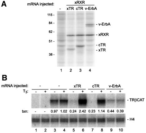
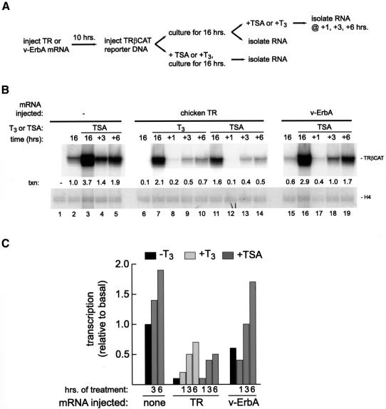
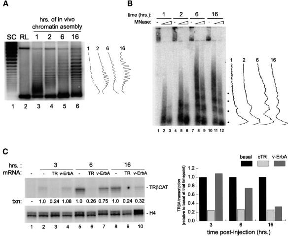
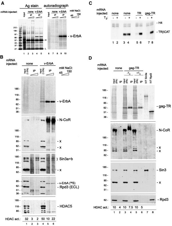
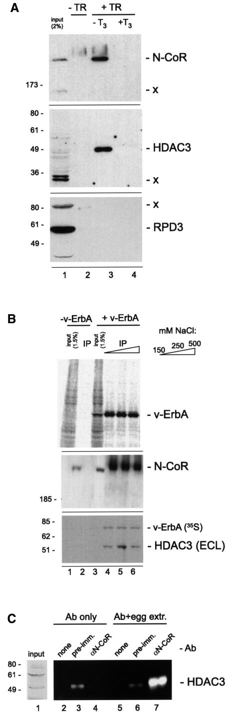
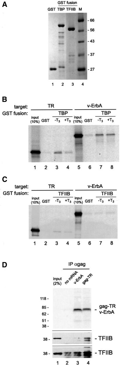
Transcriptional repression by nuclear hormone receptors is thought to result from a unison of targeting chromatin modification and disabling the basal transcriptional machinery. We used Xenopus oocytes to compare silencing effected by the thyroid hormone receptor (TR) and its mutated version, the oncoprotein v-ErbA, on partly and fully chromatinized TR-responsive templates in vivo. Repression by v-ErbA was not as efficient as that mediated by TR, was significantly more sensitive to histone deacetylase (HDAC) inhibitor treatment and, unlike TR, v-ErbA required mature chromatin to effect repression. We find that both v-ErbA and TR can recruit the corepressor N-CoR, but, in contrast to existing models, show a concomitant enrichment for HDAC3 that occurs without an association with Sin3, HDAC1/RPD3, Mi-2 or HDAC5. We propose a requirement for chromatin infrastructure in N-CoR/HDAC3-effected repression and suggest that the inability of v-ErbA to silence on partly chromatinized templates may stem from its impaired capacity to interfere with basal transcriptional machinery function. In support of this notion, we find v-ErbA to be less competent than TR for binding to TFIIB in vitro and in vivo.


Figures

None
Fig. 1. Chicken TRα (c-ErbA) and v-ErbA can be expressed in the Xenopus oocyte and can repress transcription driven by the TRβA gene promoter. (AIn vivo synthesis of chicken TRα and v-ErbA. Oocytes were left uninjected (lane 1), or injected with 5 ng of Xenopus RXRα mRNA along with 5 ng of mRNA for Xenopus TRβ (lane 2), chicken TRα (lane 3) or v-ErbA (lane 4) mRNA. The oocytes were cultured in the presence of [35S]methionine for 10 h, and oocyte protein extract was prepared as described in Materials and methods. Proteins synthesized in the oocyte during the incubation period were visualized by SDS–PAGE and autoradiography. The positions of the proteins derived from the injected mRNAs are indicated to the right of the autoradiograph; molecular weight marker size is indicated to the left. (B) Transcriptional regulation by chicken TRα and v-ErbA. Groups of 20 oocytes were injected into the cytoplasm with 5 ng of Xenopus RXRα mRNA (lanes 5–10) along with 5 ng of Xenopus TRβ (lanes 5 and 6), chicken TRα (lanes 7 and 8) or v-ErbA (lanes 9 and 10) mRNA; the oocytes in lanes 1–4 were not injected with mRNA. After a 12 h incubation, the oocytes were left uninjected (lanes 1 and 2), or injected into the nucleus with 1 ng (lanes 3–10) of double-stranded reporter plasmid DNA (pTRβA); the oocytes were then cultured for an additional 16 h in the absence (–) or presence (+) of 100 nM thyroid hormone (T3), following which total RNA was extracted and levels of H4 and TRβA mRNA were measured by primer extension as described in Materials and methods. The primer against histone H4 mRNA that is stored in the oocyte was included in all reactions to serve as a loading control. Transcriptional activity (txn) was quantitated by normalizing the signal for TRβA in each sample against that for H4 in that sample, and expressing the result in arbitrary units where 1 unit = transcriptional activity in the absence of injected mRNA (the average of signals from lanes 3 and 4).
None
Fig. 2. Analysis of the effects of an HDAC inhibitor (TSA) on the establishment and maintenance of transcriptional silencing by TR and v-ErbA. (A) Schematic representation of the two experimental regimens used. (B) Groups of 20 oocytes were left uninjected (lanes 1–5), injected into the cytoplasm with 1 ng each of Xenopus RXRα and chicken TRα mRNA (lanes 6–14), or 3 ng each of Xenopus RXRα and wild-type v-ErbA mRNA (lanes 15–19). Following incubation for 10 h, 500 pg of pTRβA double-stranded reporter plasmid DNA were injected into the nucleus (all lanes except lane 1). Following injection, oocytes in lanes 2, 6 and 15 were cultured for 16 h and total RNA extracted for analysis (these served to measure basal transcription and the repressed state in the presence of TR or v-ErbA, respectively). Oocytes in lanes 3, 11 and 16 were cultured for the same length of time in the presence of 33 nM TSA prior to total RNA extraction; this regimen revealed the effects on transcriptional activity of an HDAC inhibitor present during chromatin assembly and maturation (the effect of 100 nM T3 is shown in lane 7). To determine the effect of TSA or T3 on transcriptional activity of a mature chromatin template, groups of oocytes were injected with receptor mRNA, cultured for 10 h to allow protein synthesis, injected with reporter plasmid DNA, and cultured for 16 h to allow full chromatin assembly. TSA or T3 (33 or 100 nM, respectively) was then added where indicated, and samples of 20 oocytes were removed for total RNA isolation at 1, 3 and 6 h following addition. Transcriptional activity of the reporter plasmid was analyzed and quantitated as described in the legend to Figure 1B. (C) A graphical representation of the data in (B).
None
Fig. 3. An analysis of requirements for template chromatinization in transcriptional repression by TR and v-ErbA. (A) Alterations in plasmid topology due to progressive chromatinization were revealed by extracting plasmid DNA from injected oocytes (see below) at the indicated timepoints and analyzing the samples on a chloroquine-containing gel along with a sample of supercoiled (lane 1) and relaxed (lane 2) plasmid. Southern blotting was performed with a 0.8 kb EcoRI–SmaI fragment corresponding to positions –1325 to –515 in the TRβA gene promoter. PhosphorImager-generated scans of data from the indicated timepoints are shown to the right of the gel. (B) A large group of oocytes was injected into the nucleus with 3 ng of pTRβA double-stranded reporter plasmid DNA. At indicated timepoints, 30 oocytes were removed, homogenized as described in Materials and methods, and 1/3 of the sample was left untreated with nuclease (lanes 1, 4, 7 and 10), or treated with 30 or 120 Worthington units of MNase (the first and second lane of each pair, respectively). DNA was extracted, fractionated on a 2% agarose gel and analyzed by Southern blotting as described in Materials and methods. PhosphorImager-generated scans of data from the right lane of each pair at the indicated timepoints are shown to the right of the gel. (C) A large group of oocytes was uninjected (lanes 1, 2, 5 and 8), injected into the cytoplasm with 1 ng each of Xenopus RXRα and chicken TRα mRNA (lanes 3, 6 and 9), or 2 ng each of Xenopus RXRα and wild-type v-ErbA mRNA (lanes 4, 7 and 10). Following incubation for 10 h, 500 pg of pTRβA double-stranded reporter plasmid DNA were injected into the nucleus (all lanes except lane 1). At 3, 6 and 16 h following DNA injection (lanes 2–4, 5–7 and 8–10, respectively), samples of 20 oocytes were removed and total RNA isolated. Transcriptional activity of the reporter was analyzed by primer extension as described in the legend to Figure 1B. For each timepoint the basal level of transcription (lanes 2, 5 and 8, respectively) was set to 1.0. A graphical representation of the data is shown to the right of the autoradiograph.
None
Fig. 4. A biochemical analysis of associations of the v-ErbA oncoprotein in the oocyte. (A) v-ErbA can be immunoprecipitated from whole oocyte extract using a monoclonal antibody against the gag domain. Oocytes (100 per sample) were left uninjected (lanes 1–3 and 6–8) or injected with 10 ng of mRNA for v-ErbA (lanes 4, 5, 9 and 10) and cultured in the presence of [35S]methionine for 16 h; immunoprecipitation with anti-gag monoclonal antibody was performed as described in Materials and methods in 45 mM NaCl-containing buffer, followed by washing in buffer containing the amount of NaCl indicated on the right. One third of the immunoprecipitate was resolved on a 4–12% Laemmli SDS–PAGE followed by silver staining (lanes 1–5) and autoradiographic analysis of the silver-stained gel (lanes 6–10). The ‘input’ lane contains 1/20 of an oocyte equivalent of whole oocyte protein extract; the position of molecular weight markers is indicated to the left of the gel, the v-ErbA-sized polypeptide—to the right of the gel, and of immunoglobulin light (L) and heavy (H) chain—between lanes 5 and 6. (B) Recruitment of N-CoR and HDAC activity to v-ErbA is not accompanied by an enrichment for Sin3, HDAC1 or 5. One third of the immunoprecipitate described in the legend to (A) was resolved on a 4–12% Laemmli SDS–PAGE along with the indicated quantity of input whole oocyte extract, transferred to a nitrocellulose membrane and analyzed by autoradiography (top panel) and sequential western blotting with antibody against Xenopus N-CoR (second panel), Sin3 (third panel), Rpd3p/HDAC1 (fourth panel) and mouse HDAC5 (fifth panel). In all panels, the position of molecular weight markers is indicated to the right, and the position of the antigen targeted by the antibody is to the left of the autoradiograph. The positions of additional polypeptides that cross-react with the antibodies are indicated by –x; longer (∼5–10 min) exposures to X-ray film used in the fourth and fifth panels reveal a band corresponding precisely to the 35S-labeled v-ErbA polypeptide along with the chemiluminescence (ECL)-derived signal from the target antigen. The numbers below the fifth panel were obtained by measuring HDAC activity in the remaining 1/3 of the immunoprecipitate exactly as described (Wade et al., 1999b) and expressing the resulting values in arbitrary units relative to a blank sample. (C) The gag tag does not affect the properties of TR as a transcriptional regulator of the Xenopus TRβA gene. Oocytes were injected with mRNA for Xenopus RXRα (lanes 5–8) along with mRNA chicken TRα (lanes 5 and 6) or chicken TRα tagged with the AEV gag moiety on its N-terminus (lanes 7 and 8), followed by injection of 1 ng of double-stranded TRβA reporter DNA (lanes 3–8) and analysis of transcription by primer extension as described in the legend to Figure 1B. (D) Ligand-regulated recruitment to TR of N-CoR and HDAC activity is not accompanied by an enrichment for Sin3 or HDAC1/RPD3. Sixty oocytes were left uninjected (lanes 1 and 2), or injected with 10 ng of mRNA for gag-tagged chicken TRα (lanes 3–6) and cultured in [35S]methionine-containing medium in the presence or absence of 100 nM T3, as indicated. Immunoprecipitation with antibody against gag was performed as described in (B); binding and washing was performed in 45 mM NaCl and 1 µM T3 where indicated. An aliquot of the immunoprecipitate along with the indicated quantity of input extract and an aliquot of in vitro translated Xenopus Sin3 (lane 7) was resolved on a 4–12% Laemmli SDS–PAGE, transferred to nitrocellulose and analyzed by autoradiography (top panel; quantitation of the gag-TR signal reveals a 2-fold greater amount in lane 4 as compared with lane 6) and by western blotting with antibody against N-CoR (middle panel) and Sin3 (bottom panel). An identical immunoprecipitation was split into two aliquots, and one was analyzed by western blotting as above with an antibody against RPD3 (bottom panel), while the other was assayed for HDAC activity as above.
None
Fig. 5. Further characterization of N-CoR/HDAC enrichment by v-ErbA and TR. (A) Immunoprecipitation from 100 uninjected oocytes (lane 2) or oocytes injected with v-ErbA mRNA (lanes 4–6) was performed as described above, except the binding buffer contained 95 mM NaCl (final concentration of monovalent cations in the reaction = 100 mM), and the washing was performed with buffer containing NaCl at the concentration indicated to the right of the gel (100 mM for lane 2). One sixth of the sample was resolved on a 6% Laemmli SDS–PAGE along with the indicated quantity of input whole oocyte extract (lanes 1 and 3) and an aliquot of in vitro translated Xenopus Sin3 (lane 7). Western blotting was performed with antibody against Sin3 (top two panels; these represent two different exposures of the same membrane), followed by stripping and reprobing with an antibody for N-CoR (bottom panel). (B) The gag moiety fails to recruit N-CoR, and ligand fails to eliminate N-CoR from v-ErbA. Oocytes were left uninjected (lanes 1 and 2), injected with an mRNA representing gag-tagged TR with the entire ligand-binding domain deleted (lanes 3 and 4), v-ErbA (lanes 5–8) or gag-TR (lanes 9–12), and cultured in medium containing [35S]methionine. Following immunoprecipitation in 50 mM NaCl in the absence or presence of 1 µM T3 (as indicated) with anti-gag antibody, one half of the immunoprecipitate was resolved on a 4–20% PAGE and autoradiographed (top panel), while the other half was resolved on a 6% PAGE and immunoblotted with antibody against N-CoR (second and third panel; the lower of the two represents a longer exposure of the upper, included to demonstrate N-CoR in all the input samples) and Mi-2 (lower panel). Lanes labeled ‘i’ contain 1% input protein.
None
Fig. 6. TR and v-ErbA recruit HDAC3. (A) TR enriches for N-CoR and HDAC3, but not RPD3. A large aliquot of Xenopus egg extract was adjusted to 50 mM NaCl and 0.1% NP-40 and incubated with a preparation of bead-immobilized GST–TR (lanes 3 and 4) or GST (lane 2). Where indicated, 2.5 µM T3 was included in the reaction. Following washing as described in Figure 4D, proteins associated with the beads were analyzed by immunoblotting against N-CoR (top panel), HDAC3 (middle panel) and RPD3 (bottom panel). (B) v-ErbA-dependent enrichment for N-CoR in the immunoprecipitate is accompanied by an enrichment for HDAC3. One half of the immunoprecipitate described in the legend to Figure 5A was resolved on a 4–12% Laemmli SDS–PAGE, and analyzed by autoradiography (top panel) and western blotting with antibody against N-CoR (middle panel) and human HDAC3 (bottom panel). As in Figure 4B, the longer (10 min) exposure used in the bottom panel reveals both the 35S-labeled antigen directly targeted in the immunoprecipitation as well as chemiluminescence-derived signal from the antibody used in the western blot. Low efficiency of the anti-HDAC3 antibody prevents the visualization of HDAC3 in the 0.5 oocyte equivalents of extract used in the input lanes (lanes 1 and 3). (C) N-CoR and HDAC3 co-immunoprecipitate. A preparation of bead-immobilized rabbit antiserum against N-CoR (lanes 4 and 7), the cognate pre-immunization serum (lanes 3 and 6) or the beads with no antibody (lanes 2 and 5) was incubated in Xenopus egg extract adjusted to 50 mM NaCl and 0.1% NP-40. The proteins associating with the antibody were eluted with glycine and analyzed for the presence of HDAC3 by immunoblotting with rabbit-derived anti-HDAC3 serum and extended duration ECL reagents, followed by visualization and quantitation on a CCD camera-equipped ChemiImager-4400 low light imaging system. The small amount of rabbit anti-N-CoR antibody released from the beads (lanes 2–4) yields Ig heavy chain signal at 50 kDa (lanes 2–4), i.e. at the molecular weight of HDAC3; quantitation indicates a 60-fold greater signal in lane 7 (anti–N-CoR antibody + egg extract) than in lane 4 (anti–N-CoR antibody only). Lane 1 contains an aliquot of egg extract analyzed on the same gel and probed with conventional ECL reagents, followed by autoradiography.
None
Fig. 7. In vitro and in vivo assays for the ability of TR and v-ErbA to interact with components of the basal transcription machinery. (A) A preparation of glutathione–Sepharose beads attached to GST (lane 1), GST–TBP (lane 2) or GST–TFIIB (lane 3) was boiled in Laemmli SDS–PAGE loading buffer, electrophoresed, and the recombinant proteins visualized by Coomassie Brilliant Blue staining. Lane ‘M’ contains molecular weight markers the sizes of which are indicated to the right of the gel. (B) Equimolar quantities of reticulocyte-lysate-translated TR (lanes 1–4) or v-ErbA (lanes 5–8) were mixed with beads fused to GST (lanes 2 and 6) or GST–TBP (lanes 3, 4, 7 and 8). T3 was included at 1 µM where indicated. Following incubation and washing in buffer containing 0.5 M NaCl and 0.1% NP-40, the proteins bound to the beads were eluted and analyzed by SDS–PAGE and autoradiography. The ‘input’ lanes contain 1/10 of the total protein used in each reaction. (C) The ability of TR (lanes 1–4) and v-ErbA (lanes 5–8) to bind GST–TFIIB was assayed exactly as described in the legend to (B). (D) v-ErbA exhibits a moderate impairment in binding to TFIIB in vivo. Oocytes were left uninjected (lane 2) or injected with mRNA for v-ErbA (lane 3) or gag-TR (lane 4); the injected oocytes were cultured in [35S]methionine-containing medium. Immunoprecipitation with antibody against gag was performed as described in Figure 4B, except the binding and washing buffers contained 5 mM NaCl and 5 mM KCl. The entire immunoprecipitate was resolved on a 4–12% Laemmli SDS–PAGE along with the indicated quantity of input extract and analyzed by autoradiography (top panel) and western blotting against TFIIB (the middle and lower panels represent different exposures of the same membrane).

Similar articles

Cited by

References

    1. Adams C.R. and Kamakaka,R.T. (1999) Chromatin assembly: biochemical identities and genetic redundancy. Curr. Opin. Genet. Dev., 9, 185–190. - PubMed
    1. Almouzni G. and Wolffe,A.P. (1993a) Nuclear assembly, structure, and function: the use of Xenopus in vitro systems. Exp. Cell Res., 205, 1–15. - PubMed
    1. Almouzni G. and Wolffe,A.P. (1993b) Replication-coupled chromatin assembly is required for the repression of basal transcription in vivo. Genes Dev., 7, 2033–2047. - PubMed
    1. Almouzni G., Mechali,M. and Wolffe,A.P. (1990) Competition between transcription complex assembly and chromatin assembly on replicating DNA. EMBO J., 9, 573–582. - PMC - PubMed
    1. Baniahmad A., Ha,I., Reinberg,D., Tsai,S., Tsai,M.J. and O’Malley,B.W. (1993) Interaction of human thyroid hormone receptor β with transcription factor TFIIB may mediate target gene derepression and activation by thyroid hormone. Proc. Natl Acad. Sci. USA, 90, 8832–8836. - PMC - PubMed

MeSH terms