. 2000 Aug 1;19(15):4111-22.
doi: 10.1093/emboj/19.15.4111.

How Stat1 mediates constitutive gene expression: a complex of unphosphorylated Stat1 and IRF1 supports transcription of the LMP2 gene

Affiliations

How Stat1 mediates constitutive gene expression: a complex of unphosphorylated Stat1 and IRF1 supports transcription of the LMP2 gene

M Chatterjee-Kishore et al. EMBO J. .

Erratum in

  • EMBO J 2000 Sep 1;19(17):4855

Abstract

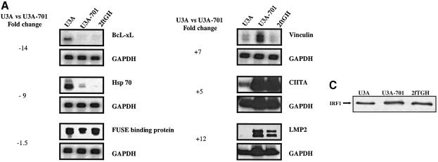
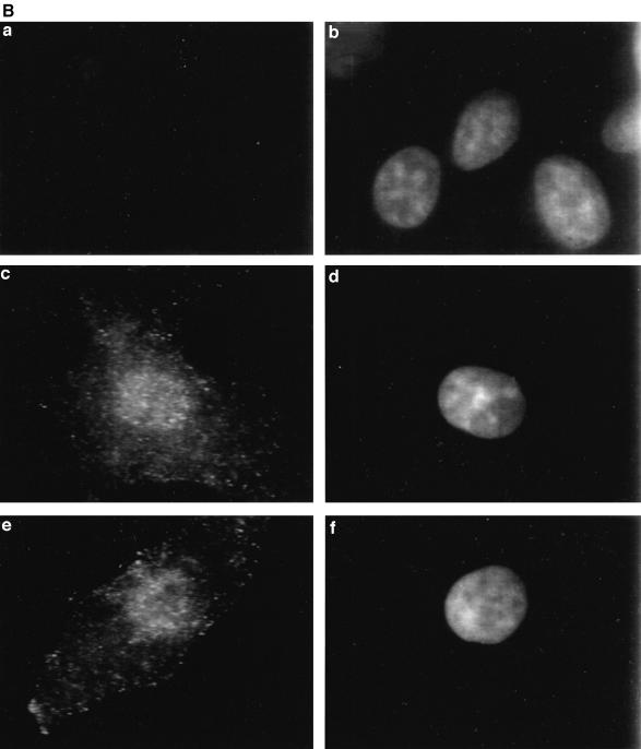
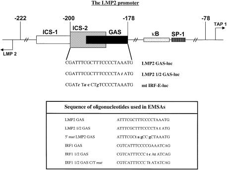
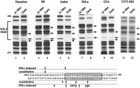
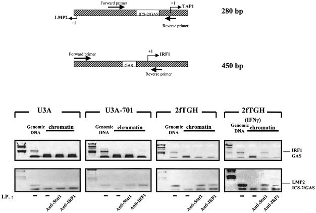
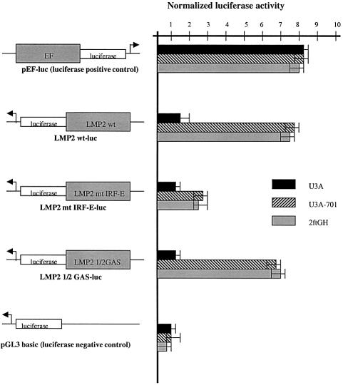
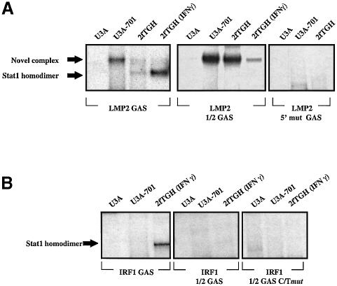
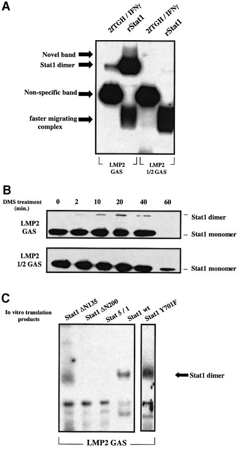
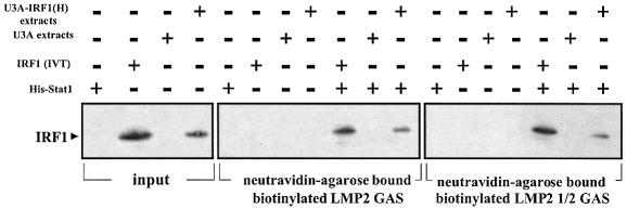
Analysis of mRNA levels in cells that express or lack signal transducers and activators of transcription 1 (Stat1) reveals that Stat1 mediates the constitutive transcription of many genes. Expression of the low molecular mass polypeptide 2 (LMP2), which requires Stat1, has been studied in detail. The overlapping interferon consensus sequence 2/gamma-interferon-activated sequence (ICS-2/GAS) elements in the LMP2 promoter bind to interferon regulatory factor 1 (IRF1) and Stat1 and are occupied constitutively in vivo. The point mutant of Stat1, Y701F, which does not form dimers involving SH2-phosphotyrosine interactions, binds to the GAS element and supports LMP2 expression. Unphosphorylated Stat1 binds to IRF1 directly and we conclude that this complex uses the ICS-2/GAS element to mediate constitutive LMP2 transcription in vivo. The promoter of the IRF1 gene, which also contains a GAS site but not an adjacent ICS-2 site, is not activated by Stat1 Y701F. The promoters of other genes whose constitutive expression requires Stat1 may also utilize complexes of unphosphorylated Stat1 with IRF1 or other transcription factors.


Figures

None
Fig. 1. Stat1 regulates constitutive gene expression. (A) Confirmation of Affymetrix data by northern and S1 nuclease analyses. The expression of some of the differentially expressed genes (in bold in Table I) was analyzed in U3A, U3A-701 and 2fTGH cells. S1 nuclease analysis was used for expression of the LMP2, CIITA and vinculin genes, with GAPDH as control. The sizes of the protected fragments were 677 bp for LMP2, 503 bp for CIITA, 450 bp for GAPDH and 286 bp for vinculin. BcL-xL, Hsp-70 and FUSE-binding protein mRNA expression was analyzed in northern transfers using PCR-derived probes. The transfers were stripped and reprobed for GAPDH expression. Fold changes were estimated by densitometry. In the case of CIITA, the change is probably underestimated due to the large signal in U3A-701 and 2fTGH cells. (B) Unphosphorylated Stat1 translocates to the nucleus. Formaldehyde-fixed U3A, 2fTGH and U3A-701 cells (a, c and e, respectively) were stained with anti-Stat1 and FITC-conjugated goat anti-rabbit antibody and DAPI was used to stain the nuclei (b, d and f, respectively). (C) IRF1 expression in U3A, U3A-701 and 2fTGH cells. Approximately 2 × 10⁶ cells were labeled in vivo using [35S]methionine. Proteins were extracted after 18 h of labeling and counts per minute were determined. Aliquots containing equal numbers of counts were immunoprecipitated with anti-IRF1 and the immunoprecipitates were separated by 12% SDS–PAGE.
None
Fig. 2. Functional diagram of the LMP2/TAP1 bi-directional promoter and the sequences of wild-type and mutant constructs representing the ICS-2/GAS region. The table shows the sequences of oligonucleotides used in EMSAs.
None
Fig. 3. Constitutive in vivo occupancy of the ICS-2/GAS. Dimethyl sulfate treatment, genomic DNA preparation and LM-PCR for the ICS-2/GAS (IRF-E) region (lower strand) of the LMP2 promoter were described by White et al. (1996). DNA samples treated with DMS in vitro or in vivo were analyzed simultaneously. Open arrows mark the bases protected from methylation and the filled arrows mark methylation enhancements. The sequence of the ICS-2/GAS region is marked with the constitutive and IFN-γ-induced protections and enhancements determined in HeLa cells by White et al. (1996).
None
Fig. 4. In vivo cross-linking of unphosphorylated Stat1 and IRF1 on the ICS-2/GAS region of the LMP2 promoter. Proteins were cross-linked to DNA in growing U3A and U3A-701 cells and in untreated and IFN-γ-treated (1000 IU/ml for 1 h) 2fTGH cells. DNA-bound Stat1 and IRF1 were immunoprecipitated. Co-immunoprecipitated DNA from uncross-linked genomic DNA (lanes marked genomic DNA) or in vivo cross-linked chromatin (lanes marked chromatin) was amplified by PCR, using primers (position shown at the top of the figure) for the LMP2 ICS/GAS or IRF1 GAS region. The positions of the 450 and 280 bp PCR products corresponding to the IRF1 GAS and the LMP2 ICS-2/GAS regions are marked.
None
Fig. 5. Activity of wild-type or mutant LMP2 promoter constructs in U3A, U3A-701 and 2fTGH cells. Cells were stably transfected with LMP2 GAS-luc, mt IRF-E-luc, LMP2 1/2GAS-luc, pEF-luc as positive control and pGL3-basic as negative control. Luciferase activity was assayed in duplicate in extracts from pools of stably transfected cells. Luciferase activity units were normalized against total protein levels, assayed spectrophotometrically.
None
Fig. 6. A novel complex binds to the LMP2 GAS in U3A-701 cells. (A) Binding activities towards the wild-type and mutant LMP2 GAS probes in extracts prepared from U3A, U3A-701 and 2fTGH cells. Extracts from untreated and IFN-γ-treated cells were assayed by EMSA. (B) Binding activities towards the wild-type and mutant IRF1 GAS probes in extracts prepared from U3A, U3A-701 and 2fTGH cells. Extracts from untreated cells were assayed in EMSAs.
None
Fig. 7. rStat1 can bind DNA as a dimer or monomer, but with low affinity. (A) EMSA analysis of the binding of rStat1 to the LMP2 GAS and LMP2 1/2 GAS probes. Approximately 200 ng of purified rStat1 and 5 µg of extracts from IFN-γ-treated 2fTGH cells were used in EMSAs with the LMP2 GAS probe. A 500 ng aliquot of rStat1 was used in EMSAs with the LMP2 1/2 GAS and 5′ mut LMP2 GAS probes. (B) Time course of DMS-mediated cross-linking of rStat1 on LMP2 GAS or LMP2 1/2 GAS probes. Neutravidin–agarose beads saturated with either the LMP2 GAS or 1/2 GAS probes were allowed to bind to rStat1 (2–5 µg). Proteins were cross-linked while bound to DNA using DMS and analyzed in western transfers using anti-Stat1. (C) The N-terminal region of unphosphorylated Stat1 is required for dimer formation in vitro. Stat1, Stat1 Y701F, Stat1 ΔN135, Stat1 ΔN200 and Stat5/1 were translated in vitro and partially purified on heparin–agarose. The DNA-binding activity of equal amounts of Stat1, Stat1 ΔN135, Stat1 ΔN200, Stat5/1 and Stat1 Y701F to the LMP2 GAS was analyzed in EMSAs.
None
Fig. 8. A complex of Stat1 and IRF1 binds to the LMP2 GAS or LMP2 1/2 GAS. Streptavidin–agarose beads were saturated with biotin-labeled oligonucleotides at 4°C for 2 h. The beads were incubated with either Stat1 (2–5 µg), IRF1 (in vitro translation products), U3A extracts or U3A-IRF1(H) extracts at 4°C for 1 h. The Stat1-saturated beads were then washed with HEM buffer and incubated with either IRF1 (in vitro translation product), U3A extracts or U3A-IRF1(H) extracts. Samples were analyzed in western transfers with anti-IRF1.

References

    1. Baden H.A., Sarma,S.P., Kapust,R.B., Byrd,R.A. and Waugh,D.S. (1998) The amino-terminal domain of human STAT4. Overproduction, purification, and biophysical characterization. J. Biol. Chem., 273, 17109–17114. - PubMed
    1. Chatterjee-Kishore M., Kishore,R., Hicklin,D.J., Marincola,F.M. and Ferrone,S. (1998) Different requirements for signal transducer and activator of transcription 1α and interferon regulatory factor 1 in the regulation of low molecular mass polypeptide 2 and transporter associated with antigen processing 1 gene expression. J. Biol. Chem., 273, 16177–16183. - PubMed
    1. Chatterjee-Kishore M., van den Akker,F. and Stark,G.R. (2000) Adenovirus E1A down-regulates LMP2 transcription by interfering with the binding of Stat1 to IRF1. J. Biol. Chem., 275, 20406–20411. - PubMed
    1. Chen X., Vinkemeier,U., Zhao,Y., Jeruzalmi,D., Darnell,J.E.,Jr and Kuriyan,J. (1998) Crystal structure of a tyrosine phosphorylated STAT-1 dimer bound to DNA. Cell, 93, 827–839. - PubMed
    1. Darnell J.E. Jr, Kerr,I.M. and Stark,G.R. (1994) Jak–STAT pathways and transcriptional activation in response to IFNs and other extracellular signaling proteins. Science, 264, 1415–1421. - PubMed

Publication types