. 2000 Aug 1;19(15):4134-43.
doi: 10.1093/emboj/19.15.4134.

Alternatively spliced hBRF variants function at different RNA polymerase III promoters

Affiliations

Alternatively spliced hBRF variants function at different RNA polymerase III promoters

V McCulloch et al. EMBO J. .

Abstract

In yeast, a single form of TFIIIB is required for transcription of all RNA polymerase III (pol III) genes. It consists of three subunits: the TATA box-binding protein (TBP), a TFIIB-related factor, BRF, and B". Human TFIIIB is not as well defined and human pol III promoters differ in their requirements for this activity. A human homolog of yeast BRF was shown to be required for transcription at the gene-internal 5S and VA1 promoters. Whether or not it was also involved in transcription from the gene-external human U6 promoter was unclear. We have isolated cDNAs encoding alternatively spliced forms of human BRF that can complex with TBP. Using immunopurified complexes containing the cloned hBRFs, we show that while hBRF1 functions at the 5S, VA1, 7SL and EBER2 promoters, a different variant, hBRF2, is required at the human U6 promoter. Thus, pol III utilizes different TFIIIB complexes at structurally distinct promoters.


Figures
None
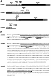
Fig. 1. Structure of hBRF variants. (A) Schematic representation of variant cDNAs. Translation start (AUG) and stop sites (STOP) are indicated. Repeat 1 and Repeat 2 refer to the regions encoding the imperfect repeats homologous to TFIIB. (B) Amino acid sequences of hBRFs 2, 3 and 4. Arrows indicate the repeat regions. Dashed lines indicate the peptides R2P and hBRF2sp used for raising antibodies. (C) The specificity of anti-peptide antibodies. hBRFs 1–4 were translated in vitro (lanes 1–4) and immunoprecipitated with purified anti-R2P (lanes 5–8) or anti-hBRF2sp antibodies (lanes 9–12).
None
Fig. 2. Characterization of interactions between hBRF variants and TBP. (A) Co-immunoprecipitation of in vitro translated hBRFs with TBP. The hBRF variants, TBP and luc were translated in vitro (lanes 1–6), incubated with TBP in either RIPA buffer (lanes 7–12) or buffer D + 300 mM KCl (lanes 13–18) or without TBP in buffer D + 300 mM KCl (lanes 23–26) and immunoprecipitated with anti-TBP mAb SL30a. (B) Identification of hBRF domains that mediate interactions with TBP. hBRF domains were translated in vitro (lanes 1–6) mixed with TBP and immunoprecipitated with SL30a (lanes 8–14). The different domains are indicated above the lanes: Rep.1, repeat 1; Rep.2, repeat 2; Rep1 + 2, repeat 1 and 2 together; hBRF1,3 sp., C-terminal domain of hBRFs 1 and 3; hBRF2 sp., C-terminal domain unique to hBRF2; hBRF4 sp., C-terminal domain unique to hBRF4.
None
Fig. 3. Reconstitution of pol III transcription using in vitro translated hBRFs, TBP and phosphocellulose fraction C. (A) Transcription of VA1 and EBER2 genes. Transcription was assayed using fraction C alone (lane 4), C + TBP (lane 1), C + luc (lane 2), luc + TBP (lane 3), the phosphocellulose B and C fractions (lane 5) and increasing amounts of luc (lanes 6–8) or each in vitro translated hBRF (lanes 9–20) with 0.1 µl of TBP and the C fraction. The amounts of hBRFs or luc added (in fmoles) are indicated above the lanes. (B) Graphical representation of the results from (A).
None
Fig. 4. Reconstitution of pol III transcription using in vitro translated hBRFs, TBP and anti-R2P antibody-depleted extract. (A) Transcription of the VA1 and U6 genes. VA1 and U6 transcription were assayed in nuclear extract (lane 1), 12CA5 mAb-depleted extract (lane 2), anti-R2P antibody-depleted extract (lane 3) or in anti-R2P antibody-depleted extract complemented with the indicated proteins. The numbers above the lanes indicate the fmoles of luc or hBRF added. (B) Graphical representation of the results from the upper panel in (A).
None
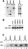
Fig. 5. Characterization of hBRF complexes assembled in human cells. (A) Detection of purified HA epitope-tagged hBRFs transiently expressed in human cells. Increasing amounts of each hBRF complex were immunoblotted with 12CA5 mAb. Arrows indicate the location of the hBRF variants on the gel. (B) TBP is present in hBRF1 and hBRF3 complexes. hBRF complexes were immunoblotted with anti-TBP mAbs. The light chain of the 12CA5 mAb and TBP are indicated. (C) Cross-linking of TBP to hBRFs expressed in human cells. Extracts from BOSC 23 cells transiently transfected with vector alone (lanes 1 and 2) or HA epitope-tagged hBRF variants (lanes 3–10) were either treated with the cross-linking agent DSP or not (indicated by + or – above the lanes) and immunoprecipitated with 12CA5 mAb under denaturing conditions. The immunoprecipitated proteins were immunoblotted with anti-TBP mAbs.
None
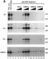
Fig. 6. Reconstitution of pol III transcription using hBRF complexes assembled in human cells. (A) Reconstitution of 5S, VA1, 7SL and EBER2 transcription. HeLa nuclear extract (lane 1) was depleted with either 12CA5 mAb (lane 2) or anti-R2P antibodies (lanes 3–15). Increasing amounts of complexes isolated from cells transfected with vector (lanes 4–6) or epitope-tagged hBRFs (lanes 7–15) were added to anti-R2P-depleted nuclear extract. The sizes (in bp) of molecular weight markers are indicated on the right. (B) Reconstitution of VA1 transcription using anti-R2P antibody-depleted extract and hBRF complexes in the presence (lanes 16–28) or absence (lanes 3–15) of exogenous TBP. Reconstitution of U6 transcription in anti-R2P antibody-depleted extracts. In the lower panel, U6 transcription was assayed in nuclear extract (lane 1), depleted extract (lane 2), depleted extract + TBP (lane 3) or with depleted extract, TBP and increasing amounts of the indicated hBRF complexes (lanes 4–15).
None
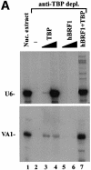
Fig. 7. U6, but not VA1, transcription is restored by the addition of recombinant TBP alone to a TBP-depleted nuclear extract. (A) Reconstitution of U6 and VA1 transcription from a TBP-depleted nuclear extract. HeLa nuclear extract was either untreated (lane 1) or immunodepleted with anti-TBP mAbs (lanes 2–7). Recombinant TBP (0.2 and 0.66 fpu; lanes 3 and 4), in vitro translated hBRF1 (3 and 5 µl; lanes 5 and 6) or 0.66 fpu of TBP in combination with 5 µl of hBRF1 (lane 7) were added to the TBP-depleted extract. (B) Anti-TBP mAbs co-deplete hBRF1 but not hBRF2 from nuclear extract. Nuclear extract depleted with anti-TBP mAbs (lanes 2, 4, 6, 8 and 10) or not (lanes 1, 3, 5, 7 and 9) was immunoblotted with pre-immune serum (lanes 1 and 2), anti-hBRF2sp antibodies (lanes 3 and 4), anti-hBRF2sp antibodies + hBRF2sp peptide (lanes 5 and 6), anti-R2P antibodies (lanes 7 and 8) or anti-R2P antibodies + R2P peptide (lanes 9 and 10). Molecular weight markers are indicated on the left.
None
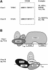
Fig. 8. Schematic showing putative transcription initiation complexes assembled on class I and class II RNA pol III promoters. B′′/? indicates the involvement of B′′ and unidentified factors.

Similar articles

Cited by

References

    1. Andrews N.C. and Faller,D.V. (1991) A rapid micropreparation technique for extraction of DNA-binding proteins from limiting numbers of mammalian cells. Nucleic Acids Res., 19, 2499. - PMC - PubMed
    1. Brow D.A. and Guthrie,C. (1990) Transcription of a yeast U6 snRNA gene requires a polymerase III promoter element in a novel position. Genes Dev., 4, 1345–1356. - PubMed
    1. Buratowski S. and Zhou,H. (1992) A suppressor of TBP mutations encodes an RNA polymerase III transcription factor with homology to TFIIB. Cell, 71, 221–230. - PubMed
    1. Carbon P. and Krol,A. (1991) Transcription of the Xenopus laevis selenocysteine tRNA(Ser)Sec gene: a system that combines an internal B box and upstream elements also found in U6 snRNA genes. EMBO J., 10, 599–606. - PMC - PubMed
    1. Colbert T. and Hahn,S. (1992) A yeast TFIIB-related factor involved in RNA polymerase III transcription. Genes Dev., 6, 1940–1949. - PubMed

Publication types

Associated data