. 2000 Aug 1;19(15):4154-63.
doi: 10.1093/emboj/19.15.4154.

TIA-1 is a translational silencer that selectively regulates the expression of TNF-alpha

Affiliations

TIA-1 is a translational silencer that selectively regulates the expression of TNF-alpha

M Piecyk et al. EMBO J. .

Abstract

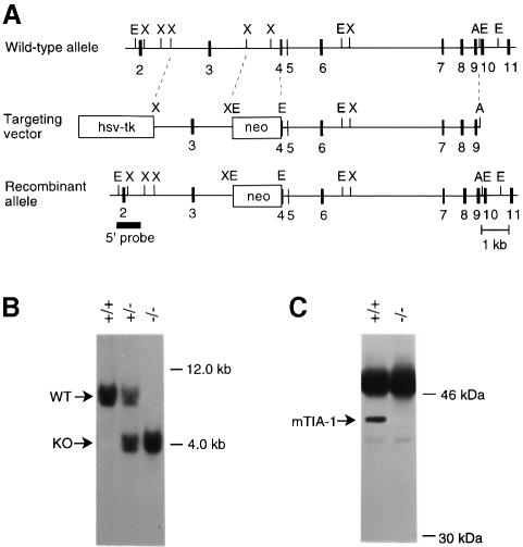
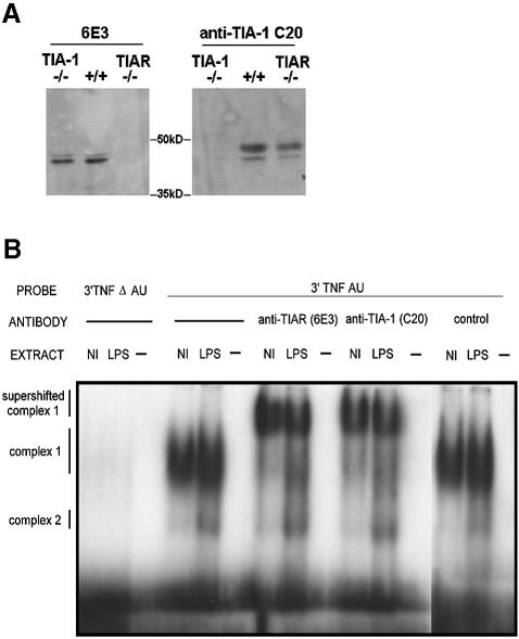
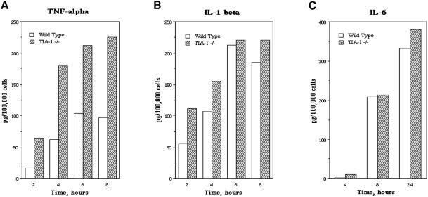
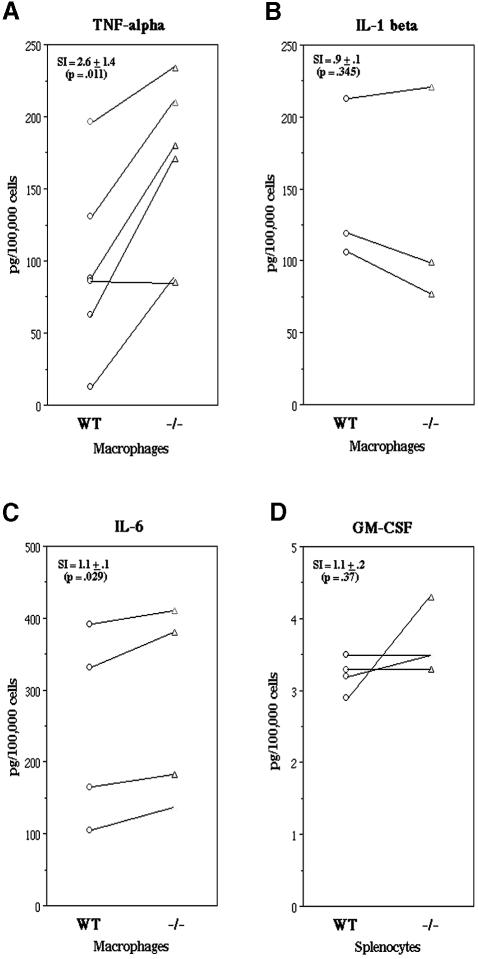
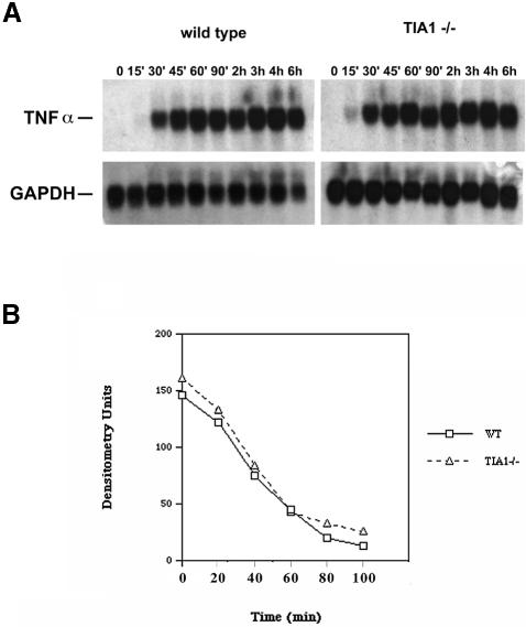
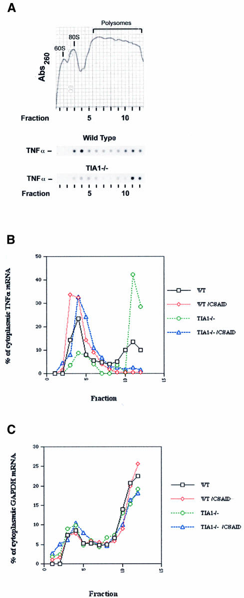
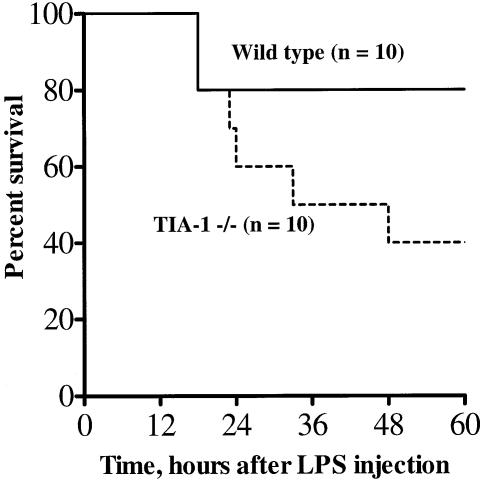
TIA-1 and TIAR are related proteins that bind to an AU-rich element (ARE) in the 3' untranslated region of tumor necrosis factor alpha (TNF-alpha) transcripts. To determine the functional significance of this interaction, we used homologous recombination to produce mutant mice lacking TIA-1. Although lipopolysaccharide (LPS)-stimulated macrophages derived from wild-type and TIA-1(-/-) mice express similar amounts of TNF-alpha transcripts, macrophages lacking TIA-1 produce significantly more TNF-alpha protein than wild-type controls. The half-life of TNF-alpha transcripts is similar in wild-type and TIA-1(-/-) macrophages, indicating that TIA-1 does not regulate transcript stability. Rather, the absence of TIA-1 significantly increases the proportion of TNF-alpha transcripts that associate with polysomes, suggesting that TIA-1 normally functions as a translational silencer. TIA-1 does not appear to regulate the production of interleukin 1 beta, granulocyte-macrophage colony-stimulating factor or interferon gamma, indicating that its effects are, at least partially, transcript specific. Mice lacking TIA-1 are hypersensitive to the toxic effects of LPS, indicating that this translational control pathway may regulate the organismal response to microbial stress.


Figures

None
Fig. 1. Construction of TIA-1–/– mouse and MEFs. (A) The expected gene replacement at the mouse tia-1 locus. Correct gene targeting should result in replacement of parts of intron 3 and exon 4 by a marker gene for positive selection (PGK–neor). The herpes simplex virus thymidine kinase expression cassette was used for negative selection. The 5′ probe used for Southern blot analysis is indicated. A, AatII; E, EcoRI, X, XbaI. (B) Southern blot analysis of DNA from offspring derived from heterozygous matings. Genomic DNA was digested with EcoRI and analyzed using the 5′ probe, yielding the 7.5 and 4.0 kb fragments expected for the wild-type and mutant allele, respectively. Southern blot analysis of ES-cell DNA using a 3′ probe and a neo probe showed proper targeting and single insertion of the transfected vector (data not shown). (C) Immunoprecipitation of lysates from tia-1+/+ and tia-1–/– embryonic fibroblasts using ML29 monoclonal antibody (mAb) and subsequent protein immunoblot with mAb 2G9 confirms the absence of TIA-1 protein in tia-1–/– cells.
None
Fig. 2. (A) Mouse embryonic fibroblasts derived from wild-type, TIA-1–/– and TIAR–/– mice. 6E3 recognizes TIAR (left panel), while anti-TIA-1 C20 recognizes TIA-1 (right panel). (B) EMSA using Raw 264.7 cells. Extracts from control or LPS-treated RAW cells were incubated with radiolabeled probes from the 3′ UTR of TNF-α with or without the ARE before separation by SDS–PAGE. Anti-TIA-1 (ML29) and anti-TIAR (6E3) supershifted complex 1, whereas a goat anti-MAD2 (Santa Cruz) control did not, indicating that both TIA-1 and TIAR are TNF-α ARE-binding proteins. The relative migration of complex 1, complex 2 and supershifted complex 1 are indicated on the left.
None
Fig. 3. Thioglycolate-elicited peritoneal macrophages collected from wild-type and TIA-1–/– BALB/c mice were treated with LPS (1 µg/ml) for the times indicated. Supernatant TNF-α and IL-6 concentrations (A and C) and combined cell lysate and supernatant IL-1β concentrations (B) were measured by ELISA. Each experiment was repeated at least three times; one representative experiment is depicted in each panel. Open bars, wild type; closed bars, TIA-1–/–.
None
Fig. 4. LPS-induced cytokine production by BALB/c mouse peritoneal macrophages. Cytokine concentrations were measured by ELISA. Each set of connected data points represents a separate experiment using age- and gender-matched mice. (A) Thioglycolate-elicited peritoneal macrophages collected from wild-type and TIA-1–/– mice were stimulated with LPS for 4 h and supernatant TNF-α concentrations were measured. (B) IL-1β concentrations in cell lysate and supernatant (combined values) of wild-type and TIA-1–/– mouse thioglycolate-elicited peritoneal macrophages after LPS stimulation for 6 h. (C) IL-6 concentrations in supernatant of wild-type and TIA-1–/– mouse thioglycolate-elicited peritoneal macrophages after stimulation with LPS for 24 h. (D) GM-CSF production by wild-type and TIA-1–/– splenocytes after LPS and anti-CD3/CD28 stimulation for 24 h. SI, stimulation index (average fold increase in cytokine production by TIA-1–/– mice compared with wild-type mice).
None
Fig. 5. (A) TNF-α mRNA expression in wild-type and TIA-1–/– peritoneal macrophages. Cells were stimulated with LPS for the times indicated and cell lysates were analyzed for TNF-α and GAPDH mRNA by northern blotting. (B) Stability of TNF-α transcripts in wild-type and TIA-1–/– peritoneal macrophages. Cells were stimulated with LPS for 2 h and then treated with 50 µM DRB to inhibit new RNA synthesis. At the times indicated, cell lysates were analyzed for TNF-α mRNA by northern blotting. Expression levels were measured by densitometry.
None
Fig. 6. Sucrose gradient analysis. Peritoneal macrophages were activated with LPS in the absence or presence of the CSAID SB202190 (5 µM) for 2 h. Cell lysates were layered over a 20–47% sucrose gradient and centrifuged at 40 000 r.p.m. for 3 h. (A) Representative profile of the 260 nm UV absorption across the gradients. The profile shown was obtained from wild-type cell lysates in the absence of CSAID; the absorption peaks corresponding to the 60S, 80S and polysome-containing fractions are indicated. Analyses from TIA-1–/– lysates were virtually identical. TNF-α mRNA levels in individual fractions from wild-type and TIA-1–/– lysates were determined by hybridization and autoradiography. Quantitation of TNF-α (B) and GAPDH (C) transcripts in individual fractions was determined by densitometry. Black squares, wild-type macrophages; red diamonds, wild-type macrophages plus CSAID; green circles, TIA-1–/– macrophages; blue triangles, TIA-1–/– macrophages plus CSAID.
None
Fig. 7. Increased susceptibility of TIA-1–/– mice to endotoxic shock. Kaplan–Meier survival plots for wild-type and TIA-1–/– BALB/c mice after intraperitoneal injection with 100 µg LPS.

Similar articles

Cited by

References

    1. Beck A.R.P., Medley,Q.G., O’Brien,S., Anderson,P. and Streuli,M. (1996) Structure, tissue distribution and genomic organization of the murine RRM-type RNA binding proteins TIA-1 and TIAR. Nucleic Acids Res., 24, 3829–3835. - PMC - PubMed
    1. Beck A.R.P., Miller,I.J., Anderson,P. and Streuli,M. (1998) RNA-binding protein TIAR is essential for primordial germ cell development. Proc. Natl Acad. Sci. USA, 95, 2331–2336. - PMC - PubMed
    1. Berlanga J., Herrero,S. and DeHaro,C. (1998) Characterization of the hemin-sensitive eukaryotic initiation factor 2α kinase from mouse nonerythroid cells. J. Cell Biol., 273, 32340–32346. - PubMed
    1. Beutler B. (1999) The role of tumor necrosis factor in health and disease. J. Rheum., 26, 16–21. - PubMed
    1. Biragyn A. and Nedospasov,S.A. (1995) Lipopolysaccharide-induced expression of TNF-α gene in the macrophage cell line ANA-1 is regulated at the level of transcription processivity. J. Immunol., 155, 674–683. - PubMed

Publication types