. 2000 Aug 1;19(15):4164-74.
doi: 10.1093/emboj/19.15.4164.

Elongation arrest is a physiologically important function of signal recognition particle

Affiliations

Elongation arrest is a physiologically important function of signal recognition particle

N Mason et al. EMBO J. .

Abstract

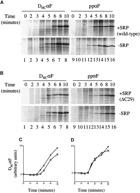
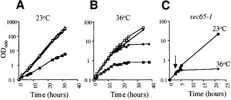
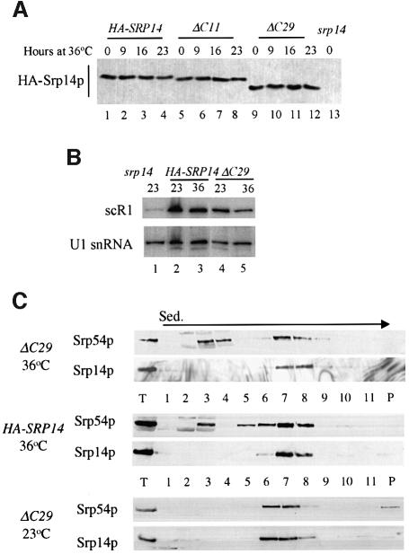
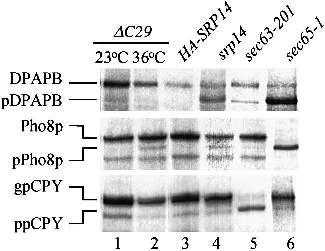
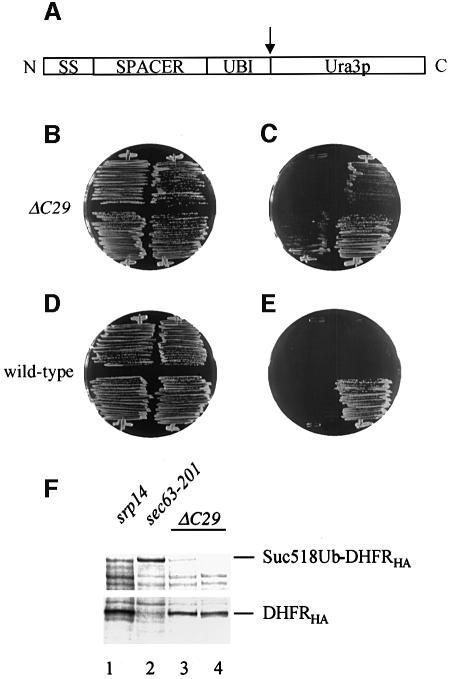
Signal recognition particle (SRP) targets proteins for co-translational insertion through or into the endoplasmic reticulum membrane. Mammalian SRP slows nascent chain elongation by the ribosome during targeting in vitro. This 'elongation arrest' activity requires the SRP9/14 subunit of the particle and interactions of the C-terminus of SRP14. We have purified SRP from Saccharomyces cerevisiae and demonstrated that it too has elongation arrest activity. A yeast SRP containing Srp14p truncated at its C-terminus (delta C29) did not maintain elongation arrest, was substantially deficient in promoting translocation and interfered with targeting by wild-type SRP. In vivo, this mutation conferred a constitutive defect in the coupling of protein translation and translocation and temperature-sensitive growth, but only a slight defect in protein translocation. In combination, these data indicate that the primary defect in SRP delta C29 is in elongation arrest, and that this is a physiologically important and conserved function of eukaryotic SRP.


Figures

None
Fig. 1. Purification of yeast SRP. (A) SRP was purified as described in Materials and methods, fractions resolved on a 10% Proseive (FMC Bioproducts) gel and stained with Coomassie Blue. Lanes 1 and 2: post-ribosomal extract and IgG–Sepharose flowthrough (1/16 000); lanes 3–5: TEV eluate, ω-aminobutyl agarose flowthrough and wash (1/80); lanes 6–12: elutions (1/20). Marker and SRP proteins are indicated. (B) Purification of SRP ΔC29 as in (A). ΔC29 contains an N-terminal triple-HA tag and thus runs above Srp21p. (C) RNA analysis. RNA isolated from purified SRP was run on a 6% acrylamide–50% urea gel and stained with ethidium bromide. Lane 1: pBR322 MspI digest markers; lanes 2 and 3: RNA from fractions shown in lanes 8 of (A) and (B), respectively (1/20).
None
Fig. 2. Activity of purified yeast SRP. (A) Confirmation of translocated species by protease protection. Translation reactions were carried out with or without addition of microsomal membranes (yRM) and subsequently incubated with 0.5 mg/ml proteinase K where indicated. (B and C) Translation reactions were carried out using extract containing (+) or immunodepleted of (–) SRP. yRM and purified SRP were added as indicated (concentrations of SRP in nanomoles). SRP was wild type except in (B) lane 13 and (C) lane 9, where SRP ΔC29 was used. (D) Translation reactions contained non-depleted extract supplemented with yRM and SRP (100 nM) as indicated. Reactions were run on 15% SDS–polyacrylamide gels and visualized by autoradiography. Untranslocated (ppL, ppαF and DHCαF) and processed, translocated (pL, gpαF and gDHCαF) species are indicated. Graphs were plotted using averaged data from three independent experiments. The translocation with non-depleted extract was set as 100%, bars, 1 SD.
None
Fig. 3. Yeast SRP mediates elongation arrest. (A and BIn vitro translation reactions were carried out as in Figure 2 using mRNAs encoding DHCαF or ppαF (left and right panels, respectively) with or without 100 nM added SRP. SRP was wild type (A) or ΔC29 (B). Samples were stopped at the times indicated. All panels are from the same experiment, and are equivalent exposures processed identically. (C and D) Quantified data from three experiments performed with DHCαF were averaged and plotted. SRP was wild type (C) or ΔC29 (D). Squares, plus SRP; diamonds, without added SRP.
None
Fig. 4. Yeast SRP contains two Srp14ps. (A) Extracts of strains expressing either wild-type and HA-Srp14p (lanes 1–5) or just HA-Srp14p (lanes 6–10) were immunoprecipitated with anti-HA (lanes 2 and 7) or anti-Sec65p antibodies (lanes 4 and 9). Material corresponding to 50% used in each immunoprecipitation (lanes 1 and 6) and of each supernate (lanes 3, 5, 8 and 10) and all immunoprecipitated material were analysed. (B and C) As (A) except that strains used expressed protein A-tagged Srp72p and wild-type Srp72p (B) or Myc-Srp21p and HA-Srp21p (C). Proteins were isolated using IgG–Sepharose or the antibody indicated. *, Ig chains recognized by secondary antibodies.
None
Fig. 5. Truncation of Srp14p results in delayed onset temperature sensitivity. (A and B) Strains expressing HA-Srp14p (open squares), DC11 (diamonds), ΔC29 (triangles) or lacking Srp14p (squares with crosses) were grown in liquid culture at either 23 (A) or 36°C (B). (Csec65-1 cells were grown at 23°C and half transferred to 36°C (arrow).
None
Fig. 6. SRP containing ΔC29 is stable at 36°C. (A and B) Protein or RNA samples were prepared from cells expressing Srp14p variants grown at 23°C or following transfer to 36°C for the time indicated. (A) Equal amounts of protein samples (assessed by Bradford assay) were analysed by imunoblotting. (B) Equal amounts of RNA (assessed by measuring OD260) were denatured, electrophoresed through a 6% acrylamide–50% urea gel, blotted on to a nylon membrane and hybridized sequentially with 32P-labelled DNA fragments of SCR1 and SNR19. 36°C samples were taken after 16 h growth at this temperature. (C) Whole-cell extracts from strains grown at the temperatures indicated for 16 h were prepared and fractionated on 10–30% w/v sucrose gradients. One quarter of each gradient fraction and 1/10th load were analysed for SRP proteins. In this experiment, SRP in srp14C29 strain extracts sedimented at slightly different rates. This was not seen in other experiments.
None
Fig. 7. Cells expressing ΔC29 have slight translocation defects for Pho8p, but not DPAPB or CPY, at 36°C. Immunoprecipitations were carried out with antibodies against the indicated proteins from extracts of cells pulse-labelled for 6 min (see Materials and methods). Immunoprecipitates were electrophoresed through 10% Proseive gels and visualized by autoradiography. Labelling was at 23°C (srp14C29, srp14), 30°C (sec63-201) or following transfer from 23 to 36°C for 30 min (sec65-1) or 16 h (srp14C29, HA-SRP14). Dipeptidyl aminopeptidase B: DPAPB, glycosylated luminal; pDPAPB, cytoplasmic. Alkaline phosphatase: Pho8p, glycosylated luminal; pPho8p, cytoplasmic. Carboxypeptidase Y: gpCPY, glycosylated lumenal; ppCPY, cytoplasmic. In this experiment a band appeared in the sec63-201 sample at the position of pDPAPB. This was not seen in other experiments.
None
Fig. 8. Translocation defects of srp14C29 cells in the UTA assay. (A) Organization of UTA constructs. The protein is depicted N- to C-terminus; signal sequence (SS), spacer (SPACER), ubiquitin (UBI) and Ura3p. The cleavage site for cytosolic ubiquitin-dependent proteases is indicated (arrow). (B and Csrp14C29 cells streaked on to a –trp (B; selecting for the plasmids) and a –ura (C; selecting for cytosolic Ura3p) plate were incubated for 2 days at 30°C. They contained empty vector (top left), Suc2277 (top right), Dap2300 (bottom left) or the control substrate Suc223, which has a 23 amino acid spacer (bottom right). (D and E) As for (B) and (C) but with wild-type cells. As seen previously (Johnsson and Varshavsky, 1994), Suc223 gave a Ura+ phenotype in wild-type cells. (F) Anti-HA immunoprecipitations from extracts of pulse-labelled cells expressing Suc2518UbDHFRHA (lanes 1–3) or Suc223UbDHFRHA control, which only yields the DHFRHA fragment (lane 4) (Johnsson and Varshavski, 1994). Labelling of cells and analysis are as in Figure 7.

Similar articles

Cited by

References

    1. Andrews D.W., Walter,P. and Ottensmeyer,F.P. (1985) Structure of the signal recognition particle by electron microscopy. Proc. Natl Acad. Sci. USA, 82, 785–789. - PMC - PubMed
    1. Birse D.E.A., Kapp,U., Strüb,K., Cusack,S. and Aberg,A. (1997) The crystal structure of the signal recognition particle Alu RNA binding heterodimer, SRP9/14. EMBO J., 16, 3757–3766. - PMC - PubMed
    1. Brown J.D., Hann,B.C., Medzihradszky,K.F., Niwa,M., Burlingame,A.L. and Walter,P. (1994) Subunits of the Saccharomyces cerevisiae signal recognition particle required for its functional expression. EMBO J., 13, 4390–4400. - PMC - PubMed
    1. Bui N. and Strüb,K. (1999) New insights into signal recognition and elongation arrest activities of the signal recognition particle. Biol. Chem., 380, 135–145. - PubMed
    1. Chang D.Y., Newitt,J.A., Hsu,K., Bernstein,H.D. and Maraia,R.J. (1997) A highly conserved nucleotide in the Alu domain of SRP RNA mediates translation arrest through high affinity binding to SRP9/14. Nucleic Acids Res., 25, 1117–1122. - PMC - PubMed

Publication types

MeSH terms