. 2000 Aug 1;14(15):1945-57.

members only encodes a Drosophila nucleoporin required for rel protein import and immune response activation

Affiliations

members only encodes a Drosophila nucleoporin required for rel protein import and immune response activation

A E Uv et al. Genes Dev. .

Abstract

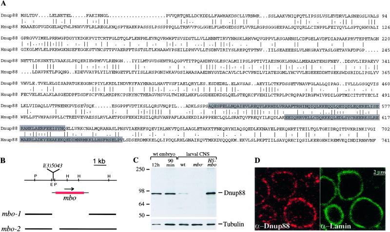
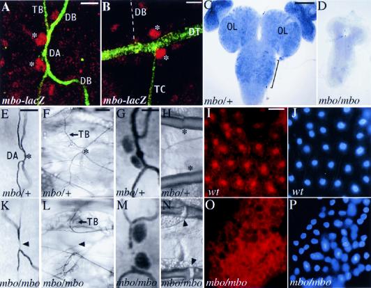
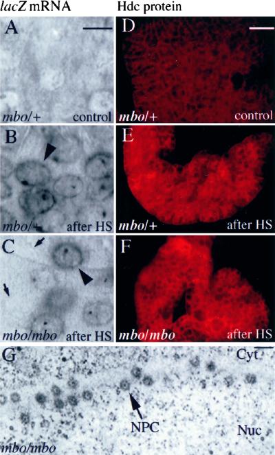
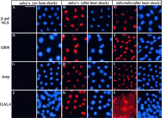
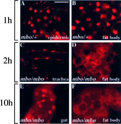
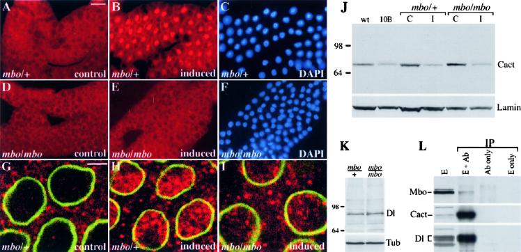
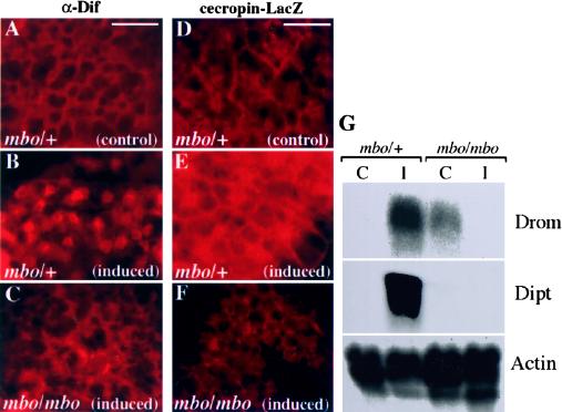
Many developmental and physiological responses rely on the selective translocation of transcriptional regulators in and out of the nucleus through the nuclear pores. Here we describe the Drosophila gene members only (mbo) encoding a nucleoporin homologous to the mammalian Nup88. The phenotypes of mbo mutants and mbo expression during development are cell specific, indicating that the nuclear import capacity of cells is differentially regulated. Using inducible assays for nucleocytoplasmic trafficking we show that mRNA export and classic NLS-mediated protein import are unaffected in mbo mutants. Instead, mbo is selectively required for the nuclear import of the yeast transcription factor GAL4 in a subset of the larval tissues. We have identified the first endogenous targets of the mbo nuclear import pathway in the Rel proteins Dorsal and Dif. In mbo mutants the upstream signaling events leading to the degradation of the IkappaB homolog Cactus are functional, but Dorsal and Dif remain cytoplasmic and the larval immune response is not activated in response to infection. Our results demonstrate that distinct nuclear import events require different nucleoporins in vivo and suggest a regulatory role for mbo in signal transduction.


Figures

Figure 1
Figure 1
The mbo product is homologous to the human nucleoporin Nup88. (A) The predicted amino acid sequence of Dnup88 (top) is aligned with human Nup88 (bottom). Identities are indicated by bars, and homologies by dots. The carboxy-terminal regions predicted to form a coiled–coil structure are shaded. Numbers indicate the positions of the respective amino acid residues. (B) Restriction map of the mbo locus (top horizontal line) for EcoRI (E), HindIII (H), and PvuII (P), showing the site of P-element insertion in l(3)5043 relative to the mbo transcript (the ORF is marked with red; the arrow shows the direction of transcription). The 1.1-kb EcoRI–HindIII fragment spanning the P-element insertion was used to screen cDNA libraries. The extent of deletions in the mbo-1 and mbo-2 alleles is depicted as gaps. (C) Western blot of extracts from 0- to-12 hr-old embryos, 0- to 90-min-old embryos, wild-type larval CNS, mbo mutant CNS, and CNSs from larvae that overexpress Dnup88 from an hsp70–mbo transgene probed with antiserum against the carboxy-terminal part of Dnup88. Staining for β-tubulin provides a loading control (bottom). (D) Confocal section of wild-type embryonic epidermis at stage 15 showing colocalization of Dnup88 (anti-Dnup88, red, left) with lamin (anti-lamin, green, right).
Figure 2
Figure 2
The expression and phenotypes of mbo are cell-specific. (A,B) Confocal images of a stage-16 embryo carrying one copy of the mbo–lacZ reporter stained with anti-β-galactosidase (red) and mAb 2A12 to visualize the tracheal lumen (green). (A) lacZ expression is detected in fusion cells (asterisks) of the dorsal branches (DB) forming the dorsal anastomosis (DA). mbo–lacZ is not expressed in the stalk cells of the DB or the cells extending terminal branches (TB). (B) mbo–lacZ is expressed in the fusion cells (asterisks) of the dorsal trunk (DT) but not in the stalk cells of the DB or in the transverse connective (TC). The DB is out of focus; its position is drawn with a broken line. Bars, 5 μm (A); 2 μm (B). (C,D) In situ hybridization to mbo mRNA in third instar larval CNS. In wild type (C) mbo is expressed in proliferating cells of the nerve cord (brackets), in the optic lobes (OL) of the brain, and the imaginal discs shown attached to the lobes. mbo RNA is not detectable in the CNS of mbo mutants (D), and the size of the CNS is reduced. Bars in C and D; 100 mm. (E,K) Dorsal anastomoses in late stage-16 wild-type (asterisk in E) and mbo mutant (K) embryos. In mbo mutant embryos, 20% of the dorsal branches fail to connect (arrowhead in K) Bar, 10 μm. (F,L) Dorsal anastomoses in third instar wild-type (asterisk in F) and mbo mutant (L) larvae. In mutants the DBs are disconnected (arrowhead), but terminal branching is not affected (arrows in F,L). Bar, 50 μm. (G,M) Dorsal anastomoses in stage-16 embryos carrying one copy of the esg–lacZ marker. esg–lacZ is expressed in the fusion cells of both wild type (G) and mbo mutants (M). Bars in G and M, 2 μm. (H,N) Segments of the dorsal trunks of wild-type and mbo third instar larvae. In mutants the cuticular lining of the dorsal trunks is disrupted at the positions of the fusion junctions (arrowheads in N) compared to junctions in the wild type (asterisks in H). Bar, 50 μm (I–P) Dnup88 expression in larval fat body detected with the antiserum against the amino-terminal part of the protein. Nuclear staining is detected in wild-type larvae (I) but absent in mbo mutants (O). The nuclei are visualized by DAPI staining in the adjacent panels J and P. Bar in I,J,O, and P, 40 μm.
Figure 3
Figure 3
mbo is not required for mRNA export. (A–C) In situ hybridization to lacZ RNA in wild-type and mbo larvae carrying the hs–GAL4 and UAS–lacZNLS transgenes. The lacZ RNA is detected in the proventriculus of wild-type (B) and mbo mutant (C) larvae after heat shock and does not accumulate in the nucleus (arrowheads). The dark spot inside each nucleus is likely to correlate with the site of transcription. lacZ expression is reduced in some of the cells of mbo mutants (arrows). Bar, 10 μm. (D–F) Heat shock-induced expression of Hdc protein in wild-type and mbo mutants. Fat bodies from untreated wild-type (D) and heat-shocked wild-type (E) and mbo (F) larvae carrying the hs–hdc transgene were stained with an antibody against the Hdc protein. Bar, 50 μm. (G) Electron micrograph of a section through the lymph gland of an mbo larva. In this tangential section, several NPCs (arrow) can be identified in the space between the cytoplasm (Cyt) and the nucleus (Nuc). Their distribution and morphology are indistinguishable from wild type at this level. Bar, 100 nm.
Figure 4
Figure 4
mbo is not required for general protein import. β-Gal–NLS, GRH, Antp, and GAL4 were expressed ectopically in wild type and mbo mutants and detected with the relevant antibodies. GRH, Antp, and GAL4 were expressed under the control of the hsp70 promoter, and β-Gal–NLS expression was activated by heat shock-induced GAL4 protein. (A,D,G,J) Background levels in wild-type fat bodies of untreated animals. Nuclei are visualized by DAPI in the adjacent panels. (B,E,H,K) In wild-type larvae all four proteins are found in the nuclei of the fat bodies after heatshock. (C,F,I,L) In fat bodies of mbo mutants all proteins become nuclear except for GAL4, which is predominantly cytoplasmic (L). The amount of β-Gal–NLS in C is reduced compared to that in B. Bar, 50 μm.
Figure 5
Figure 5
GAL4 nuclear import in mutant and mbo heterozygous larvae. GAL4 was expressed under the control of the hsp70 promoter and detected with an anti-GAL4 antibody. (A,B) One hour after heat shock treatment GAL4 is nuclear in heterozygous larvae, (A) epidermis; (B) fat body]. (C–F) In mbo mutants, GAL4 remains cytoplasmic in the fat body, both at 2 and 10 hr after heat treatment (D,F), whereas it is nuclear in the trachea (C; 2 hr after heat shock) and the gut (E; 10 hr after heat shock) of the same animals.
Figure 6
Figure 6
mbo is required for nuclear translocation of Dorsal. (A–F) Fat bodies from control wild-type (A) and mbo mutant (D) larvae stained for Dorsal. One hour after bacterial infection Dorsal is predominantly nuclear in wild type (B) but remains cytoplasmic in mbo mutants (E). Nuclei where visualized with DAPI (C,F). (G–I) Confocal images of lymph glands from control wild-type (G), induced wild-type (H), and induced mbo (I) larvae stained for lamin (green) and Dorsal (red). In mbo mutants, the inducible Dorsal translocation is reduced severely. (J) Cactus becomes degraded in mbo mutants. Protein extracts from control (C) and infected (I) wild-type and mbo larvae and untreated Toll 10b larvae were obtained in parallel with in situ experiments, blotted, and probed with antisera against Cactus. The amount of Cactus is similarly reduced in untreated Toll 10b and induced wild-type and mbo larvae. The filter was probed with an anti-lamin antibody as loading control. (K) The amount of Dorsal in wild-type and mbo mutant larvae is similar. Shown is a Western blot of larval extracts probed with the Dorsal antiserum followed by staining with an anti-tubulin antibody for loading control. (L) Dnup88 and Dorsal coimmunoprecipitate from protein extracts of 0- to 3-hr wild-type embryos. Embryo extract (E) and precipitates produced from incubations of extract with anti-Dorsal and beads (E + Ab), anti-Dorsal and beads without extract (Ab only), and extract and beads without anti-Dorsal (E only) were analyzed by Western blot. The blot was probed with antisera against Dnup88 (top), Cactus (middle) and Dorsal (bottom).
Figure 7
Figure 7
Nuclear import of Dif and induction of antimicrobial gene expression is impaired in mbo larvae. (A–C) Fat bodies from wild-type (A,B) and mbo mutant (C) larvae stained for Dif protein. One hour after bacterial infection Dif is nuclear in wild-type fat body (B) but remains cytoplasmic in mbo mutants (C). (D–F) Expression of cec–lacZ in fat bodies from wild-type (D,E) and mbo mutant (F) was revealed by staining against β-galactosidase. Two hours after bacterial challenge wild-type larvae express high levels of cec–lacZ (E), whereas mbo mutants do not express the reporter (F). (G) Northern blot analysis of the expression of drosomycin (Drom) and diptericin (Dipt) in wild-type and mbo mutant larvae. RNA samples from control (C) and induced (I) larvae from both genotypes were probed. In two of three experiments, the level of drosomycin RNA was elevated in untreated animals, but both genes were always strongly induced upon bacterial infection in wild-type larvae. This induction was severely impaired in mbo mutants. The blots were hybridized with an actin probe for loading control.

Similar articles

Cited by

References

    1. Askjaer P, Bachi A, Wilm M, Bischoff FR, Weeks DL, Ogniewski V, Ohno M, Niehrs C, Kjems J, Mattaj IW, et al. RanGTP-regulated interactions of CRM1 with nucleoporins and a shuttling DEAD-box helicase. Mol Cell Biol. 1999;19:6276–6285. - PMC - PubMed
    1. Aspland SE, White RA. Nucleocytoplasmic localisation of extradenticle protein is spatially regulated throughout development in Drosophila. Development. 1997;124:741–747. - PubMed
    1. Bastos R, Ribas de Pouplana LR, Enarson M, Bodoor K, Burke B. Nup84, a novel nucleoporin that is associated with CAN/Nup214 on the cytoplasmic face of the nuclear pore complex. J Cell Biol. 1997;137:989–1000. - PMC - PubMed
    1. Bray SJ, Kafatos FC. Developmental function of Elf-1: An essential transcription factor during embryogenesis in Drosophila. Genes & Dev. 1991;5:1672–1683. - PubMed
    1. Briggs LJ, Stein D, Goltz J, Corrigan VC, Efthymiadis A, Hübner S, Jans DA. The cAMP-dependent protein kinase site (Ser312) enhances dorsal nuclear import through facilitating nuclear localization sequence/importin interaction. J Biol Chem. 1998;273:22745–22752. - PubMed

Publication types

MeSH terms