

Duplex PCR for differential identification of Mycobacterium bovis, M. avium, and M. avium subsp. paratuberculosis in formalin- fixed paraffin-embedded tissues from cattle
- PMID: 10921976
- PMCID: PMC87183
- DOI: 10.1128/JCM.38.8.3048-3054.2000
Duplex PCR for differential identification of Mycobacterium bovis, M. avium, and M. avium subsp. paratuberculosis in formalin- fixed paraffin-embedded tissues from cattle
Abstract
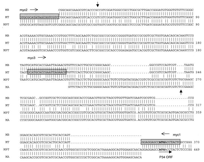
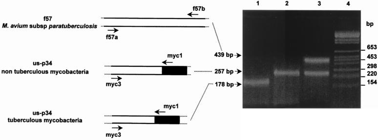
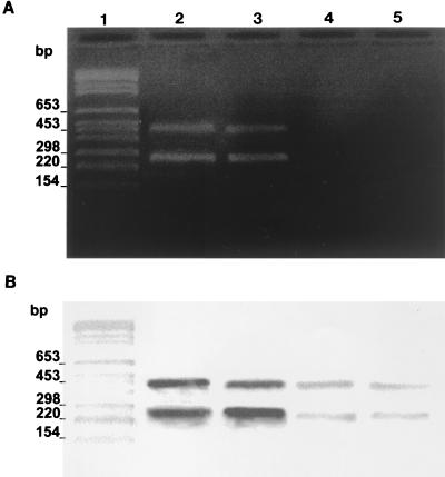
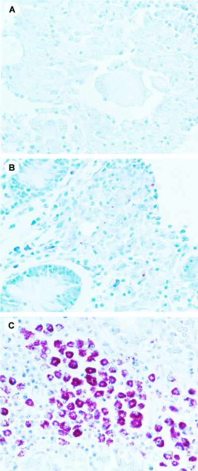
We previously isolated and sequenced two genomic segments of Mycobacterium avium subsp. paratuberculosis, namely, f57, a species-specific sequence, and the p34 gene, coding for a 34-kDa antigenic protein. Comparison of sequences upstream of the p34 open reading frame (us-p34) from M. avium subsp. paratuberculosis and M. tuberculosis showed a 79-base deletion in M. tuberculosis. Sequence analysis of the p34 genes in another two species, M. bovis (strain BCG) and M. avium (strain D4), confirmed the differences observed between tuberculous and nontuberculous species. A duplex diagnostic PCR strategy based on coamplification of nonhomologous us-p34 and species-specific f57 sequences was therefore developed. Duplex PCR yielded three different patterns, specific either for tuberculous bacilli (M. tuberculosis, M. bovis, and M. africanum), for both nontuberculous mycobacteria M. avium and M. intracellulare, or for M. avium subsp. paratuberculosis. The specificity of this single-step DNA-based assay was assessed on DNA from cultured mycobacterial strains, as well as on a panel of formalin-fixed and paraffin-embedded tissues from cattle. Molecular assay results from tissular DNA were compared to conventional bacteriological and histological test results, including those obtained by Ziehl-Neelsen staining on tissue biopsy specimens. Molecular discrimination was successful and confirmed the value of duplex us-p34 and f57 sequence amplification for differential diagnosis of tuberculosis, paratuberculosis, or infections caused by other members of the M. avium complex.
Figures
Similar articles
-
Definitive differentiation between single and mixed mycobacterial infections in red deer (Cervus elaphus) by a combination of duplex amplification of p34 and f57 sequences and Hpy188I enzymatic restriction of duplex amplicons.J Clin Microbiol. 2005 Sep;43(9):4640-8. doi: 10.1128/JCM.43.9.4640-4648.2005. J Clin Microbiol. 2005. PMID: 16145120 Free PMC article.
-
PCR-restriction endonuclease analysis for identification and strain typing of Mycobacterium avium subsp. paratuberculosis and Mycobacterium avium subsp. avium based on polymorphisms in IS1311.Mol Cell Probes. 1999 Apr;13(2):115-26. doi: 10.1006/mcpr.1999.0227. Mol Cell Probes. 1999. PMID: 10208802
-
Evaluation of the accuracy and reproducibility of a practical PCR panel assay for rapid detection and differentiation of Mycobacterium avium subspecies.Mol Cell Probes. 2000 Jun;14(3):153-61. doi: 10.1006/mcpr.2000.0299. Mol Cell Probes. 2000. PMID: 10860713
-
Non-tuberculous mycobacterial infections of veterinary relevance.Res Vet Sci. 2014 Oct;97 Suppl:S69-77. doi: 10.1016/j.rvsc.2014.08.007. Epub 2014 Aug 27. Res Vet Sci. 2014. PMID: 25256964 Review.
-
Paratuberculosis.Clin Microbiol Rev. 1994 Jul;7(3):328-45. doi: 10.1128/CMR.7.3.328. Clin Microbiol Rev. 1994. PMID: 7923053 Free PMC article. Review.
Cited by
-
Tuberculosis Epidemiology and Spatial Ecology at the Cattle-Wild Boar Interface in Northern Spain.Transbound Emerg Dis. 2023 Feb 23;2023:2147191. doi: 10.1155/2023/2147191. eCollection 2023. Transbound Emerg Dis. 2023. PMID: 40303683 Free PMC article.
-
Detection of Mycobacterium tuberculosis and Mycobacterium bovis in Sahiwal cattle from an organized farm using ante-mortem techniques.Vet World. 2016 Apr;9(4):383-7. doi: 10.14202/vetworld.2016.383-387. Epub 2016 Apr 15. Vet World. 2016. PMID: 27182134 Free PMC article.
-
Diagnostic Application of IS900 PCR Using Blood as a Source Sample for the Detection of Mycobacterium avium Subspecies Paratuberculosis in Early and Subclinical Cases of Caprine Paratuberculosis.Vet Med Int. 2010;2010:748621. doi: 10.4061/2010/748621. Epub 2009 Nov 16. Vet Med Int. 2010. PMID: 20445791 Free PMC article.
-
Histopathological Investigations and Molecular Confirmation Reveal Mycobacterium bovis in One-Horned Rhinoceros (Rhinoceros unicorns).Biomed Res Int. 2022 May 18;2022:5816986. doi: 10.1155/2022/5816986. eCollection 2022. Biomed Res Int. 2022. Retraction in: Biomed Res Int. 2024 Jan 9;2024:9858936. doi: 10.1155/2024/9858936. PMID: 35647178 Free PMC article. Retracted.
-
Prevalence of Mycobacterium avium Subsp. paratuberculosis in Feral Pigeons (Columba livia) Associated with Difficulties Controlling Paratuberculosis in a Bovine Herd (Fighting Bull Breed).Animals (Basel). 2022 Nov 27;12(23):3314. doi: 10.3390/ani12233314. Animals (Basel). 2022. PMID: 36496835 Free PMC article.
References
-
- Catanzano A, Davidson B L, Fujiwara P I, Coldberger M J, Gordin F, Salfinger M, Spodoro J, Schluger N W, Sierra M F, Woods G L. Rapid diagnostic tests for tuberculosis: what is the appropriate use? American Thoracic Society Workshop. Am J Respir Crit Care Med. 1997;155:1804–1814. - PubMed
-
- Coetsier C, Havaux X, Mattelard F, Sadatte S, Cormont F, Buergelt K, Limbourg B, Latinne D, Bazin H, Denef J-F, Cocito C. Detection of Mycobacterium avium subsp. paratuberculosis in infected tissues by new species-specific immunohistological procedures. Clin Diagn Lab Immunol. 1998;5:446–451. - PMC - PubMed
-
- Cousins D V, Dawson D J. Tuberculosis due to Mycobacterium bovis in the Australian population: cases recorded during 1970-1994. Int J Tuberc Lung Dis. 1999;3:715–721. - PubMed
Publication types
MeSH terms
Substances
LinkOut - more resources
Full Text Sources
Other Literature Sources
Medical
Molecular Biology Databases
