

Recovery and identification of West Nile virus from a hawk in winter
- PMID: 10921991
- PMCID: PMC87202
- DOI: 10.1128/JCM.38.8.3110-3111.2000
Recovery and identification of West Nile virus from a hawk in winter
Abstract
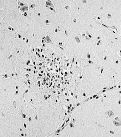
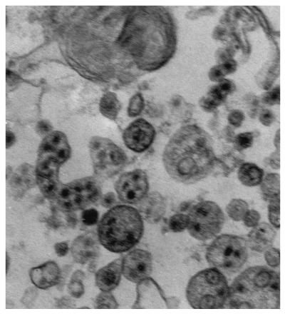
West Nile virus was recovered from the brain of a red-tailed hawk that died in Westchester County, N.Y., in February 2000. Multiple foci of glial cells, lymphocytes, and a few pyknotic nuclei were observed in the brain. Three to 4 days after inoculation of Vero cells with brain homogenates, cytopathic changes were detected. The presence of West Nile virus antigen in fixed cells or cell lysates was revealed by fluorescent antibody testing or enzyme-linked immunosorbent assay, respectively. Furthermore, Reverse transcriptase-PCR with primers specific for the NS3 gene of West Nile virus resulted in an amplicon of the expected size (470 bp). Electron microscopy of thin sections of infected Vero cells revealed the presence of viral particles approximately 40 nm in diameter, within cytoplasmic vesicles. The demonstration of infection with the West Nile virus in the dead of the winter, long after mosquitoes ceased to be active, is significant in that it testifies to the survival of the virus in the region beyond mosquito season and suggests another route of transmission: in this case, prey to predator.
Figures
References
-
- Anderson J F, Andreadis T G, Vossbrinck C R, Tirrel S, Wakem E M, French R A, Garmendia A E, Van Kruiningen H J. Isolation of West Nile virus from mosquitoes, crows and a Cooper's hawk in Connecticut. Science. 1999;286:2331–2333. - PubMed
-
- Asnis D, Conetta R, Waldman G, Texeira A, McNamara T, et al. Outbreak of West Nile-like viral encephalitis New York, 1999. Morbid Mortal Weekly Rep. 1999;48:845–849. - PubMed
-
- Briese T, Jia X-Y, Huang C, Grady L I, Lipkin W I. Identification of a Kunjin/West Nile like flavivirus in brains of patients with New York encephalitis. Lancet. 1999;354:1261–1262. - PubMed
-
- Lancioti R S, Roehrig J T, Deubel V, Smith J, Parker M, Steele K, Crise B, Volpe K E, Crabtree M B, Scherret J H, Hall R A, MacKenzie J S, Cropp C B, Panigraphy B, Ostlund E, Schmitt B, Malkinson M, Banet C, Weissman J, Komar N, Savage H M, Stone W, McNamara T, Gubler D J. Origin of West Nile virus responsible for an outbreak of encephalitis in the northeastern United States. Science. 1999;286:2333–2337. - PubMed
-
- Oderola H A, Oduye O O. West Nile virus infection of adult mice by oral route. Arch Virol. 1977;54:251–253. - PubMed
MeSH terms
LinkOut - more resources
Full Text Sources
Other Literature Sources
Medical
