

Review
doi: 10.1073/pnas.97.16.8762.
Structure and function of pectic enzymes: virulence factors of plant pathogens
Affiliations
- PMID: 10922032
- PMCID: PMC34009
- DOI: 10.1073/pnas.97.16.8762
Item in Clipboard
Review
Structure and function of pectic enzymes: virulence factors of plant pathogens
Proc Natl Acad Sci U S A.
.
Abstract
The structure and function of Erwinia chrysanthemi pectate lysase C, a plant virulence factor, is reviewed to illustrate one mechanism of pathogenesis at the molecular level. Current investigative topics are discussed in this paper.
Figures
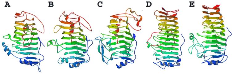
Five examples of plant cell wall degradative enzymes that fold into a
parallel β helix motif. The predominant secondary structural features
of the proteins are illustrated as β strands, and the coils represent
α helices. (A) E. chrysanthemi pectate
lyase C. (B) E. chrysanthemi pectate
lyase E. (C) A. niger pectin lyase B.
(D) E. carotovora polygalacturonase.
(E) A. aculeatus rhamnogalacturonase A.
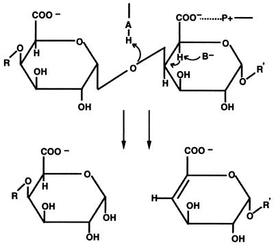
Schematic diagram of α-1,4-polygalacturonic acid cleavage by a
β-elimination mechanism. The pectate lyases are expected to
contribute a minimum of three groups to the catalytic mechanism:
P+, which neutralizes the charge on the carboxylic acid
group; B, a general base that abstracts the proton from C-5; and A,
which is involved in the transfer of the final proton to the glycosidic
oxygen, leaving a double bond between C-4 and C-5.
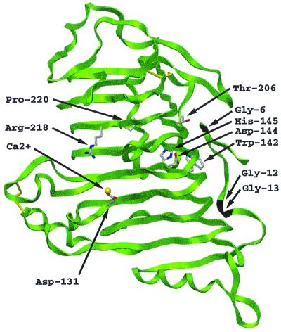
Stereoview of the PelC locations of 10 invariant amino acids within the
extracellular pectate lyase superfamily. The two clusters of invariant
amino acids are located on opposite sides of the parallel β helix,
separated by approximately 25 Å across the diameter and by
approximately 90 Å around the circumference of the parallel β helix.
The α-carbon backbone of PelC is illustrated as a green ribbon and
the Ca2+ ions as yellow spheres. The invariant amino acids
are labeled at the α-carbon and are represented by rods using the
International Union of Pure and Applied Chemistry coloring code: carbon
atoms are gray; oxygen atoms, red; and nitrogen atoms, blue.
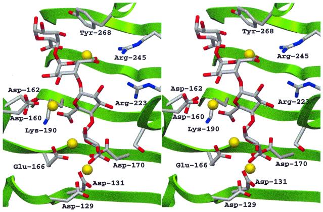
Stereoview of the active site of the PelC
R218K-(Ca2+)4-pentaGalpA
complex. The color code is the same as that used in Fig. 3.
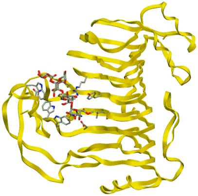
Overview of the PLB-(mGalpA)4 model. Pectin
lyase B is shown as yellow ribbons. The substrate and the interacting
amino acids are represented by rods using the color code described in
Fig. 3.
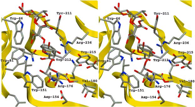
Stereoview of the active site of the
PLB-(mGalpA)4 model. The view of the active
site is the same as that used in Fig. 4 to allow for comparisons. The
hydrophobic pockets around three of the four methyl groups esterified
to the uronate moieties are visible. The color code is the same as that
used in Fig. 6.
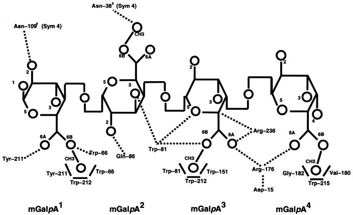
Schematic representation of the pectin lyase B with
(GalpA)4 at a distance of ≤3.0 Å.
mGalpA (1) is the reducing saccharide and
mGalpA (4) is the nonreducing terminus. Hydrogen bonds
are represented with dotted lines and hydrophobic interactions, with
boldface lines. Oxygen atoms and methyl groups are represented by
circles, with the corresponding number, and the carbon atoms are
assumed at the intersection of bonds designated in boldface lines.
References
-
- Carpita N C, Gibeaut D M. Plant J. 1993;3:1–30. - PubMed
-
- Albersheim P, Darvill A, O'Neill M, Schols H A, Voragen A G J. In: Progress in Biotechnology: Pectins and Pectinases. Visser J, Voragen A G J, editors. Vol. 14. Amsterdam: Elsevier; 1996. pp. 47–53.
-
- Davies G, Henrissat B. Structure (London) 1995;3:853–859. - PubMed
-
- Kiss J. Adv Carbohydr Chem Biochem. 1974;29:229–230.
-
- He S Y, Lindeberg M, Collmer A. In: Biotechnology in Plant Disease Control. Chet I, editor. New York: Wiley–Liss; 1993. pp. 39–64.
Publication types
MeSH terms
Substances
LinkOut - more resources
Full Text Sources
Other Literature Sources
Molecular Biology Databases
