Review
. 2000 Aug 1;97(16):8778-83.
doi: 10.1073/pnas.97.16.8778.

Molecular and cell biology aspects of plague

Affiliations
Review

Molecular and cell biology aspects of plague

G R Cornelis. Proc Natl Acad Sci U S A. .

Abstract

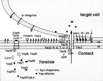
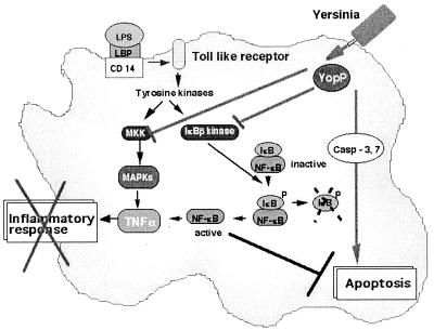
A 70-kb virulence plasmid (sometimes called pYV) enables Yersinia spp. to survive and multiply in the lymphoid tissues of their host. It encodes the Yop virulon, a system consisting of secreted proteins called Yops and their dedicated type III secretion apparatus called Ysc. The Ysc apparatus forms a channel composed of 29 proteins. Of these, 10 have counterparts in almost every type III system. Secretion of some Yops requires the assistance, in the bacterial cytosol, of small individual chaperones called the Syc proteins. These chaperones act as bodyguards or secretion pilots for their partner Yop. Yop proteins fall into two categories. Some are intracellular effectors, whereas the others are "translocators" needed to deliver the effectors across the eukaryotic plasma membrane, into eukaryotic cells. The translocators (YopB, YopD, LcrV) form a pore of 16-23 A in the eukaryotic cell plasma membrane. The effector Yops are YopE, YopH, YpkA/YopO, YopP/YopJ, YopM, and YopT. YopH is a powerful phosphotyrosine phosphatase playing an antiphagocytic role by dephosphorylating several focal adhesion proteins. YopE and YopT contribute to antiphagocytic effects by inactivating GTPases controlling cytoskeleton dynamics. YopP/YopJ plays an anti-inflammatory role by preventing the activation of the transcription factor NF-kappaB. It also induces rapid apoptosis of macrophages. Less is known about the role of the phosphoserine kinase YopO/YpkA and YopM.


Figures

Figure 1
Figure 1
The Yop virulon. When Yersinia are placed at 37°C in a rich environment, the Ysc secretion channel is installed. Proteins YscD, -R, -S, -T, -U, and -V are localized in the inner membrane (IM), whereas YscC and YscP are exposed at the bacterial surface. Lipoprotein YscW stabilizes YscC. YscN belongs to the family of ATPases. A stock of Yop proteins is synthesized, and some of them are capped with their specific Syc chaperone. As long as there is no contact with a eukaryotic cell, a stop-valve, possibly made of YopN, TyeA, and LcrG, blocks the Ysc secretion channel. On contact with a eukaryotic target cell, the bacterium attaches tightly by interaction between its YadA and Inv adhesins and β-integrins, and the secretion channel opens. The Yops are then transported through the Ysc channel, and the Yop effectors are translocated across the plasma membrane, guided by the translocators YopB, YopD, and LcrV.
Figure 2
Figure 2
Inhibition of phagocytosis by YopE, YopH, and YopT. (A) Phagocytosis of an invading bacterium by a macrophage. The process involves phosphorylation of focal adhesion proteins (p130cas, Fak, Fyn, paxillin) and actin polymerization controlled by GTPases such as RhoA and Rac. Phagocytosis is followed by killing of the bacterium. (B) Resistance to phagocytosis by Yersinia. On contact, Yersinia injects Yop effectors. YopH dephosphorylates proteins from the focal adhesion (PTPase, phosphotyrosine phosphatase); YopE inactivates Rac and cdc42 by stimulating their GTPase activity (GAP, GTPase-activating protein); YopT deactivates RhoA.
Figure 3
Figure 3
Effects of YopP/YopJ. Bacterial lipopolysaccharide (LPS), bound to the LPS-binding protein (LBP), interacts with its receptor CD14 and coreceptor from the Toll-like family, which leads to phosphorylation cascades resulting in the activation of mitogen-activated protein kinases (MAPKs) and of the kinase of the inhibitor of NF-κB (IκB). Phosphorylation of IκB is followed by its degradation, and NF-κB migrates to the nucleus and activates transcription of proinflammatory cytokines, including TNF-α. Translocated YopP/YopJ prevents the activation of the two phosphorylation cascades, and thus blocks the release of TNF-α. YopP/YopJ also induces macrophage apoptosis. See text for details and references.

References

    1. Simonet M, Richard S, Berche P. Infect Immun. 1990;58:841–845. - PMC - PubMed
    1. Nakajima R, Brubakker R R. Infect Immun. 1993;61:23–31. - PMC - PubMed
    1. Cornelis G R, Boland A, Boyd A P, Geuijen C, Iriarte M, Neyt C, Sory M-P, Stainier I. Microbiol Mol Biol Rev. 1998;62:1315–1352. - PMC - PubMed
    1. Hueck C J. Microbiol Mol Biol Rev. 1998;62:379–433. - PMC - PubMed
    1. Michiels T, Wattiau P, Brasseur R, Ruysschaert J M, Cornelis G R. Infect Immun. 1990;58:2840–2849. - PMC - PubMed

Publication types

Substances