Review
. 2000 Aug 1;97(16):8807-14.
doi: 10.1073/pnas.97.16.8807.

Genetic complexity of pathogen perception by plants: the example of Rcr3, a tomato gene required specifically by Cf-2

Affiliations
Review

Genetic complexity of pathogen perception by plants: the example of Rcr3, a tomato gene required specifically by Cf-2

M S Dixon et al. Proc Natl Acad Sci U S A. .

Abstract

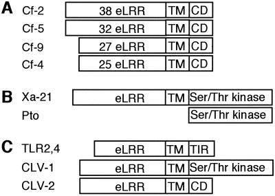
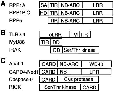
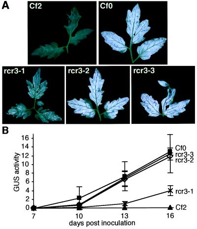
Genetic analysis of plant-pathogen interactions has demonstrated that resistance to infection is often determined by the interaction of dominant plant resistance (R) genes and dominant pathogen-encoded avirulence (Avr) genes. It was postulated that R genes encode receptors for Avr determinants. A large number of R genes and their cognate Avr genes have now been analyzed at the molecular level. R gene loci are extremely polymorphic, particularly in sequences encoding amino acids of the leucine-rich repeat motif. A major challenge is to determine how Avr perception by R proteins triggers the plant defense response. Mutational analysis has identified several genes required for the function of specific R proteins. Here we report the identification of Rcr3, a tomato gene required specifically for Cf-2-mediated resistance. We propose that Avr products interact with host proteins to promote disease, and that R proteins "guard" these host components and initiate Avr-dependent plant defense responses.


Figures

Figure 1
Figure 1
Tomato Cf proteins and structurally related proteins. (A) Cf proteins contain different numbers of N-terminal eLRRs, a transmembrane (TM) domain, and a short cytoplasmic domain (CD). (B) Rice Xa-21 and tomato Pto bacterial R proteins. (C) eLRR-TM domain-containing proteins with different cytoplasmic domains (CD). See text for descriptions.
Figure 2
Figure 2
Arabidopsis RPP1, RPP5, and structurally related proteins. (A) RPP1 and RPP5 have an N-terminal TIR domain, a nucleotide-binding Apaf-1, R proteins and CED4 homology (NB-ARC) domain, and a LRR domain. The RPP1 family differ by their N-terminal domains, which are absent in RPP5: RPP1A has a putative signal anchor (SA) domain, RPP1B,C have hydrophobic domains (HD). (B) Human proteins that function in the NF-κB pathway. eLRR, extracellular LRR domain; TM, transmembrane domain. (C) Human proteins with CARDs that function in NF-κB and/or apoptosis pathways. See text for descriptions.
Figure 3
Figure 3
Polymorphic R gene families revealed by DNA gel blot analysis. Comparisons of tomato lines with introgressed chromosome segments containing different Cf genes show interspecific polymorphisms (44). Comparisons of different Arabidopsis landraces show intraspecific polymorphisms at the RPP1 (47) and RPP5 loci (48).
Figure 4
Figure 4
Infection phenotypes of rcr3 mutants. Seedlings were immersed in a solution containing 3.5 × 10⁵ ml−1 spores of the appropriate C. fulvum race. Inoculated plants were kept for 3 to 6 days in a phytochamber at saturated humidity and subsequently at 70% relative humidity, 24°C, during the 16-hr day–light, 18°C overnight. (A) Comparison of infection phenotype of the rcr3 mutants and the Cf0-sensitive and Cf2-resistant control lines. Plants were inoculated with C. fulvum race 5 15 days after sowing, and leaves were photographed 17 days after inoculation. Fungal colonization is visible on the abaxial surface of the leaves as white mycelium. (B) Comparison of fungal biomass accumulation in rcr3–1 (X), rcr3–2 (⋄), rcr3–3 (●), Cf0 (■), and Cf2 (▴) lines by using a fluorometric GUS assay (56) over a time course after inoculation. Twelve-day-old plants were inoculated with C. fulvum race 4 GUS (57). No GUS activity (4-methyl umbelliferone mg−1 protein min−1) was detected on Cf2 controls in contrast to Cf0 controls. Three cotyledons were harvested and analyzed separately for each sample at each time point (the mean value for each set of triplicate samples is shown and error bars indicate the standard deviation).
Figure 5
Figure 5
Comparison of fungal development and plant cell death response during the C. fulvum infection of rcr3 mutants and control lines. At least three cotyledons and two leaves were sampled every 3 days from each genotype during the experiment described in Fig. 4A and were stained with lactophenol-trypan blue (58). Photomicrographs of cotyledons were taken 7 days after inoculation at the same magnification. Scale bar = 100 μm. (A) Fungal growth in the Cf0 line: C. fulvum hyphae (h) tend to localize close to plant veins (v) where they start swelling. (B) Characteristic early resistance response in the Cf2 line: patches of dead mesophyll cells (p) develop at the vicinity of the plant veins, whereas the fungus is undetectable. (C) Infection phenotype of rcr3–3, and rcr3–2 (not shown): once the fungus has penetrated the leaf apoplast, the fungus develops as in Cf0. However, these fungal foci appear to occur at a lower frequency on rcr3–3 and rcr3–2 than on Cf0. (D) rcr3–1 intermediate phenotype: although rcr3–1 allows sparse fungal growth in some areas; the fungus remains undetectable on the major part of the cotyledon. Discrete dead cells (d) were scattered around the upper mesophyll of the mutants but not in wild type, irrespective of C. fulvum infection (noninoculated material not shown). This cell death phenotype was poorly reproducible. Calcium oxalate crystals (c) in some mesophyll cells are common in tomato leaves. These phenotypes were observed in three independent infections with C. fulvum race 5 and in an infection with C. fulvum race 4 GUS, where over 300 infection sites were analyzed in cotyledon and leaf samples.
Figure 6
Figure 6
Rcr3 maps to chromosome 2. AFLP analysis was carried out as previously described (59). (A) Identification of AFLP markers linked to Rcr3 (arrows). Susceptible (S) and resistant (R) pools of the BC2 mapping populations were subjected to AFLP analysis. Pairs of lanes (1 to 7) represent analysis with different AFLP primer combinations. (B) Mapping of a linked AFLP marker by using L. pennellii introgression lines (1–2 to 4–4) with primer combination 4. Susceptible and resistant pools of the BC2 mapping populations were analyzed as controls. The arrows indicate the AFLP fragment linked to L. pennellii Rcr3 present in introgression lines 2–3 and 2–4 that localizes that marker to chromosome 2.

Similar articles

Cited by

References

    1. Flor H H. Annu Rev Phytopathol. 1971;9:275–296.
    1. Galan E J, Collmer A. Science. 1999;284:1322–1328. - PubMed
    1. Kearney B, Staskawicz B J. Nature (London) 1990;346:385–386. - PubMed
    1. Ritter C, Dangl J L. Mol Plant-Microbe Interact. 1995;8:444–453. - PubMed
    1. Erickson F L, Holzberg S, Calderon-Urrea A, Handley V, Axtell M, Corr C, Baker B. Plant J. 1999;18:67–75. - PubMed

Publication types

LinkOut - more resources