. 2000 Aug 1;97(16):8973-8.
doi: 10.1073/pnas.97.16.8973.

The RING-H2 finger protein APC11 and the E2 enzyme UBC4 are sufficient to ubiquitinate substrates of the anaphase-promoting complex

Affiliations

The RING-H2 finger protein APC11 and the E2 enzyme UBC4 are sufficient to ubiquitinate substrates of the anaphase-promoting complex

M Gmachl et al. Proc Natl Acad Sci U S A. .

Abstract

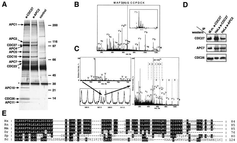
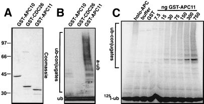
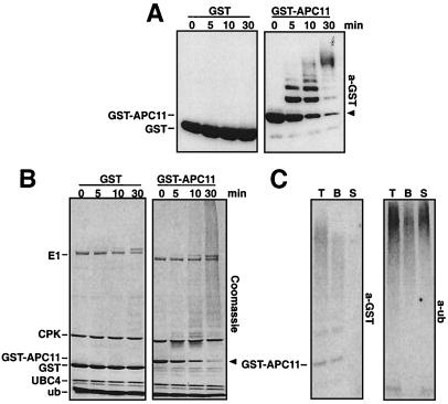
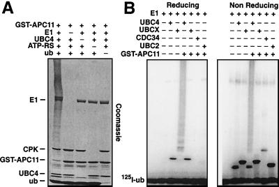
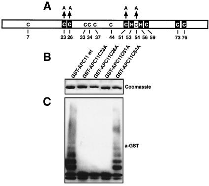
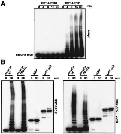
The anaphase-promoting complex (APC) is a cell cycle-regulated ubiquitin-protein ligase that targets cyclin B, securin and other destruction box containing proteins for proteolysis. Nine APC subunits have been identified in vertebrates and eleven in yeast, but for none of them it is known how they contribute to the catalysis of ubiquitination reactions. Here we report the mass spectrometric identification of CDC26 and of the RING-H2 finger protein APC11 in the human APC. We have expressed these proteins and several other APC subunits in Escherichia coli and have tested their activities in vitro. We find that APC11 alone is sufficient to allow the synthesis of multiubiquitin chains in the presence of E1 and UBC4. These multiubiquitin chains are partly unanchored and partly bound to APC11 itself. APC11 and UBC4 are also able to ubiquitinate securin and cyclin B, but these reactions show a decreased dependency on the destruction box. The integrity of the putative zinc binding RING-H2 finger is required for the ability of APC11 to support ubiquitination reactions. These results suggest that APC11 and UBC4 catalyze the formation of isopeptide bonds in APC-mediated ubiquitination reactions.


Figures

Figure 1
Figure 1
Characterization of CDC26 and APC11 as subunits of the vertebrate APC. (A) Identification of APC subunits by silver staining. Immunoprecipitates obtained from logarithmically growing HeLa cells with CDC27 or APC2 antibodies were analyzed by SDS/PAGE and silver staining. The positions of APC subunits are indicated. The IgG light and heavy chains are marked by stars. (B and C) Identification of APC11 and CDC26 by nanoelectrospray tandem mass spectrometry. (B) Mass spectrometric sequencing of APC11. Tandem mass spectrum of a tryptic peptide (m/z 680.9) obtained after in-gel digestion of p10. The presence of peaks shifted by 1 Da (marked by asterisks) allowed to localize the site of deamidation. (C) Mass spectrometric sequencing of CDC26. Mass spectrum of in-gel digested human p14 (Upper left). The peaks marked with T correspond to trypsin autolysis products. The peak marked by an arrow (Lower left) was sequenced by nanoelectrospray mass spectrometry. The right panel shows the tandem mass spectrum of the selected peak. (D) APC was immunoprecipitated from logarithmically growing HeLa cells (HeLa) and from mouse myeloma Ag8.653 cells (Mm) with CDC27 or APC2 antibodies. Precipitates were subsequently analyzed by SDS/PAGE and immunoblotting by using CDC27, APC7, and CDC26 antibodies. (E) Alignment of the amino acid sequences of CDC26 orthologs from various species. CDC26 orthologs were identified by psi-blast by using the human CDC26 sequence as a query. Amino acid residues that are identical or similar (according to the Blosum 62 substitution matrix) are highlighted by black boxes. Amino acid residues are given in the one-letter code. For Sc Cdc26 insertions of 18 and 16 amino acids are indicated. Ce, Caenorhabditis elegans; Dr, Danio rerio; Hs, Homo sapiens; Mm, Mus musculus; Rn, Rattus norwegicus; Sc, Saccharomyces cerevisiae; Sp, Schizosaccharomyces pombe.
Figure 2
Figure 2
Formation of polyubiquitin chains in the presence of GST-APC11. (A) Bacterial lysates expressing GST-APC10, GST-CDC26, and GST-APC11 were incubated with glutathione beads, were washed, were separated by SDS/PAGE, and were stained with Coomassie blue. (B) Identical amounts of GST-APC10, GST-CDC26, and GST-APC11 lysates as in A were bound to glutathione beads, were washed, and were used for in vitro ubiquitination. Samples were separated by SDS/PAGE (15 + 8%), were transferred to poly(vinylidene difluoride) membrane, and were immunodecorated with anti-ubiquitin antibodies. (C) Purified holo-APC, buffer QA, GST (750 ng), or the indicated amounts of GST-APC11 were incubated with E1, UBC4, and ATP as in B in the presence of 125I-ubiquitin. Samples were incubated for 10 min at 37°C, were separated by SDS/PAGE, and were visualized by phosphorimaging.
Figure 3
Figure 3
Characterization of ubiquitin conjugates formed in the presence of GST-APC11. (A) Purified GST (Left) or GST-APC11 (Right) were incubated for the indicated time with E1, UBC4, an ATP regenerating system, and ubiquitin as in Fig. 2B. Samples were separated by 12% SDS/PAGE. GST and GST-APC11 were detected by immunodecoration with anti-GST antibodies. The position of unmodified GST-APC11 is indicated by an arrowhead. (B) Identical samples as in A were separated by 15 + 8% SDS/PAGE and were stained with Coomassie blue. Identified proteins are indicated on the left (CPK, creatine phosphokinase; for all others, see text). The arrowhead indicates the position of GST-APC11. (C) A GST-APC11-mediated ubiquitination reaction as described in Fig. 2B was split into three aliquots as described in Materials and Methods. Identical amounts of total (T), beads (B), and supernatant (S) fractions were loaded on a 15 + 8% polyacrylamide gel and were transferred to poly(vinylidene difluoride) membrane. One part (Left) was decorated with anti-GST antibodies, the other part (Right) with anti-ubiquitin antibodies.
Figure 4
Figure 4
Ubiquitination reactions occurring in the presence of APC11 depend on E1, UBC4, ATP, and ubiquitin. (A) Coomassie-stained gel of reactions containing (+) or missing (−) the indicated components. All reactions were incubated for 30 min at 37°C, and missing components were substituted with buffer. (B) Autoradiogram of reactions separated under reducing (Left) or nonreducing (Right) conditions. All reactions were performed in the presence of E1, ATP, 125I-ubiquitin, and with either UBC4, UBCx, CDC34, UBC2, GST-APC11, or combinations of these. Reactions were incubated for 5 min at room temperature, were stopped by addition of SDS/sample buffer with or without DTT, and were analyzed by SDS/PAGE.
Figure 5
Figure 5
The formation of multiubiquitin chains depends on the integrity of the APC11 RING-H2 finger. (A) Schematic representation of APC11. Cysteines (C) and two histidine residues (H) are indicated. Boxed residues represent the predicted Zn coordinating amino acids. The cysteine residues that were changed to alanine (A) in this study are indicated by arrows. (B) Bacterial lysates containing GST-APC11 wild type and the indicated GST-APC11 mutants were immobilized on glutathione-beads, were washed, and were eluted with SDS/sample buffer. Shown is a 12% polyacrylamide gel stained with Coomassie blue. (C) Identical amounts of bacterial lysates as in B were bound to glutathione beads, were washed, and were incubated with a reaction mixture as in Fig. 2B for 30 min at 37°C. Proteins were visualized by immunodecoration with anti-GST antibodies followed by enhanced chemiluminescence.
Figure 6
Figure 6
GST-APC11 mediates the ubiquitination of the APC substrate securin. (A) Time course of reactions containing either purified GST-APC10 (Left) or GST-APC11 (Right), E1 and UBC4, an ATP regenerating system, ubiquitin and securin-myc-His. At the indicated time points, reactions were stopped by addition of SDS/sample buffer. Separation on 15 + 8% SDS/PAGE was followed by Western blotting and immunodecoration with an anti-myc antibody (9E10). (B) Purified GST-APC11 (Left) or holo-APC plus CDH1 (Right) were incubated with E1, UBC4, ATP, and ubiquitin as in A for 0 and 30 min at 37°C in the presence of either 20 ng securin-myc-His wild type, 20 ng of securin-myc-His D box mutant, 50 ng MBP, or 50 ng of GST-p53. After separation on SDS/PAGE and transfer to poly(vinylidene difluoride) membranes, proteins were detected by using anti-myc (9E10) antibodies (securin), anti-MBP antibodies, or anti p53 antibodies.

References

    1. Morgan D O. Nat Cell Biol. 1999;1:E47–E53. - PubMed
    1. Zachariae W, Nasmyth K. Genes Dev. 1999;13:2039–2058. - PubMed
    1. Pfleger C M, Kirschner M W. Genes Dev. 2000;14:655–665. - PMC - PubMed
    1. Peters J-M. Exp Cell Res. 1999;248:339–349. - PubMed
    1. Gieffers C, Schleiffer A, Peters J M. Protoplasma. 2000;211:20–28.

Publication types

MeSH terms

Substances