. 2000 Aug 1;97(16):9052-7.
doi: 10.1073/pnas.97.16.9052.

The NF-kappa B signal transduction pathway in aortic endothelial cells is primed for activation in regions predisposed to atherosclerotic lesion formation

Affiliations

The NF-kappa B signal transduction pathway in aortic endothelial cells is primed for activation in regions predisposed to atherosclerotic lesion formation

L Hajra et al. Proc Natl Acad Sci U S A. .

Abstract

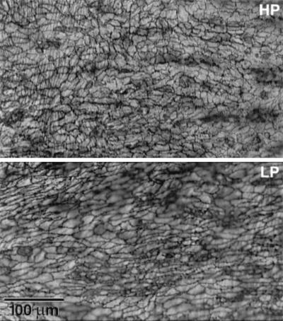
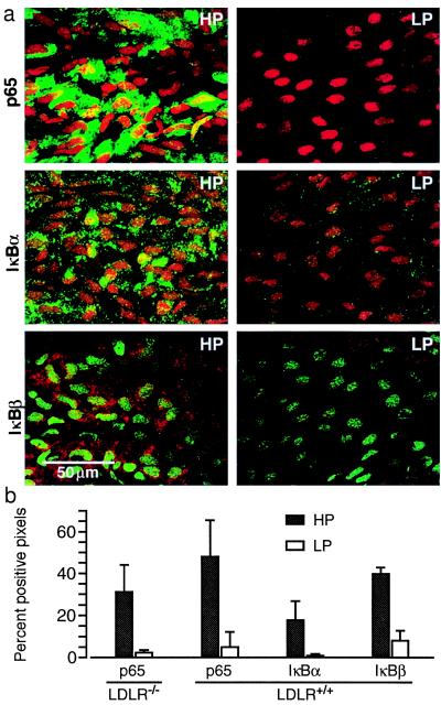
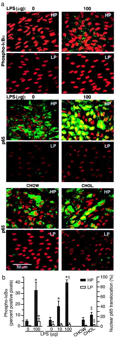
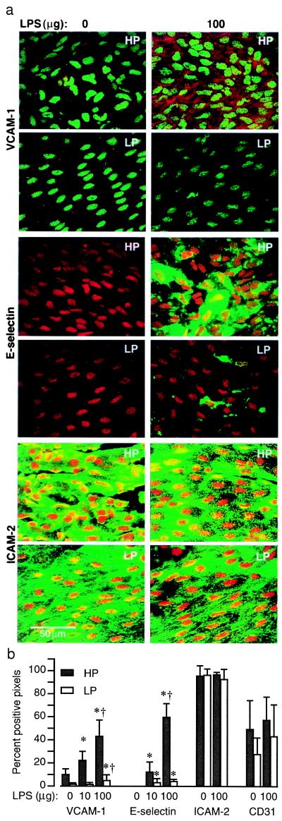
Atherosclerotic lesions form at distinct sites in the arterial tree, suggesting that hemodynamic forces influence the initiation of atherogenesis. If NF-kappaB plays a role in atherogenesis, then the activation of this signal transduction pathway in arterial endothelium should show topographic variation. The expression of NF-kappaB/IkappaB components and NF-kappaB activation was evaluated by specific antibody staining, en face confocal microscopy, and image analysis of endothelium in regions of mouse proximal aorta with high and low probability (HP and LP) for atherosclerotic lesion development. In control C57BL/6 mice, expression levels of p65, IkappaBalpha, and IkappaBbeta were 5- to 18-fold higher in the HP region, yet NF-kappaB was activated in a minority of endothelial cells. This suggested that NF-kappaB signal transduction was primed for activation in HP regions on encountering an activation stimulus. Lipopolysaccharide treatment or feeding low-density lipoprotein receptor knockout mice an atherogenic diet resulted in NF-kappaB activation and up-regulated expression of NF-kappaB-inducible genes predominantly in HP region endothelium. Preferential regional activation of endothelial NF-kappaB by systemic stimuli, including hypercholesterolemia, may contribute to the localization of atherosclerotic lesions at sites with high steady-state expression levels of NF-kappaB/IkappaB components.


Figures

Figure 1
Figure 1
Silver nitrate staining in HP and LP regions. Representative images were obtained from a C57BL/6 mouse aorta. Endothelial cells in the HP region have variable shapes and random orientation, whereas in the LP region they are elongated and aligned in the direction of blood flow (Left to Right).
Figure 2
Figure 2
High expression levels of p65, IκBα, and IκBβ in the HP region of untreated standard chow-fed C57BL/6 mice. (a) For each antibody, representative confocal images of HP and LP regions obtained from the same mouse are shown (p65 and IκBα, green and IκBβ, red fluorescence). (b) Fluorescent staining is quantified for each antibody in histograms that illustrate the percent of pixels with a positive signal after subtraction of background fluorescence. Similar expression levels of p65 were found in standard chow-fed LDLR−/− mice bred into the C57BL/6 background (LDLR−/−) and wild-type C57BL/6 mice (LDLR+/+). Data are presented as means ± SD. Significant differences (P < 0.05) were found between all HP and LP regions, determined by using a paired t test (n = 5).
Figure 3
Figure 3
LPS treatment or feeding LDLR−/− mice an atherogenic diet activates NF-κB predominantly in the HP region. (a) Representative immunoconfocal images from HP and LP regions are shown. Punctate green staining for phospho-IκBα (P-IκBα) is abundant in the cytoplasm of HP region endothelium after administration of 100 μg LPS. Nuclear translocation of p65 (green) is seen in HP regions 30 min after i.p. injection of LPS. Nuclei are counterstained with propidium iodide. Increased p65 nuclear translocation occurs in HP regions of LDLR−/− mice 2 weeks after ingestion of a 1.25% cholesterol-enriched diet (CHOL.) relative to standard chow-fed (CHOW) mice. (b) Fluorescent staining is quantified in histograms showing percent of pixels positive for phospho-IκBα and percent of nuclei with translocated p65 (mean ± SD, n = 3 to 5). Significant differences (P < 0.05) are found between all HP and LP regions (paired t test). Significant differences from 0 and 10 μg LPS and chow groups are denoted by *, †, and ‡, respectively (ANOVA).
Figure 4
Figure 4
Constitutive and LPS-induced expression of VCAM-1, E-selectin, and ICAM-2 in HP and LP regions. (a) Representative immunoconfocal images of permeabilized endothelium illustrate LPS-induced VCAM-1 (red) and E-selectin (green) but not ICAM-2 (green) expression. The magnitude of LPS-induced expression is significantly greater in HP regions. In control mice (0 μg LPS), VCAM-1, but not E-selectin, expression is detected in the HP region. Expression of ICAM-2 and PECAM-1 (CD 31) (supplementary Fig. 7; see www.pnas.org) is abundant in HP and LP regions of control and LPS-treated mice and is not induced by LPS. (b) The means ± SD of percent pixels with fluorescent staining are plotted for each group (n = 3–5). There are significant differences (P < 0.05, paired t test) between HP and LP regions for VCAM-1 and E-selectin but not for ICAM-2 and PECAM-1. Significant differences from 0 and 10 μg LPS groups are denoted by * and †, respectively (ANOVA).

Similar articles

Cited by

References

    1. Fuster V, Ross R, Topol E J. Atherosclerosis and Coronary Artery Disease. Philadelphia: Lippincott–Raven; 1996.
    1. Ross R. N Engl J Med. 1999;340:115–126. - PubMed
    1. Munro J M, Cotran R S. Lab Invest. 1988;58:249–261. - PubMed
    1. Ku D N, Zhu C. In: Hemodynamic Forces and Vascular Cell Biology. Sumpio B E, editor. Austin, TX: Landes; 1993. pp. 1–23.
    1. Ishida T, Takahashi M, Corson M A, Berk B C. Ann NY Acad Sci. 1997;811:12–23. - PubMed

Publication types