. 2000 Aug 1;97(16):9064-9.
doi: 10.1073/pnas.97.16.9064.

The junctional multidomain protein AF-6 is a binding partner of the Rap1A GTPase and associates with the actin cytoskeletal regulator profilin

Affiliations

The junctional multidomain protein AF-6 is a binding partner of the Rap1A GTPase and associates with the actin cytoskeletal regulator profilin

B Boettner et al. Proc Natl Acad Sci U S A. .

Abstract

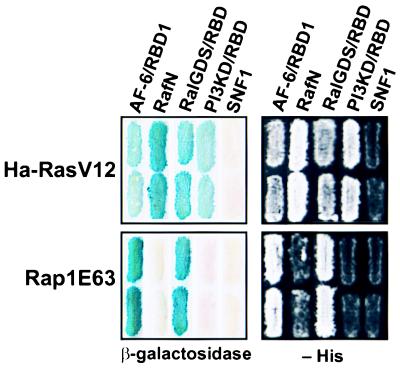
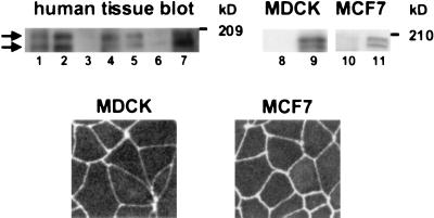
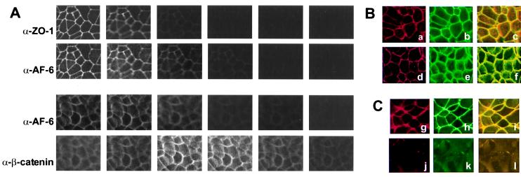
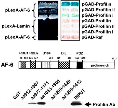
The AF-6 protein is a multidomain protein that contains two potential Ras-binding domains within its N terminus. Because of this feature, AF-6 has been isolated in both two-hybrid and biochemical approaches and is postulated to be a potential Ras-effector protein. Herein, we show that it is specifically the first Ras-binding domain of AF-6 that mediates this interaction and that the Ras-related Rap1A protein can associate with this motif even more efficiently than the oncogenic Ha-, K-, and N-Ras GTPases. We further demonstrate that both Ras and Rap1 interact with full-length AF-6 in vivo in mammalian cells and that a fraction of Rap1 colocalizes with AF-6 at the membrane. Dominant active Rap1A, in contrast to Ras, when introduced into epithelial MDCK and MCF-7 cells, does not perturb AF-6-specific residency in cell-cell adhesion complexes. In a pursuit to gain further understanding of the role of AF-6 in junctions, we identified profilin as an AF-6-binding protein. Profilin activates monomeric actin units for subsequent polymerization steps at barbed ends of actin filaments and has been shown to participate in cortical actin assembly. To our knowledge, AF-6 is the only integral component in cell-cell junctions discovered thus far that interacts with profilin and thus could modulate actin modeling proximal to adhesion complexes.


Figures

Figure 1
Figure 1
Interaction between Ras/Rap1 and their potential targets. The LexA two-hybrid tester strain L40 was transformed with plasmids expressing CA Ras and Rap1 fused to LBD, and their potential targets AF-6, c-Raf, RalGDS, and δ-PI3-kinase (PI3KD) fused to GAD. Transformants were assayed for β-galactosidase expression (Left) and for their ability to grow on medium lacking histidine (−His; Right).
Figure 2
Figure 2
(A) Association of Rap1 and full-length AF-6 in vivo in mammalian cells. Cos1 cells were transfected with myc-tagged full-length AF-6 together with empty vector, HA-tagged Rap1E63, or HA-tagged RasV12. Protein expression was confirmed by immunoblotting cell lysates with anti-HA Ab for detection of Ras and Rap1 and anti-myc Ab for detection of AF-6. Ha-tagged GTPases were immunoprecipitated with anti-HA Ab, and associated AF-6 was probed with anti-myc Ab on a Western blot (WB). (B) Comparative localization of overexpressed Rap1AE63 and endogenous AF-6 in epithelial cells. A cDNA encoding green fluorescent protein (GFP)-Rap1AE63 was introduced into MCF7 cells, and the distribution of the GTPase was assessed in vivo by confocal microscopy. The distribution of endogenous AF-6 was visualized with an AF-6-specific Ab and rhodamine-conjugated anti-rabbit IgGs.
Figure 3
Figure 3
(Upper) Distribution of AF-6 in multiple tissues and cell lines. (Lanes 1–7) A multiple human tissue blot from lung, kidney, spleen, testis, ovary, heart, and pancreas (Geno Technology, St. Louis) was immunoblotted with anti-AF-6 Ab. (Lanes 8–11) Immunoblot analysis of MDCK and MCF7 cell lysates with preimmune serum (lanes 8 and 10) and anti-AF-6 Ab (lanes 9 and 11), respectively. Two bands with molecular masses of approximately 195 and 180 kDa (kD) can be seen (arrows). (Lower) Localization of AF-6 in MDCK and MCF7 cells. Confluent MDCK and MCF7 cells were stained with rabbit polyclonal Ab against AF-6, followed by Texas red-conjugated anti-rabbit IgGs.
Figure 4
Figure 4
(A) Confocal images of MCF7 cells comparing the localization of AF-6, ZO-1, and β-catenin. Confluent MCF7 cells were doubly stained with a rabbit polyclonal Ab against AF-6 and a mouse mAb against ZO-1 or a mouse mAb against β-catenin. As secondary Abs, Texas red-conjugated anti-rabbit IgGs and FITC-conjugated anti-mouse IgGs were used. Six serial optical sections (one section every 2 μm) are shown for each staining. From left to right, apical to basolateral side. (B) Localization of AF-6, ZO-1, and β-catenin in confluent control MCF7 cells. As described above, MCF7 cells were costained with a rabbit polyclonal Ab against AF-6 (a and d) and a mouse mAb against ZO-1 (b) or a mouse mAb against β-catenin (e). AF-6 is shown in red; ZO-1 and β-catenin are shown in green; and overlay is shown in yellow (c and f). (C) Localization of AF-6 and ZO-1 in Rap1E63-expressing (g–i) and Ha-RasV12-expressing (j–l) MDCK cells. The indicated cells were costained with a rabbit polyclonal Ab against AF-6 (g and j) and a mouse mAb against ZO-1 (h and k).
Figure 5
Figure 5
Interaction of the C terminus of AF-6 and profilin. (Top) Two-hybrid interaction. An isolated profilin I cDNA containing an additional leader sequence and PCR-amplified profilin I and profilin II cDNAs fused to GAD were cotransformed with LBD AF-6N into L40 and were assessed for β-galactosidase expression and for their ability to grow on medium lacking histidine. pLex-Lamin and pGADGH were used as negative controls. (Middle) Structure of AF-6. (Bottom) AF-6–profilin association examined in GST pull-down assays. Overlapping fragments of the AF-6 C-terminal 700 amino acids were expressed as GST fusions and used to bind profilin from a whole-cell MDCK lysate. Bound profilin was visualized with an α-profilin Ab in a subsequent Western blot analysis.

References

    1. Katz M E, McCormick F. Curr Opin Genet Dev. 1997;7:75–79. - PubMed
    1. Kinch M S, Burridge K. Biochem Soc Trans. 1995;23:446–450. - PubMed
    1. Potempa S, Ridley A J. Mol Biol Cell. 1998;9:2185–2200. - PMC - PubMed
    1. Bos J L. EMBO J. 1998;23:6776–6782. - PMC - PubMed
    1. Kitayama H, Sugimoto Y, Matsuzaki T, Ikawa Y, Noda M. Cell. 1989;56:77–84. - PubMed

Publication types

LinkOut - more resources