

doi: 10.1073/pnas.97.16.9093.
Gastrulation defective is a serine protease involved in activating the receptor toll to polarize the Drosophila embryo
Affiliations
- PMID: 10922064
- PMCID: PMC16827
- DOI: 10.1073/pnas.97.16.9093
Item in Clipboard
Gastrulation defective is a serine protease involved in activating the receptor toll to polarize the Drosophila embryo
Proc Natl Acad Sci U S A.
.
Abstract
The dorsoventral axis of the Drosophila embryo is induced by a ventrally restricted ligand for the receptor Toll. The Toll ligand is generated by a proteolytic processing reaction, which occurs at the end of a proteolytic cascade and requires the gastrulation defective (gd), nudel, pipe, and windbeutel genes. Here we demonstrate that the GD protein is a serine protease and that the three other genes act to restrict GD activity to the ventral side of the embryo. Our data support a model in which the GD protease catalyzes the ventral activation of the proteolytic cascade that produces the Toll ligand.
Figures
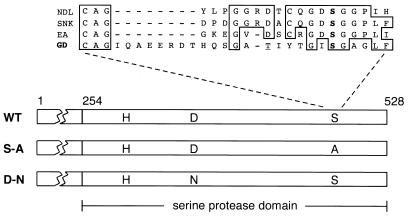
Structure of GD protein. Within the C-terminal domain of GD having sequence homology to serine proteases, the positions of the putative catalytic triad residues (H, D, and S) are indicated. Two of these residues were altered by site-directed mutagenesis to generate the S-A and D-N mutants. At the top is a comparison of the region around the catalytic serine (boldface type) in the Nudel (NDL), Snake (SNK), and Easter (EA) proteases with a similar region in GD. The candidate catalytic serine in GD is unusual, as it is positioned 8 aa further C terminal in the sequence and lacks a conserved aspartic acid as a neighbor. Our DNA sequence analysis reveals a threonine (underlined) instead of a serine in the GD sequence as previously reported (21). WT, wild type.

Rescue of ventral and lateral development in embryos by injection of gd RNA. After injection, live embryos at the gastrulation stage (A, C, E, and G) or cuticles produced by the embryos (B, D, F, and H) were examined. Embryos are oriented anterior end to the left and dorsal side up. (A and B) Embryos lacking maternal gd develop the dorsalized phenotype, symmetric folds both dorsally and ventrally at gastrulation, and a cuticle lacking all ventral and lateral structures. (C and D) After injection with 0.03 mg/ml gd RNA, the majority of gd mutant embryos are completely rescued, as evident by the lateral head fold (*) and anterior-ward displacement of pole cells (arrowhead) from original posterior position. These embryos produce a normal dorsoventral cuticle pattern showing ventral denticles (inverted v) and the dorsolateral Filzkörper (arrow). (E and F) Injection of ≥0.75 mg/ml gd RNA causes gd mutant embryos to develop the ventralized phenotype, recognizable by head fold on dorsal side (*) and retention of pole cells at posterior. These embryos produce a cuticle showing mainly ventral denticles in a disorganized pattern. (G and H) After injection with ≥0.75 mg/ml gd RNA, embryos from a nudel or pipe mutant develop the lateralized phenotype, as evident by prominence of head fold both dorsally and ventrally (*). These embryos develop a cuticle pattern similar to the ventralized phenotype. Embryos from the nudel mutant were injected without prior removal of the outer eggshell layer. The bright structure surrounding the cuticles in B, F, and H is the inner eggshell layer.
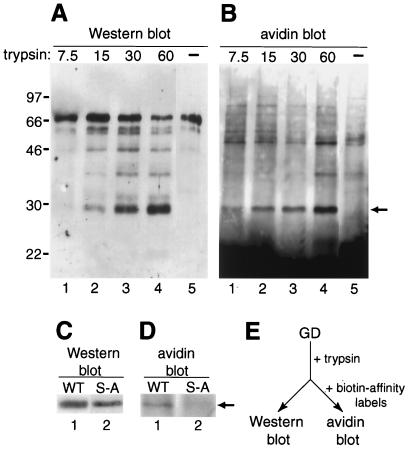
Identification of active GD protease in vitro. Recombinant GD protein in S2 cell culture medium was treated with immobilized trypsin and then analyzed by Western blot (A and C) or by avidin blot after further incubation with biotinylated affinity reagents specific for active serine proteases (B and D). (A) Increasing amounts of trypsin (nM) converts full-length GD (lane 5) into smaller polypeptides, including a prominent 29-kDa form (lanes 1–4). (B) The 29-kDa polypeptide (arrowhead, lanes 1–4), but not full-length GD (lane 5), is biotinylated. Free reagents are likely the cause of an intense signal toward the bottom of the avidin blot. Molecular masses of markers in kDa are indicated on the left. (C) Trypsin treatment generates a 29-kDa polypeptide from both wild-type GD (WT, lane 1) and mutant GD lacking putative catalytic serine (S-A, lane 2). (D) The 29-kDa polypeptide from wild type (lane 1), but not mutant (lane 2), is biotinylated (arrow). (E) Basic scheme of experiment.
Similar articles
-
A ventrally localized protease in the Drosophila egg controls embryo dorsoventral polarity.Curr Biol. 2012 Jun 5;22(11):1013-8. doi: 10.1016/j.cub.2012.03.065. Epub 2012 May 10. Curr Biol. 2012. PMID: 22578419 Free PMC article.
-
Localized serine protease activity and the establishment of Drosophila embryonic dorsoventral polarity.Fly (Austin). 2013 Jul-Sep;7(3):161-7. doi: 10.4161/fly.25141. Epub 2013 Jun 10. Fly (Austin). 2013. PMID: 24047959 Free PMC article.
-
Signal transduction by a protease cascade.Trends Cell Biol. 1999 Mar;9(3):102-7. doi: 10.1016/s0962-8924(98)01494-9. Trends Cell Biol. 1999. PMID: 10201075 Review.
-
Regulation of translation and proteolysis during the development of embryonic dorso-ventral polarity in Drosophila. Homology of easter proteinase with Limulus proclotting enzyme and translational activation of Toll receptor synthesis.Biochim Biophys Acta. 1992 Oct 20;1132(3):290-6. doi: 10.1016/0167-4781(92)90163-t. Biochim Biophys Acta. 1992. PMID: 1420309
-
Axis determination. Proteolytic generation of a morphogen.Curr Biol. 1994 Aug 1;4(8):755-7. doi: 10.1016/s0960-9822(00)00170-6. Curr Biol. 1994. PMID: 7953570 Review.
Cited by
-
Profiling ATM regulated genes in Drosophila at physiological condition and after ionizing radiation.Hereditas. 2022 Oct 21;159(1):41. doi: 10.1186/s41065-022-00254-9. Hereditas. 2022. PMID: 36271387 Free PMC article.
-
Serine proteolytic pathway activation reveals an expanded ensemble of wound response genes in Drosophila.PLoS One. 2013 Apr 24;8(4):e61773. doi: 10.1371/journal.pone.0061773. Print 2013. PLoS One. 2013. PMID: 23637905 Free PMC article.
-
Three-dimensional morphology and gene expression in the Drosophila blastoderm at cellular resolution II: dynamics.Genome Biol. 2006;7(12):R124. doi: 10.1186/gb-2006-7-12-r124. Genome Biol. 2006. PMID: 17184547 Free PMC article.
-
Spatially Restricted Regulation of Spätzle/Toll Signaling during Cell Competition.Dev Cell. 2018 Sep 24;46(6):706-719.e5. doi: 10.1016/j.devcel.2018.08.001. Epub 2018 Aug 23. Dev Cell. 2018. PMID: 30146479 Free PMC article.
-
No requirement for localized Nudel protein expression in Drosophila embryonic axis determination.Fly (Austin). 2008 Jul-Aug;2(4):220-8. doi: 10.4161/fly.6794. Fly (Austin). 2008. PMID: 18776742 Free PMC article.
References
-
- Morisato D, Anderson K V. Annu Rev Genet. 1995;29:371–399. - PubMed
-
- Morisato D, Anderson K V. Cell. 1994;76:677–688. - PubMed
-
- Schneider D S, Jin Y, Morisato D, Anderson K V. Development (Cambridge, UK) 1994;120:1243–1250. - PubMed
-
- DeLotto R, Spierer P. Nature (London) 1986;323:688–692. - PubMed
-
- Chasan R, Anderson K V. Cell. 1989;56:391–400. - PubMed
Publication types
MeSH terms
Substances
Grants and funding
LinkOut - more resources
Full Text Sources
Molecular Biology Databases
