. 2000 Aug 1;97(16):9264-9.
doi: 10.1073/pnas.97.16.9264.

Pseudorabies virus expressing enhanced green fluorescent protein: A tool for in vitro electrophysiological analysis of transsynaptically labeled neurons in identified central nervous system circuits

Affiliations

Pseudorabies virus expressing enhanced green fluorescent protein: A tool for in vitro electrophysiological analysis of transsynaptically labeled neurons in identified central nervous system circuits

B N Smith et al. Proc Natl Acad Sci U S A. .

Abstract

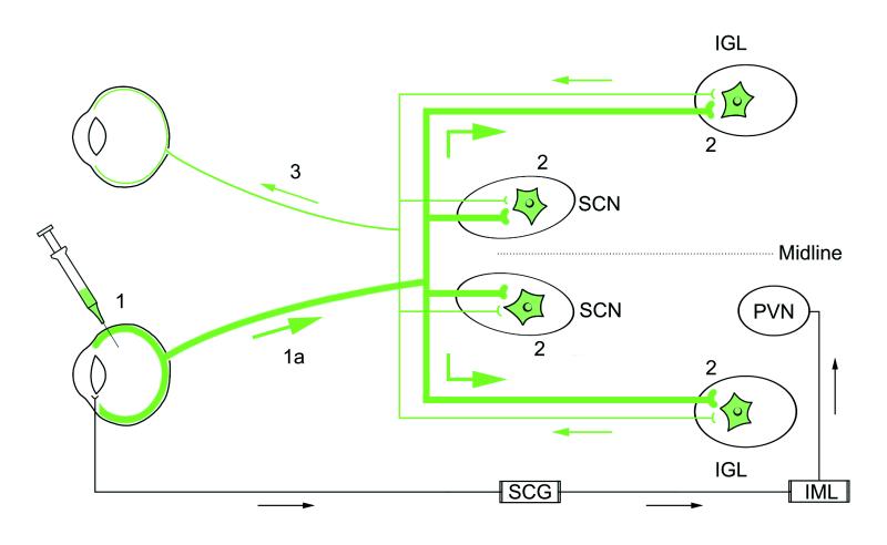
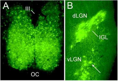
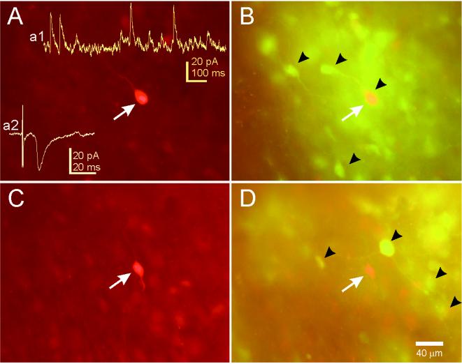
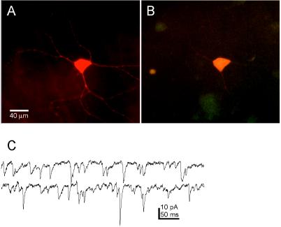
Physiological properties of central nervous system neurons infected with a pseudorabies virus were examined in vitro by using whole-cell patch-clamp techniques. A strain of pseudorabies virus (PRV 152) isogenic with the Bartha strain of PRV was constructed to express an enhanced green fluorescent protein (EGFP) from the human cytomegalovirus immediate early promoter. Unilateral PRV 152 injections into the vitreous body of the hamster eye transsynaptically infected a restricted set of retinorecipient neurons including neurons in the hypothalamic suprachiasmatic nucleus (SCN) and the intergeniculate leaflet (IGL) of the thalamus. Retinorecipient SCN neurons were identified in tissue slices prepared for in vitro electrophysiological analysis by their expression of EGFP. At longer postinjection times, retinal ganglion cells in the contralateral eye also expressed EGFP, becoming infected after transsynaptic uptake and retrograde transport from infected retinorecipient neurons. Retinal ganglion cells that expressed EGFP were easily identified in retinal whole mounts viewed under epifluorescence. Whole-cell patch-clamp recordings revealed that the physiological properties of PRV 152-infected SCN neurons were within the range of properties observed in noninfected SCN neurons. Physiological properties of retinal ganglion cells also appeared normal. The results suggest that PRV 152 is a powerful tool for the transsynaptic labeling of neurons in defined central nervous system circuits that allows neurons to be identified in vitro by their expression of EGFP, analyzed electrophysiologically, and described in morphological detail.


Figures

Figure 1
Figure 1
A schematic diagram illustrating the neuronal circuits labeled after the intraocular injection of PRV 152. Primary infection of retinal ganglion cells occurs after intravitreal injection of PRV 152 (step 1). Viral replication and anterograde transport of virus (step 1a, thick green line) in axons of RGCs follows primary infection. Transsynaptic passage of virus from RGC axons produces secondary infection in a restricted set of retinorecipient central nuclei [i.e., SCN, IGL (shown), PT, and lateral terminal nucleus (not shown) as indicated by green (EGFP-labeled) neurons (2)]. Axon terminals of retinal ganglion cells in the contralateral retina, presynaptic to PRV 152-infected neurons in the SCN, IGL, PT, and lateral terminal nucleus, take up the virus via transsynaptic passage. The virus is retrogradely transported (thin green line) to the contralateral retina, where PRV 152-expressing ganglion cells are easily identified in whole-mount preparations (step 3). Intravitreal PRV 152 injection also infects autonomic afferents to the eye, resulting in retrograde transport of virus to the superior cervical ganglion (SCG), followed by transsynaptic uptake and retrograde transport to preganglionic neurons in the intermediolateral nucleus (IML) of the spinal cord (18). Neurons in the hypothalamic paraventricular nucleus (PVN) are subsequently infected after transsynaptic uptake of the virus from infected IML neurons and retrograde transport.
Figure 2
Figure 2
Micrographs demonstrating transsynaptic EGFP-neuronal labeling after unilateral intravitreal injection of PRV 152 116 h after inoculation. (A) Bilateral transsynaptic infection was evident in the retinorecipient hypothalamic SCN. (B) Bilateral PRV 152 transsynaptic labeling was also evident in the retinorecipient IGL and the ventral lateral geniculate nucleus (vLGN), but not the dorsal LGN. OC, optic chiasm; III, third ventricle.
Figure 3
Figure 3
Examples of neurons recorded in the SCN after PRV 152 intravitreal injection. (A) This SCN neuron was filled with biocytin during a whole-cell patch-clamp recording and visualized with an avidin-rhodamine conjugate. (Inset a1) An example of spontaneous IPSCs in this neuron while voltage clamped at −15 mV. (Inset a2) An example of an EPSC evoked in this neuron by stimulation of the optic nerve. (B) PRV 152 labeling in the same tissue viewed under optics that permits visualization of rhodamine and EGFP concurrently (double-labeled cells appear yellow-orange). The arrow indicates the filled cell in A was also infected with PRV 152. Arrowheads point to examples of EGFP-labeled SCN neurons in the hypothalamic slice. (C) Another biocytin-filled and recorded neuron in the contralateral SCN from the same animal. (D) PRV 152 labeling in the same tissue. The arrow indicates the position of the filled cell in C, which was not infected. Arrowheads point to examples of nearby EGFP-labeled SCN neurons.
Figure 4
Figure 4
Biocytin-filled EGFP-expressing retinal ganglion cell viewed in whole mount. (A) Intravitreal PRV 152 injection results in EGFP-expressing ganglion cells in the contralateral retina. This neuron was targeted for recording, filled with biocytin, and reacted with avidin-rhodamine. (B) The same neuron as in A viewed with optics demonstrating double labeling (i.e., EGFP and rhodamine fluorescence). (C) Examples of spontaneous EPSCs recorded from this retinal ganglion cell. Holding potential = −65 mV; traces are continuous.

Similar articles

Cited by

References

    1. Card J P. Anat Rec. 1998;253:176–185. - PubMed
    1. Card J P, Enquist L W, Moore R Y. J Comp Neurol. 1999;407:438–452. - PubMed
    1. Chen S, Yang M, Miselis R R, Aston-Jones G. Brain Res. 1999;838:171–183. - PubMed
    1. Enquist L W, Husak P J, Banfield B W, Smith G A. Adv Virus Res. 1999;51:237–347. - PubMed
    1. Kuypers H G J M, Ugolini G. Trends Neurosci. 1990;13:71–75. - PubMed

Publication types

Substances

LinkOut - more resources