. 2000 Aug;106(3):385-92.
doi: 10.1172/JCI9896.

Persistence of platelet thrombus formation in arterioles of mice lacking both von Willebrand factor and fibrinogen

Affiliations

Persistence of platelet thrombus formation in arterioles of mice lacking both von Willebrand factor and fibrinogen

H Ni et al. J Clin Invest. 2000 Aug.

Abstract

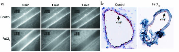
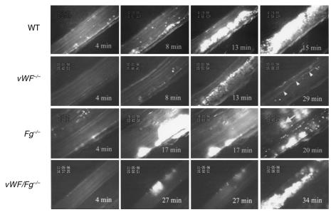
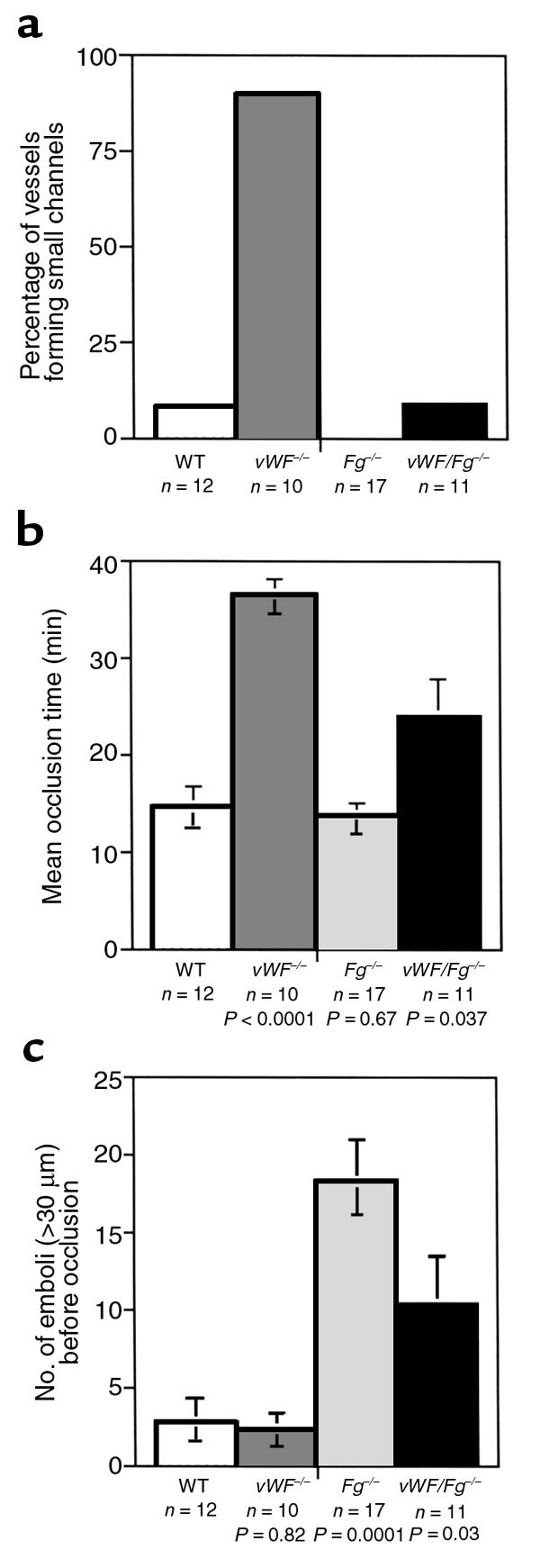
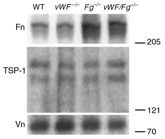
We used intravital microscopy to observe the formation of platelet plugs in ferric chloride-injured arterioles of live mice. With this model, we evaluated thrombus growth in mice lacking von Willebrand factor (vWF) and fibrinogen (Fg), the two key ligands known to mediate platelet adhesion and aggregation. In vWF(-/-) mice, despite the presence of arterial shear, delayed platelet adhesion occurred and stable thrombi formed. In many mice, a persisting high-shear channel never occluded. Abundant thrombi formed in Fg(-/-) mice, but they detached from the subendothelium, which ultimately caused downstream occlusion in all cases. Surprisingly, mice deficient in both vWF and Fg successfully formed thrombi with properties characteristic of both mutations, leading to vessel occlusion in the majority of vessels. Platelets of these doubly deficient mice specifically accumulated fibronectin in their alpha-granules, suggesting that fibronectin could be the ligand supporting the platelet aggregation.


Figures

Figure 1
Figure 1
Arteriolar injury and denudation of endothelium induced by FeCl3. (a) Progression of FeCl3-induced injury. Fluorescence intensity of endothelium of mesenteric arterioles (n = 14/group) from GFP-transgenic mice was blindly ranked from 5 to 0. FeCl3-treated or untreated arterioles were then compared by Mann-Whitney U test. Before FeCl3 treatment (0 min), all arterioles had a similar intensity (∼5). Significant differences were found after 1 minute of FeCl3 exposure (P = 0.0006). After 2 minutes (not shown), the intensity in control vessels decreased only slightly (mean = 4.1), whereas in FeCl3-treated vessels, fluorescence was greatly decreased (mean = 1.4; P = 0.0001). After 4 minutes, the fluorescence intensity in the control group was still high (mean = 3.4), but in the FeCl3 group was almost undetectable (mean = 0.4; P < 0.0001). Representative photographs of one vessel from each group are shown. (b) Denudation of endothelium by FeCl3. Control (∼130 μm diameter) and FeCl3-treated (∼100 μm diameter) sections of mesenteric arterioles from wild-type mice were stained with rabbit anti-human vWF polyclonal Ab, which cross-reacts with mouse vWF. Endothelial cells in the control arteriole were heavily stained, whereas only a few areas were stained with this Ab (arrow) after 5-minute FeCl3 treatment. These areas may be remaining endothelial cells or small thrombi. The brown ring at the inner side of the injured vessel wall may result from plasma vWF deposition or from subendothelial vWF.
Figure 2
Figure 2
Characteristics of thrombus growth differ with mouse genotype. Times after FeCl3-induced injury are indicated (min). In wild-type mice (WT), numerous adherent fluorescently labeled platelets can be seen 4 minutes after FeCl3 treatment. Thrombi usually grow fast and cause vessels to occlude at the site of injury (15 minutes). In vWF–/– mice, very few platelet–vessel wall interactions were seen at the early time points, but thrombus formation occurred (13 minutes). Thrombi usually stopped growing at the later times, leaving a small channel open (arrowheads, 29 minutes) with high shear flow. The dark thrombus at this time point indicates that few new platelets were recruited, and platelet fluorescence was bleached after a long exposure to UV light. In Fg–/– mice, early platelet depositions were similar to wild-type mice (4 minutes), and thrombi grew very efficiently (bright thrombi). However, thrombi were not stable. They were often stripped off the vessel wall by blood flow (two sequential panels at 17 minutes) and ultimately led to a downstream occlusion (20 minutes with fluorescent platelets suspended in stagnant blood). The arrow in this panel (Fg–/–, 20 minutes) shows a thrombus formed in a neighboring venule. In the double-knockout mice (vWF/Fg–/–), very few fluorescently labeled platelets were seen on the vessel wall at an early time point (4 minutes), but delayed thrombus formation occurred with frequent embolization (two sequential panels at 27 minutes). The majority of the double-deficient arterioles eventually occluded.
Figure 3
Figure 3
Quantitative analysis of thrombi formation in wild-type and mutant arterioles. (a) Frequent occurrence of narrow flow channels in thrombi of arterioles in vWF–/– mice. Small channels within thrombi lasting more than 5 minutes were recorded and their frequency established for each genotype. Thrombi in nine out of 10 vessels in vWF–/– mice formed these channels in the late stage, which was a frequency significantly higher than in wild-type mice (1/12) (χ2 = 14.66, P < 0.005) or in other genotypes of mice. (b) The effects of vWF or Fg deficiency on vessel occlusion time. The time before blood flow completely stopped in each vessel was determined. If blood flow did not cease during the 40-minute observation period, 40 minutes was used as the occlusion time. Surprisingly, the time needed to stop blood flow in Fg–/– arterioles was similar to wild-type. The occlusion time in vWF-knockout mice was longer than that of double-knockout mice (P < 0.01), where embolization led to a more rapid thrombus accumulation, and both were prolonged in comparison with wild-type. The P values shown in this figure were determined by comparison with wild-type arterioles. (c) The roles of vWF or Fg in thrombus embolization. The number of large emboli (diameter > 30 μm) generated in the period before vessel occlusion was determined. Although this period was short in Fg–/– mice, the number of emboli formed in these mice was the highest. However, no statistically significant difference was found between Fg–/– mice and double knockouts (P = 0.07). The P values shown in this figure were determined by comparison with wild-type arterioles.
Figure 4
Figure 4
Increased fibronectin content in platelets of Fg–/– and vWF/Fg–/– mice. For each genotype, platelets were isolated from pooled blood of six mice. Gel-filtered platelets (2 × 10⁷) were analyzed by Western blot. The position of migration of molecular mass standards is shown on the right (in kiloDaltons). Platelet vitronectin (Vn) and thrombospondin-1 (TSP-1) levels were the same in all samples. In contrast, the amount of fibronectin in platelets from either the Fg–/– or vWF/Fg–/– mice was three times greater than from the wild-type mice or vWF–/– mice. The doublet seen with anti–thrombospondin-1 is a consequence of proteolytic cleavage of thrombospondin-1.

Similar articles

Cited by

References

    1. von Willebrand E. Hereditar psseudohemofili. Fin Lakaresallsk Handl. 1926;67:87–112.
    1. Gugler E, Lüscher EF. Platelet function in congenital afibrinogenemia. Thromb Diath Haemorrh. 1965;14:361–373. - PubMed
    1. Ewenstein BM. von Willebrand’s disease. Annu Rev Med. 1997;48:525–542. - PubMed
    1. Martinez J. Congenital dysfibrinogenemia. Curr Opin Hematol. 1997;4:357–365. - PubMed
    1. Tschopp TB, Weiss HJ, Baumgartner HR. Decreased adhesion of platelets to subendothelium in von Willebrand’s disease. J Lab Clin Med. 1974;83:296–300. - PubMed

Publication types

MeSH terms