. 2000 Aug;11(8):2543-51.
doi: 10.1091/mbc.11.8.2543.

The tumor suppressor p53 can both stimulate and inhibit ultraviolet light-induced apoptosis

Affiliations
Free PMC article

The tumor suppressor p53 can both stimulate and inhibit ultraviolet light-induced apoptosis

B C McKay et al. Mol Biol Cell. 2000 Aug.
Free PMC article

Abstract

We have previously shown that the tumor suppressor p53 can play a protective role against UV-induced apoptosis in human fibroblasts. In the present study, we investigated whether the protective function of p53 expression is established before or after UV irradiation. Using a stable human cell line expressing a murine temperature-sensitive p53 in which p53 function could be tightly and reversibly regulated, we found that functional p53 stimulated the induction of apoptosis when expressed for as little as 4-12 h after UV irradiation and that this induction was not dependent on de novo protein synthesis. In contrast, expression of p53 for 12 h or more before UV irradiation reduced the extent of apoptosis even when functional p53 expression was maintained after irradiation. The protection conferred by p53 required ongoing protein synthesis and correlated with enhanced recovery of mRNA synthesis. Together, these results suggest that p53 induces distinct proapoptotic and antiapoptotic signals and that these opposing activities can be separated both temporally and by their requirement for de novo protein synthesis. These findings may have important implications for the refinement of gene therapy approaches combining p53 with pharmacological agents that target transcription or translation.


Figures
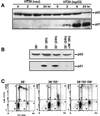
Figure 1
Figure 1
p53 function can be rapidly turned on and off in HT29-tsp53 cells by temperature switching. (A) Western blot showing the expression of p53 and p21WAF1 in HT29-neo and HT29-tsp53 cells after incubation of cells for different periods at 32°C. (B) Western blots showing stable expression of p53 and induction of p21WAF1 within 6 h at 32°C and the rapid loss of p21WAF1 when switching the HT29-tsp53 cells back to 38°C. (C) Two-parameter flow cytometry diagram of cells pulse labeled for 15 min with BrdU immediately before collection. BrdU incorporation (replicative DNA synthesis) is plotted on the Y axis and DNA content is plotted on the X axis. HT29-tsp53 cells were grown at 38°C (left panels), incubated at 32°C for 24 h (middle panels), or incubated at 32°C for 24 h and then returned to 38°C for 24 h (right panels).
Figure 2
Figure 2
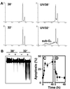
Post-UV expression of functional p53 enhances UV-induced apoptosis. (A) Flow cytometry diagram of propidium iodide–stained HT29-tsp53 cells maintained at 38°C (upper panels) or switched to 32°C (lower panels) at the time of mock irradiation (left panels) or UV irradiation with 20 J/m2 (right panels). Forty-eight hours after UV irradiation, cells were subjected to flow cytometry analysis, and cells with a sub-G1 DNA content were considered to be apoptotic. (B) HT29-tsp53 cells treated as in A were lysed in the wells of the agarose gel, and the extent of DNA fragmentation was assessed by electrophoresis. The black-and-white image of the ethidium bromide–stained gel has been reversed for clarity. (C) HT29-tsp53 cells were switched to 32°C at the time of UV irradiation and returned to 38°C at various times after UV treatment. Apoptosis was scored as in A and plotted as a function of post-UV incubation time at 32°C. (D) HT29-tsp53 cells were UV irradiated and maintained at 38°C for various periods before switching them to 32°C. Apoptosis was scored as in A and plotted as a function of time between UV irradiation and the switch to 32°C. In C and D, each point represents the mean ± SEM of two to eight independent experiments with background values subtracted. Mean background sub-G1 values varied between 6 and 11%.
Figure 3
Figure 3
Previous expression of functional p53 protects cells against UV-induced apoptosis. HT29-neo (A) or HT29-tsp53 (B) cells were mock irradiated (white bars) or UV irradiated with 20 J/m2 (black bars), and apoptosis was scored 48 h later by flow cytometry. The numbers below the bars represent the temperature-shift protocols described in Table 1. (C) The effect of UV dose on the induction of apoptosis in HT29-tsp53 cells was determined for the same temperature-shift protocols: 1 (▪), 2 (●), 3 (▴), and 4 (▾). Each value represents the mean ± SEM of at least three independent experiments. (D) HT29-tsp53 cells were incubated at 32°C for various periods before being exposed to UV irradiation (20 J/m2). The cells were maintained at 32°C after UV irradiation, and apoptosis was scored as the percentage of cells with a sub-G1 DNA content measured 48 h after treatment. Each point represents the mean ± SEM of three to seven independent experiments with background values subtracted. The mean of the background values from the different experiments varied between 7 and 16%.
Figure 4
Figure 4
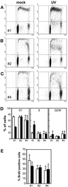
Protection conferred by p53 does not appear to be related to sustained G1 arrest. (A–D) Twenty-four hours after either mock irradiation or UV irradiation with 20 J/m2, BrdU pulse-labeled HT29-tsp53 cells (15-min pulse) were subjected to two-parameter flow cytometry analysis as described in Figure 1. (A) Cells were maintained at 38°C throughout the experiment (protocol 1; Table 1). (B) Cells were switched to 32°C immediately after UV irradiation (protocol 2). (C) Cells were switched to 32°C 24 h before UV irradiation and maintained at 32°C during the post-UV incubation period (protocol 4). (D) The cell cycle distributions (as in A–C) of mock-irradiated (white bars) or UV-irradiated (black bars) HT29-tsp53 cells subjected to protocols 1, 2, and 4 were determined from multiple experiments. (E) HT29-tsp53 cells were subjected to protocols 1, 2, and 4 (Table 1), BrdU labeled continuously for 12 h after mock irradiation or UV irradiation with 20 J/m2, and subjected to two-parameter flow cytometry analysis. Results are expressed as the percentage of cells that entered S phase within 12 h after UV irradiation. Each point in D and E represents the mean ± SE of two to four experiments.
Figure 5
Figure 5
Protection conferred by p53 does not correlate with previous establishment of G1 arrest. Two-parameter flow cytometry analysis of the cell cycle distribution of unirradiated HT29-tsp53 cells at different times after switching to the permissive temperature of 32°C. Numbers represent the percentages of cells in S phase.
Figure 6
Figure 6
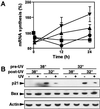
p53 stimulates the recovery of transcription after UV irradiation. (A) Cells were subjected to the temperature-shift protocols described in Table 1: 1 (▪), 2 (●), 3 (▴), and 4 (▾). At various times after UV irradiation, nascent RNA was labeled for 30 min with [3H]uridine. Polyadenylated RNA was isolated with the use of oligo-dT beads and quantified with a scintillation counter. (B) The expression of p21WAF1 and Bax was assessed 6 h after UV irradiation by Western blot analysis in HT29-tsp53 cells subjected to the different temperature-shift protocols (Table 1). Expression of actin was used as a loading control.
Figure 7
Figure 7
CHX blocks p21WAF1 expression and potentiates p53-mediated apoptosis. (A) Cells were switched to 32°C at the time of UV or CHX treatment, and p53, p21WAF1, Bax, and actin levels were assessed by Western blot analysis 6 h later. (B) HT29-neo and HT29-tsp53 cells were subjected to protocols 1 (white bars), 2 (black bars), and 4 (gray bars) with CHX for 48 h. The induction of apoptosis in mock-treated controls was subtracted from that determined for each treatment. CHX significantly stimulated the induction of apoptosis in HT29-tsp53 cells subjected to protocol 2. Values are expressed as means ± SEM from two to five independent experiments.
Figure 8
Figure 8
In this model, the apoptosis-promoting functions of high levels of functional p53 are balanced by the p53-mediated transactivation of survival factors. When expression of these survival factors is attenuated by either UV light or CHX, p53 will induce transactivation-independent apoptosis. In addition, p53 expression before UV irradiation increases the efficiency of survival-promoting functions such as the recovery of mRNA synthesis. We also suggest that previous expression of p53 protects cells against UV- or CHX-induced apoptosis by increasing the expression of p53-regulated survival-promoting factors before cellular stress. These protective functions are not fully independent because stimulating the recovery of transcription after UV irradiation will also permit the recovery of post-UV expression of p53-regulated survival factors, thus decreasing the induction of apoptosis. TDF, transactivation-dependent function; TIF, transactivation-independent function; RRS, recovery of mRNA synthesis.

Similar articles

Cited by

References

    1. Aragane Y, Kulms D, Metze D, Wilkes G, Pöppelmann B, Luger T, Schwarz T. Ultraviolet light induces apoptosis via direct activation of CD95 (fas/APO-1) independently of its ligand CD95L. J Cell Biol. 1998;140:171–182. - PMC - PubMed
    1. Aranda-Anzaldo A, Orozco-Velasco F, Garcia-Villa E, Gariglio P. p53 is a rate-limiting factor in the repair of higher-order DNA structure. Biochim Biophys Acta. 1999;1446:181–192. - PubMed
    1. Barley RDC, Enns L, Paterson MC, Mirzayans R. Aberrant p21(WAF1)-dependent growth arrest as the possible mechanism of abnormal resistance to ultraviolet light cytotoxicity in Li-Fraumeni syndrome fibroblast strains heterozygous for TP53 mutations. Oncogene. 1998;17:533–543. - PubMed
    1. Bissonnette N, Hunting DJ. p21-induced cycle arrest in G1 protects cells from apoptosis induced by UV-irradiation or RNA polymerase II blockage. Oncogene. 1998;16:3461–3469. - PubMed
    1. Bunz F, Dutriaux A, Lengauer C, Waldman T, Zhou S, Brown JP, Sedivy JM, Kinzler KW, Vogelstein B. Requirement for p53 and p21 to sustain G2 arrest after DNA damage. Science. 1998;282:1497–1501. - PubMed

Publication types

Substances