. 2000 Aug;11(8):2617-29.
doi: 10.1091/mbc.11.8.2617.

The interaction between Ran and NTF2 is required for cell cycle progression

Affiliations
Free PMC article

The interaction between Ran and NTF2 is required for cell cycle progression

B B Quimby et al. Mol Biol Cell. 2000 Aug.
Free PMC article

Abstract

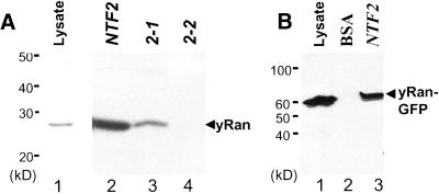
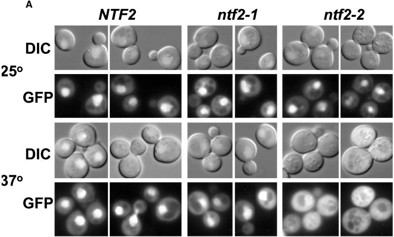
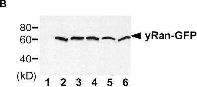
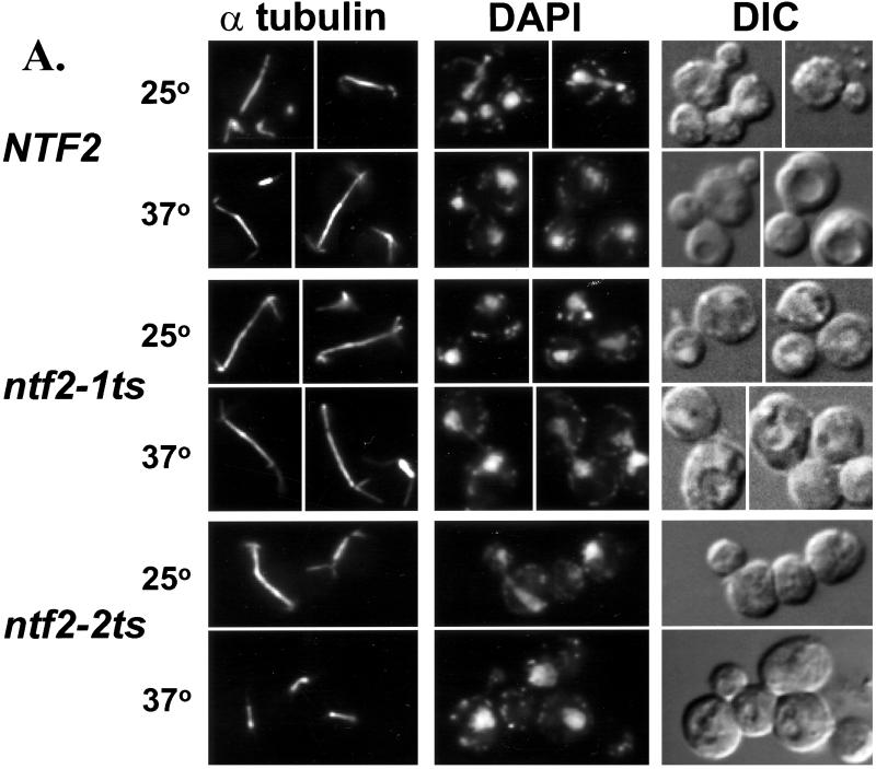
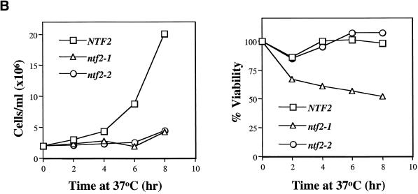
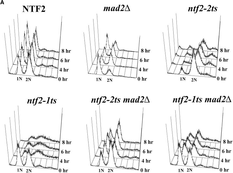
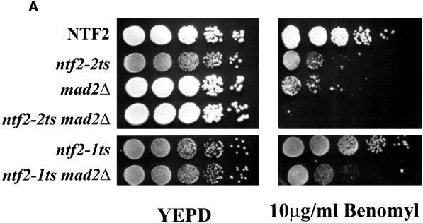
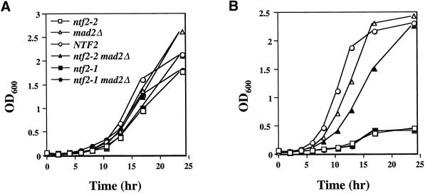
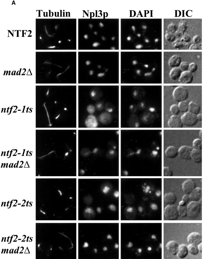
The small GTPase Ran is required for the trafficking of macromolecules into and out of the nucleus. Ran also has been implicated in cell cycle control, specifically in mitotic spindle assembly. In interphase cells, Ran is predominately nuclear and thought to be GTP bound, but it is also present in the cytoplasm, probably in the GDP-bound state. Nuclear transport factor 2 (NTF2) has been shown to import RanGDP into the nucleus. Here, we examine the in vivo role of NTF2 in Ran import and the effect that disruption of Ran imported into the nucleus has on the cell cycle. A temperature-sensitive (ts) mutant of Saccharomyces cerevisiae NTF2 that does not bind to Ran is unable to import Ran into the nucleus at the nonpermissive temperature. Moreover, when Ran is inefficiently imported into the nucleus, cells arrest in G(2) in a MAD2 checkpoint-dependent manner. These findings demonstrate that NTF2 is required to transport Ran into the nucleus in vivo. Furthermore, we present data that suggest that depletion of nuclear Ran triggers a spindle-assembly checkpoint-dependent cell cycle arrest.


Figures

Figure 1
Figure 1
Interaction of scRan with Ntf2p. Interactions were examined using bead-binding assays as described in Materials and Methods. Immunoblots detecting scRan are shown. (A) Interaction of scRan with Ntf2–1p and Ntf2–2p. Lane 1, yeast cell lysate; lane 2, wild-type Ntf2p beads; lane3, Ntf2–1p beads; and lane 4, Ntf2–2p beads. (B) Interaction of scRan-GFP with Ntf2p. Lane 1, yeast cell lysate; lane 2, BSA beads; and lane 3, Ntf2p beads.
Figure 2
Figure 2
Localization of scRan. (A) NTF2, ntf2–1ts, and ntf2–2ts cells were transformed with a 2μ plasmid encoding scRan-GFP (pAC410). Transformants were grown in liquid media lacking uracil to log phase at 25°C, were split, and were grown at 25°C or 37°C for 3 h. scRan-GFP was viewed directly in living cells. (B) Levels of scRan-GFP are the same in NTF2, ntf2–1ts, and ntf2–2ts transformants grown at 25°C and 37°C. Cells were lysed as described in Materials and Methods, 10 μg of each lysate was resolved on SDS-polyacrylamyide gel, transferred to nitrocellulose, and detected with anti-GFP. Lane 1, vector alone (no GFP) 37°C; lanes 2–4, 25°C (lane 2, NTF2; lane 3, ntf2–1ts; and lane 4, ntf2–2ts); lanes 5 and 6, 37°C (lane 5, ntf2–1ts; lane 6, ntf2–2ts).
Figure 2
Figure 2
Localization of scRan. (A) NTF2, ntf2–1ts, and ntf2–2ts cells were transformed with a 2μ plasmid encoding scRan-GFP (pAC410). Transformants were grown in liquid media lacking uracil to log phase at 25°C, were split, and were grown at 25°C or 37°C for 3 h. scRan-GFP was viewed directly in living cells. (B) Levels of scRan-GFP are the same in NTF2, ntf2–1ts, and ntf2–2ts transformants grown at 25°C and 37°C. Cells were lysed as described in Materials and Methods, 10 μg of each lysate was resolved on SDS-polyacrylamyide gel, transferred to nitrocellulose, and detected with anti-GFP. Lane 1, vector alone (no GFP) 37°C; lanes 2–4, 25°C (lane 2, NTF2; lane 3, ntf2–1ts; and lane 4, ntf2–2ts); lanes 5 and 6, 37°C (lane 5, ntf2–1ts; lane 6, ntf2–2ts).
Figure 3
Figure 3
Cell cycle arrest in the ntf2–2ts mutant. (A) Visualization of spindles in NTF2 mutants. NTF2, ntf2–1ts, and ntf2–2ts cells were grown to log phase at 25°C, were split, and were shifted at 25°C or 37°C for 3 h. Cells were stained with antitubulin, to visualize microtubules, and with DAPI, to visualize DNA, as described in Materials and Methods. (B) Growth and viability of NTF2 mutants at 37°C. Cells were grown overnight at 25°C, diluted to 2 × 10⁶ cells/ml, and shifted to 37°C. Samples were removed every 2 h, counted, and 200 cells were plated onto YEPD at 25°C. The results are plotted as cell number versus time at 37°C (left panel) or percent of viability versus time at 37°C (right panel).
Figure 3
Figure 3
Cell cycle arrest in the ntf2–2ts mutant. (A) Visualization of spindles in NTF2 mutants. NTF2, ntf2–1ts, and ntf2–2ts cells were grown to log phase at 25°C, were split, and were shifted at 25°C or 37°C for 3 h. Cells were stained with antitubulin, to visualize microtubules, and with DAPI, to visualize DNA, as described in Materials and Methods. (B) Growth and viability of NTF2 mutants at 37°C. Cells were grown overnight at 25°C, diluted to 2 × 10⁶ cells/ml, and shifted to 37°C. Samples were removed every 2 h, counted, and 200 cells were plated onto YEPD at 25°C. The results are plotted as cell number versus time at 37°C (left panel) or percent of viability versus time at 37°C (right panel).
Figure 4
Figure 4
Analysis of the cell cycle arrest in ntf2–2ts mutant cells. (A) DNA content in NTF2 mutants. NTF2, ntf2–1ts, ntf2–2ts, MAD2Δ, ntf2–2ts MAD2Δ, and ntf2–1ts MAD2Δ cells were grown overnight at 25°C, were diluted to 2 × 10⁶ cells/ml in YEPD, and were shifted to 37°C, and samples were taken at 0, 4, 6, and 8 h. The DNA content of individual NTF2, ntf2–1ts, and ntf2–2ts cells was determined by flow cytometry. (B) Spindle pole body localization in NTF2 mutants. NTF2, ntf2–1ts and ntf2–2ts cells were transformed with a plasmid encoding Nuf2-GFP (Kahana et al., 1995) to visualize spindle pole bodies. Transformed cells were grown to log phase at 25°C and then shifted to 37°C for 3 h. GFP-labeled spindle pole bodies were visualized by direct fluorescent microscopy.
Figure 4
Figure 4
Analysis of the cell cycle arrest in ntf2–2ts mutant cells. (A) DNA content in NTF2 mutants. NTF2, ntf2–1ts, ntf2–2ts, MAD2Δ, ntf2–2ts MAD2Δ, and ntf2–1ts MAD2Δ cells were grown overnight at 25°C, were diluted to 2 × 10⁶ cells/ml in YEPD, and were shifted to 37°C, and samples were taken at 0, 4, 6, and 8 h. The DNA content of individual NTF2, ntf2–1ts, and ntf2–2ts cells was determined by flow cytometry. (B) Spindle pole body localization in NTF2 mutants. NTF2, ntf2–1ts and ntf2–2ts cells were transformed with a plasmid encoding Nuf2-GFP (Kahana et al., 1995) to visualize spindle pole bodies. Transformed cells were grown to log phase at 25°C and then shifted to 37°C for 3 h. GFP-labeled spindle pole bodies were visualized by direct fluorescent microscopy.
Figure 5
Figure 5
The ntf2–2ts MAD2Δ double mutant is hypersensitive to microtubule-destabilizing drugs. (A) ntf2–2ts is a unique benomyl-sensitive allele of NTF2. NTF2, ntf2–2ts, ntf2–1ts, ntf2–2ts MAD2Δ, and ntf2–1ts MAD2Δ cells were grown at 25°C overnight, and 100,000, 10,000, 1000, 100, and 10 cells were spotted onto YEPD (left) and 10 μg/ml benomyl plates (right). Plates were incubated at 25°C for 4 days. (B) The ntf2–2ts MAD2Δ double mutant is hypersensitive to nocodazole. NTF2, MAD2Δ, ntf2–2ts, and ntf2–2ts MAD2Δ were grown at 25°C overnight, were diluted to 0.2 × 10⁶ cells/ml into YEPD containing 15 μg/ml nocodazole, and growth was monitored by OD600.
Figure 5
Figure 5
The ntf2–2ts MAD2Δ double mutant is hypersensitive to microtubule-destabilizing drugs. (A) ntf2–2ts is a unique benomyl-sensitive allele of NTF2. NTF2, ntf2–2ts, ntf2–1ts, ntf2–2ts MAD2Δ, and ntf2–1ts MAD2Δ cells were grown at 25°C overnight, and 100,000, 10,000, 1000, 100, and 10 cells were spotted onto YEPD (left) and 10 μg/ml benomyl plates (right). Plates were incubated at 25°C for 4 days. (B) The ntf2–2ts MAD2Δ double mutant is hypersensitive to nocodazole. NTF2, MAD2Δ, ntf2–2ts, and ntf2–2ts MAD2Δ were grown at 25°C overnight, were diluted to 0.2 × 10⁶ cells/ml into YEPD containing 15 μg/ml nocodazole, and growth was monitored by OD600.
Figure 6
Figure 6
MAD2Δ suppresses the temperature sensitivity of the ntf2–2ts but not of the ntf2–1ts allele. Cells were grown overnight at 25°C, and 0.2 × 10⁶ cells/ml were inoculated into 50 ml of YEPD, split, and (A) grown at 25°C and (B) grown at 37°C. Growth was monitored by OD600.
Figure 7
Figure 7
The ntf2–2ts cell cycle arrest is MAD2 dependent. (A) MAD2Δ suppresses the ntf2–2ts cell cycle arrest. NTF2, MAD2Δ, ntf2–1ts, ntf2–1ts MAD2Δ, ntf2–2ts, and ntf2–2ts MAD2Δ cells were grown overnight at 25°C and were shifted to 37°C for 3 h. Cells were stained with an antitubulin antibody followed by antimouse Texas Red to visualize microtubules, anti-Npl3p was followed by staining with antirabbit FITC to analyze nuclear protein localization and with DAPI to visualize DNA. (B) ntf2–1ts cells exhibit a more severe protein import defect than ntf2–2ts cells. A standard import assay (Shulga et al., 1996) was used to analyze nuclear import rates. Results are plotted as the percent cells showing nuclear signal versus time for NTF2 (□), ntf2–1ts (▵), and ntf2–2ts (○).
Figure 7
Figure 7
The ntf2–2ts cell cycle arrest is MAD2 dependent. (A) MAD2Δ suppresses the ntf2–2ts cell cycle arrest. NTF2, MAD2Δ, ntf2–1ts, ntf2–1ts MAD2Δ, ntf2–2ts, and ntf2–2ts MAD2Δ cells were grown overnight at 25°C and were shifted to 37°C for 3 h. Cells were stained with an antitubulin antibody followed by antimouse Texas Red to visualize microtubules, anti-Npl3p was followed by staining with antirabbit FITC to analyze nuclear protein localization and with DAPI to visualize DNA. (B) ntf2–1ts cells exhibit a more severe protein import defect than ntf2–2ts cells. A standard import assay (Shulga et al., 1996) was used to analyze nuclear import rates. Results are plotted as the percent cells showing nuclear signal versus time for NTF2 (□), ntf2–1ts (▵), and ntf2–2ts (○).

Similar articles

Cited by

References

    1. Adams A, Gottschling DE, Kaiser CA, Stearns T. Methods in Yeast Genetics. Cold Spring Harbor NY: Cold Spring Harbor Laboratory Press; 1997.
    1. Bischoff FR, Ponstingl H. Catalysis of guanine nucleotide exchange on Ran by the mitotic regulator RCC1. Nature. 1991;354:80–82. - PubMed
    1. Bullock TL, Clarkson WD, Kent HM, Stewart M. The 1.6Å resolution crystal structure of nuclear transport factor 2 (NTF2) J Mol Biol. 1996;260:422–431. - PubMed
    1. Carazo-Salas RE, Guarguaglini G, Gruss OJ, Segref A, Karsenti E, Mattaj IW. Generation of GTP-bound Ran by RCC1 is required for chromatin-induced mitotic spindle formation. Nature. 1999;400:178–181. - PubMed
    1. Chalfie M, Tu Y, Euskirchen W, Ward W, Prasher DC. Green fluorescent protein as a marker for gene expression. Science. 1994;263:802–805. - PubMed

Publication types

MeSH terms

LinkOut - more resources