. 2000 Aug;11(8):2673-89.
doi: 10.1091/mbc.11.8.2673.

Distinct roles for the yeast phosphatidylinositol 4-kinases, Stt4p and Pik1p, in secretion, cell growth, and organelle membrane dynamics

Affiliations
Free PMC article

Distinct roles for the yeast phosphatidylinositol 4-kinases, Stt4p and Pik1p, in secretion, cell growth, and organelle membrane dynamics

A Audhya et al. Mol Biol Cell. 2000 Aug.
Free PMC article

Abstract

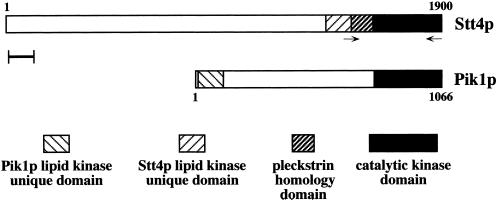
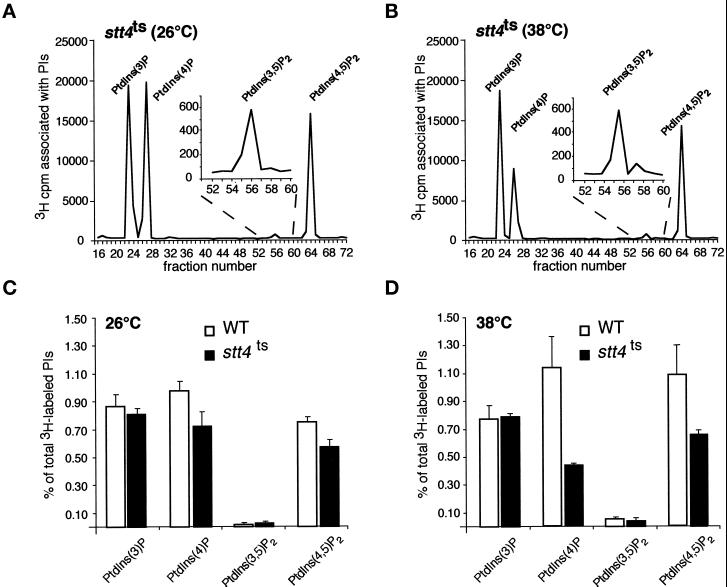
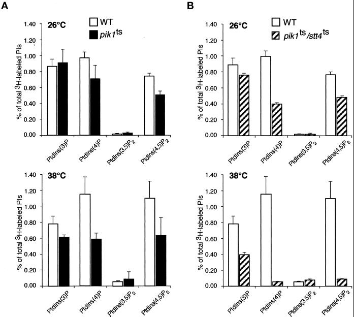
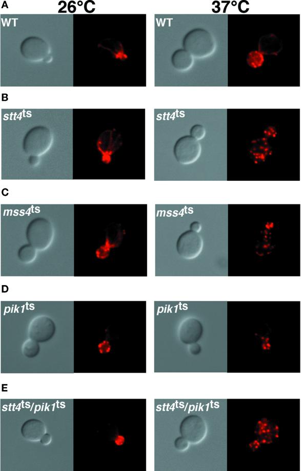
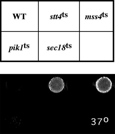
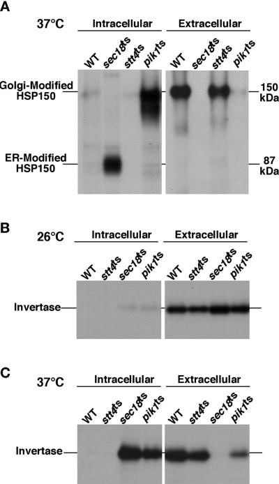
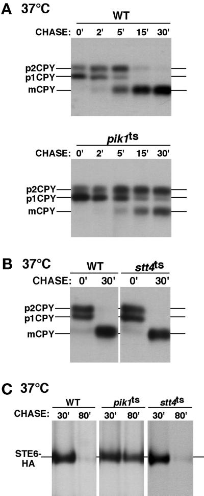
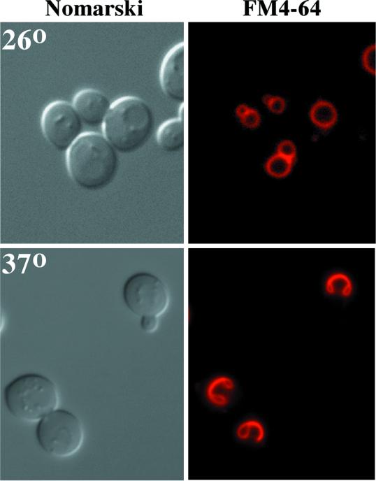
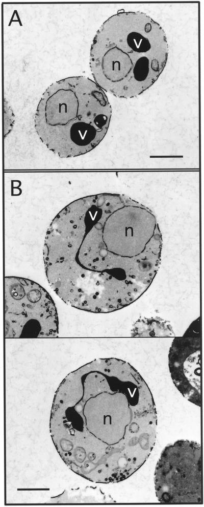
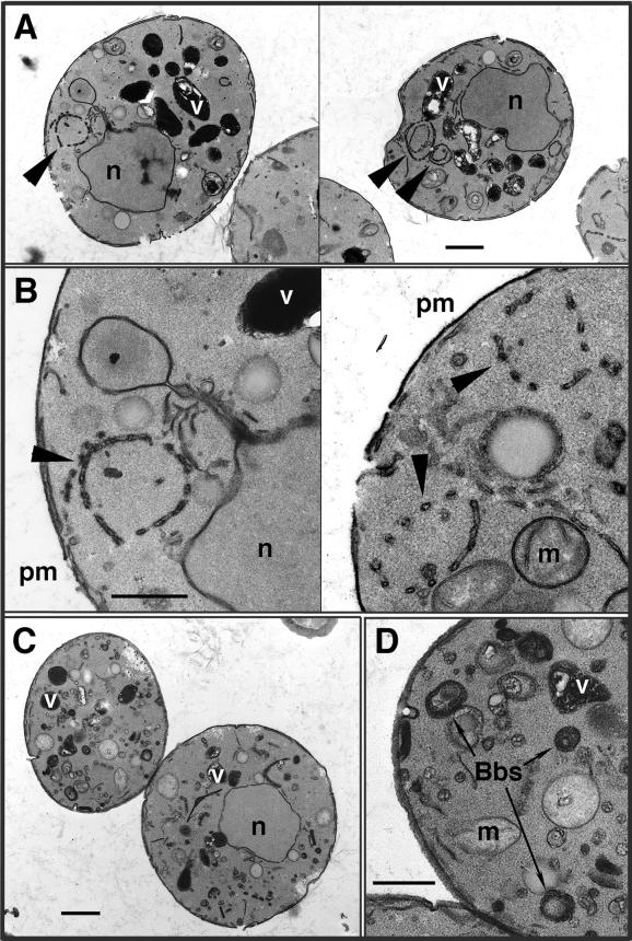
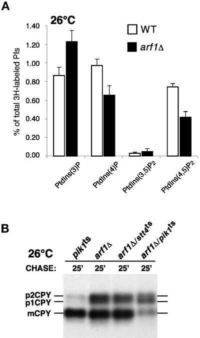
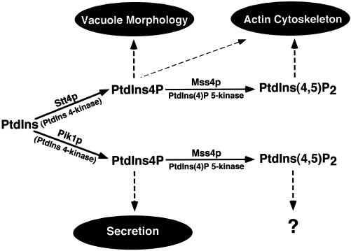
The yeast Saccharomyces cerevisiae possesses two genes that encode phosphatidylinositol (PtdIns) 4-kinases, STT4 and PIK1. Both gene products phosphorylate PtdIns at the D-4 position of the inositol ring to generate PtdIns(4)P, which plays an essential role in yeast viability because deletion of either STT4 or PIK1 is lethal. Furthermore, although both enzymes have the same biochemical activity, increased expression of either kinase cannot compensate for the loss of the other, suggesting that these kinases regulate distinct intracellular functions, each of which is required for yeast cell growth. By the construction of temperature-conditional single and double mutants, we have found that Stt4p activity is required for the maintenance of vacuole morphology, cell wall integrity, and actin cytoskeleton organization. In contrast, Pik1p is essential for normal secretion, Golgi and vacuole membrane dynamics, and endocytosis. Strikingly, pik1(ts) cells exhibit a rapid defect in secretion of Golgi-modified secretory pathway cargos, Hsp150p and invertase, whereas stt4(ts) cells exhibit no detectable secretory defects. Both single mutants reduce PtdIns(4)P by approximately 50%; however, stt4(ts)/pik1(ts) double mutant cells produce more than 10-fold less PtdIns(4)P as well as PtdIns(4,5)P(2). The aberrant Golgi morphology found in pik1(ts) mutants is strikingly similar to that found in cells lacking the function of Arf1p, a small GTPase that is known to regulate multiple membrane trafficking events throughout the cell. Consistent with this observation, arf1 mutants exhibit reduced PtdIns(4)P levels. In contrast, diminished levels of PtdIns(4)P observed in stt4(ts) cells at restrictive temperature result in a dramatic change in vacuole size compared with pik1(ts) cells and persistent actin delocalization. Based on these results, we propose that Stt4p and Pik1p act as the major, if not the only, PtdIns 4-kinases in yeast and produce distinct pools of PtdIns(4)P and PtdIns(4,5)P(2) that act on different intracellular membranes to recruit or activate as yet uncharacterized effector proteins.


Figures

Figure 1
Figure 1
Alignment of Stt4p and Pik1p. The STT4 gene encodes a high-molecular-weight protein possessing three conserved domains in its carboxyl terminus. Only the catalytic kinase domains of Stt4p and Pik1p share similarity, whereas the lipid kinase–unique domains of each protein diverge extensively. Additionally, Stt4p appears to contain a pleckstrin homology domain, whereas Pik1p does not. Neither protein contains obvious transmembrane domains. Oligonucleotides for PCR mutagenesis of STT4 are shown. Scale bar, 100 amino acids.
Figure 2
Figure 2
Levels of PtdIns(4)P and PtdIns(4,5)P2 decrease in stt4ts cells at restrictive temperature. (A and C) stt4ts cells (AAY102) were labeled with myo-[2-3H]inositol for 10 min at 26°C, chased with excess unlabeled myo-inositol for 30 min, and glass bead lysed. Cellular lipids were extracted, deacylated, and separated by HPLC (see MATERIALS AND METHODS). Peaks corresponding to glycero-Ins(3)P, glycero-Ins(4)P, glycero-Ins(3,5)P2, and glycero-Ins(4,5)P2 are indicated. Quantitative comparisons of glycero-phosphoinositols generated by wild-type and stt4ts cells at 26°C are shown below the elution profile. (B and D) stt4ts cells were preincubated at 38°C for 10 min, labeled with myo-[2-3H]inositol for 10 min, and chased with excess unlabeled myo-inositol for 30 min. Quantitative comparisons of glycero-phosphoinositols generated by wild-type and stt4ts cells at 38°C are shown below the elution profile. These data represent means ± ranges of at least three independent experiments.
Figure 3
Figure 3
pik1ts cells show defects in both PtdIns(4)P and PtdIns(4,5)P2 synthesis. Wild-type (SEY6210), pik1ts (AAY104), and stt4ts/pik1ts double mutant (AAY105) cells were grown to mid log phase, shifted to the appropriate temperature for 10 min, labeled with myo-[2-3H]inositol for 10 min, and chased in the presence of excess unlabeled myo-inositol for 30 min. Cellular lipids were recovered, deacylated, and separated by HPLC as described in Figure 1. Levels of deacylated products corresponding to the indicated phosphoinositides are shown. (A) A comparison of phosphoinositide levels in wild-type and pik1ts cells at 26 and 38°C. (B) A comparison of phosphoinositide levels in wild-type and stt4ts/pik1ts double mutant cells at 26 and 38°C. These data represent means ± ranges of at least three independent experiments.
Figure 4
Figure 4
Cells lacking Stt4 kinase activity fail to reorganize their actin cytoskeleton after heat shock. Cells were grown to early log phase, shifted to the appropriate temperature for 2.5 h, and then fixed with 3.7% formaldehyde. Cells were labeled with rhodamine–phalloidin (see MATERIALS AND METHODS) and visualized. Pictures shown are representative of more than 200 cells observed (see Table 2). (A) Wild-type (SEY6210) cells at 26 and 37°C. (B) stt4ts (AAY102) cells at 26 and 37°C. (C) mss4ts (AAY107) cells at 26 and 37°C. (D) pik1ts (AAY104) cells at 26 and 37°C. (E) stt4ts/pik1ts double mutant (AAY105) cells harboring temperature-sensitive alleles of both PtdIns 4-kinases at 26 and 37°C.
Figure 5
Figure 5
stt4ts cells exhibit a cell lysis defect. Equivalent numbers of wild-type (SEY6210), stt4ts (AAY102), mss4ts (AAY107), pik1ts (AAY104), and sec18ts (SEY5188) cells were incubated on YPD plates for 2 d at 26°C and then shifted for 15 h to 37°C. Plates were then overlayed with the 5-bromo-4-chloro-3-indolyl-phosphate solution described in MATERIALS AND METHODS, and pictures were taken after 30 min.
Figure 6
Figure 6
pik1ts but not stt4ts cells exhibit defects in protein secretion. Wild-type and mutant yeast cells were preincubated at 26 or 37°C for 10 min, metabolically labeled with a 35S-protein labeling mix during a 10-min pulse, and then chased in the presence of excess unlabeled methionine and cysteine for the times indicated. Each protein was immunoprecipitated with the appropriate antibody and resolved on SDS-PAGE. For Hsp150p secretion, labeled cells were spun down and Hsp150p was immunoprecipitated from both the media (external) fraction and the cellular (internal) fraction. Both the Golgi-modified and ER-modified forms of Hsp150p are shown. For invertase secretion, cellular transport was stopped with the addition of NaN3 and NaF after the pulse chase, and the cells were converted to spheroplasts. Internal and external fractions were separated by centrifugation and analyzed for the presence of invertase by immunoprecipitation. Before separation by SDS-PAGE, invertase was deglycosylated by endoglycosidase H treatment (see MATERIALS AND METHODS). (A) Analysis of Hsp150p secretion in wild-type (SEY6210), sec18ts (SEY5188), stt4ts (AAY102), and pik1ts (AAY104) cells. (B) Analysis of invertase secretion in wild-type, sec18ts, stt4ts, and pik1ts cells at 26°C. (C) Analysis of invertase secretion in wild-type, sec18ts, stt4ts, and pik1ts cells at 37°C. All data are representative of multiple experiments.
Figure 7
Figure 7
pik1ts cells exhibit defects in vacuole protein sorting and endocytosis. (A and B) Yeast cells were treated as described in Figure 5. The migration positions of precursor and mature forms of CPY are indicated on the left side of each panel. (A) A kinetic experiment showing the maturation of CPY in wild-type (SEY6210) and pik1ts (AAY104) cells at 37°C. (B) The maturation of CPY in wild-type and stt4ts (AAY102) cells at 37°C. (C) For Ste6p-HA endocytosis, cells were metabolically labeled for 10 min and chased for 10 min at 26°C. Cells were then shifted to 37°C, and samples were taken 10 and 50 min after the shift. A time course comparing the degradation of Ste6p-HA in wild-type, stt4ts, and pik1ts cells at 37°C is shown. Times shown indicate total time of label and chase. All data are representative of multiple experiments.
Figure 8
Figure 8
Stt4 kinase activity is required for normal vacuole morphology. stt4ts cells (AAY102) were grown to early log phase at 26°C, and vacuoles were labeled with the vital dye FM4-64 for 15 min (see MATERIALS AND METHODS). After labeling, cells were chased for 45 min and shifted to the indicated temperature for 1 h. Samples were taken every 15 min to examine vacuole morphology. On the left, cells were observed by Nomarski optics. On the right, identical fields are shown under fluorescent illumination (rhodamine channel). stt4ts cells at 26°C are shown on the top, and stt4ts cells after a 1-h shift to 37°C are shown on the bottom. These cells are representative of >80% of cells observed.
Figure 9
Figure 9
Ultrastructure of stt4ts cells. stt4ts cells (AAY102) were grown to early log phase, shifted to the appropriate temperature for 3 h, and fixed with 3% glutaraldehyde (see MATERIALS AND METHODS). (A) stt4ts cells at 26°C. (B) stt4ts cells at 38°C. These cells are representative of >65% of cells observed. v, vacuole; n, nucleus. Bars, 1 μm.
Figure 10
Figure 10
pik1ts cells accumulate morphologically abnormal Golgi and vacuole compartments. pik1ts cells (AAY104) were incubated at either 26 or 37°C for 90 min, fixed in 3% glutaraldehyde, and processed for electron microscopy (see MATERIALS AND METHODS). (A) pik1ts cells at 26°C. Bar, 1.0 μm. (B) Enlarged view of membrane ring structures found in pik1ts cells at 26°C. Bar, 0.4 μm. (C) pik1ts cells at 37°C. Bar, 1.0 μm. (D) Enlarged view of Berkeley bodies observed in pik1ts cells at 37°C. Bar, 0.4 μm. All cells shown are representative of >85% of cells observed. Arrowheads indicate atypical ring structures. Vacuoles (v), mitochondria (m), Berkeley bodies (Bbs), the plasma membrane (pm), and nuclei (n) are indicated.
Figure 11
Figure 11
Cells lacking Arf1p produce diminished levels of PtdIns(4)P and PtdIns(4,5)P2. (A) arf1 mutant cells were treated as described in Figure 2A. Quantitative comparisons of glycerophosphoinositols generated by wild-type (SEY6210) and arf1Δ cells at 26°C are shown. These data represent means ± ranges of at least three independent experiments. (B) pik1ts (AAY104), arf1Δ, stt4/arf1 double mutant (AAY114), and pik1/arf1 double mutant (AAY116) cells were metabolically labeled with a 35S-protein labeling mix during a 10-min pulse at 26°C and then chased in the presence of excess unlabeled methionine and cysteine for the times indicated. Each protein was immunoprecipitated with an antibody specific to CPY and resolved on SDS-PAGE.
Figure 12
Figure 12
The pathways for biosynthesis of PtdIns(4)P and PtdIns(4,5)P2 in yeast, indicating cellular role(s) for each phosphoinositide produced. The Stt4p- or Pik1p-dependent phosphorylation of PtdIns leads to the production of distinct pools of PtdIns(4)P. Arrows indicate the proposed roles for each phosphorylated lipid product. Stt4p-dependent PtdIns(4)P is required for maintenance of vacuole morphology and the actin cytoskeleton, whereas Pik1p-dependent PtdIns(4)P is essential for secretory vesicle formation and Golgi/endosome maintenance. Both Stt4 and Pik1 kinase activities appear to be required for generating the cellular pool of PtdIns(4,5)P2. Although we propose that Stt4p-dependent generation of PtdIns(4,5)P2 is required for actin polarization, a role for Pik1p-dependent PtdIns(4,5)P2 synthesis is not yet apparent.

Similar articles

Cited by

References

    1. Benedetti H, Raths S, Crausaz F, Riezman H. The END3 gene encodes a protein that is required for the internalization step of endocytosis and for actin cytoskeleton organization in yeast. Mol Biol Cell. 1994;5:1023–1037. - PMC - PubMed
    1. Bottomley MJ, Salim K, Panayotou G. Phospholipid-binding protein domains. Biochim Biophys Acta. 1998;1436:165–183. - PubMed
    1. Conibear E, Stevens TH. Multiple sorting pathways between the late Golgi and the vacuole in yeast. Biochim Biophys Acta. 1998;1404:211–230. - PubMed
    1. Cowles CR, Synder WB, Burd CG, Emr SD. Novel Golgi to vacuole delivery pathway in yeast: identification of a sorting determinant and required transport component. EMBO J. 1997;16:2769–2782. - PMC - PubMed
    1. Cutler NS, Heitman J, Cardenas ME. STT4 is the essential phosphatidylinositol 4-kinase that is a target of wortmannin in Saccharomyces cerevisiae. J Biol Chem. 1997;272:27671–27677. - PubMed

Publication types

MeSH terms