. 2000 Aug;11(8):2821-31.
doi: 10.1091/mbc.11.8.2821.

Cell cycle-dependent expression of mammalian E2-C regulated by the anaphase-promoting complex/cyclosome

Affiliations
Free PMC article

Cell cycle-dependent expression of mammalian E2-C regulated by the anaphase-promoting complex/cyclosome

A Yamanaka et al. Mol Biol Cell. 2000 Aug.
Free PMC article

Abstract

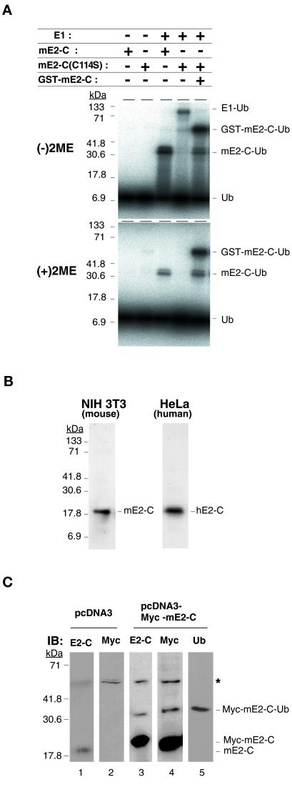
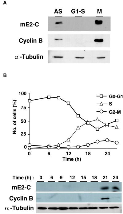
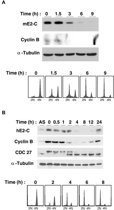
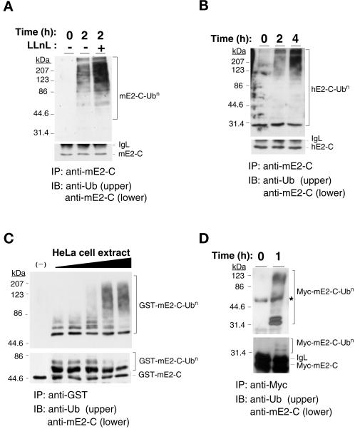
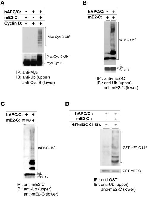
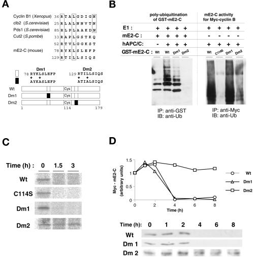
Progression through mitosis requires the precisely timed ubiquitin-dependent degradation of specific substrates. E2-C is a ubiquitin-conjugating enzyme that plays a critical role with anaphase-promoting complex/cyclosome (APC/C) in progression of and exit from M phase. Here we report that mammalian E2-C is expressed in late G(2)/M phase and is degraded as cells exit from M phase. The mammalian E2-C shows an autoubiquitinating activity leading to covalent conjugation to itself with several ubiquitins. The ubiquitination of E2-C is strongly enhanced by APC/C, resulting in the formation of a polyubiquitin chain. The polyubiquitination of mammalian E2-C occurs only when cells exit from M phase. Furthermore, mammalian E2-C contains two putative destruction boxes that are believed to act as recognition motifs for APC/C. The mutation of this motif reduced the polyubiquitination of mammalian E2-C, resulting in its stabilization. These results suggest that mammalian E2-C is itself a substrate of the APC/C-dependent proteolysis machinery, and that the periodic expression of mammalian E2-C may be a novel autoregulatory system for the control of the APC/C activity and its substrate specificity.


Figures

Figure 1
Figure 1
Autoubiquitination activity of mE2-C. (A) Recombinant wild-type (wt) or mutant (C114S) mE2-C (20 kDa) was incubated with 125I-ubiquitin in the absence or presence of E1 (116 kDa). The reaction mixtures then were boiled in SDS sample buffer, without (upper panel) or with (lower panel) 2-mercaptoethanol (2ME), and were subjected to SDS-PAGE and autoradiography. The positions of free ubiquitin (Ub), E1 conjugated with ubiquitin (E1-Ub), mE2-C conjugated with ubiquitin (mE2-C-Ub), and GST-mE2-C conjugated with ubiquitin (GST-mE2-C-Ub) are indicated on the right, and those of molecular size standards (in kilodaltons) are shown on the left. (B) Cell lysates prepared from mouse NIH 3T3 and human HeLa cells were subjected to immunoblot analysis with anti-mE2-C. The positions of the immunoreactive mE2-C and hE2-C proteins are indicated. (C) NIH 3T3 cells were transfected with either an expression plasmid encoding Myc-tagged mE2-C or the empty vector (pcDNA3). Cell lysates were subsequently prepared and subjected to immunoblot analysis (IB) with anti-mE2-C, anti-Myc, or antiubiquitin. The positions of mE2-C, Myc-mE2-C, and monoubiquitinated Myc-mE2-C (Myc-mE2-C-Ub) are shown. The asterisk indicates a nonspecific band. Because the exposure time was shorter for lane 3 than for lane 1, the endogenous mE2-C is not visible in lane 3.
Figure 2
Figure 2
Expression of mammalian E2-C during G2–M phase of the cell cycle. (A) Cell lysates were prepared from asynchronous (AS) NIH 3T3 cells or from cells synchronized at either G1–S phase by aphidicolin treatment or M phase by nocodazole treatment and then were subjected to immunoblot analysis with anti-mE2-C (top panel), anti-cyclin B (middle panel), or anti-α-tubulin (bottom panel). (B) NIH 3T3 cells were arrested at G0–G1 phase by contact inhibition and then were induced to resume the cell cycle by replating. At the indicated times thereafter, the proportion of cells in G0–G1 phase (squares), S phase (triangles), or G2–M phase (circles) was determined by flow cytometry and was expressed as a percentage of the total number of cells (upper panel). Cell lysates were also prepared, adjusted to equal protein concentrations, and subjected to immunoblot analysis (lower panel), as in (A).
Figure 3
Figure 3
Down-regulation of mammalian E2-C at early G1 phase. Mouse NIH 3T3 (A) and human HeLa (B) cells were arrested at metaphase by nocodazole treatment and then were incubated in nocodazole-free medium for the indicated times. Cell lysates were prepared from asynchronous (AS) or synchronized cells and then were subjected to immunoblot analysis with anti-mE2-C, anti-cyclin B, anti-CDC27, and anti-α-tubulin (upper panels), and the DNA content of cells was determined by flow cytometry (lower panels).
Figure 4
Figure 4
Cell cycle-dependent polyubiquitination of mammalian E2-C. (A) Mouse NIH 3T3 cells were arrested at metaphase by nocodazole treatment and then were incubated in nocodazole-free medium for the indicated times in the absence or presence of the proteasome inhibitor LLnL at a concentration of 250 μM. Cell lysates were prepared and subjected to immunoprecipitation (IP) with anti-mE2-C, and the resulting precipitates were subjected to immunoblot analysis (IB) with either antiubiquitin (upper panel) or anti-mE2-C (lower panel). The positions of mE2-C, polyubiquitinated mE2-C (mE2-C-Ubn), and immunoglobulin light chain (IgL) are indicated. (B) Human HeLa cells were subjected to metaphase arrest, were incubated for the indicated times in nocodazole-free medium in the presence of LLnL, and were subjected to immunoprecipitation and immunoblot analysis, as in (A). The positions of hE2-C, monoubiquitinated hE2-C (hE2-C-Ub1), polyubiquitinated hE2-C (hE2-C-Ubn), and IgL are indicated. (C) A recombinant GST fusion protein of mE2-C was incubated for 1 h at 37°C in the absence (–) or presence of various concentrations (0.25 μg/μl, 0.5 μg/μl, 1.0 μg/μl, 2.5 μg/μl, and 5.0 μg/μl) of cell extract prepared from HeLa cells 2 h after release from nocodazole-induced arrest. The fusion protein then was immunoprecipitated with anti-GST and ws subjected to immunoblot analysis with antiubiquitin (upper panel) or anti-mE2-C (lower panel). (D) Cell lysates were prepared from NIH 3T3 cells expressing Myc-tagged mE2-C that had been arrested at metaphase by nocodazole treatment and incubated for 0 or 1 h in nocodazole-free medium. The lysates were subjected to immunoprecipitation with anti-Myc, and the resulting precipitates were boiled in SDS-containing buffer and were subjected to reimmunoprecipitation with anti-Myc. The final precipitates then were subjected to immunoblot analysis with antiubiquitin (upper panel) or anti-mE2-C (lower panel). The asterisk indicates a nonspecific band.
Figure 5
Figure 5
Polyubiquitination of mammalian E2-C by the APC/C. (A) Recombinant Myc-tagged cyclin B was subjected to an in vitro ubiquitination assay by incubation with rabbit E1, bovine ubiquitin, and either immunopurified human APC/C (hAPC/C) or recombinant mE2-C, as indicated. The reaction mixtures were subjected to immunoprecipitation with anti-Myc, and the resulting precipitates were subjected to immunoblot analysis with antiubiquitin (upper panel) or anti-cyclin B (lower panel). Cyc B, cyclin B. (B) Recombinant mE2-C was subjected to in vitro ubiquitination assay, as in (A), in the absence or presence of purified human APC/C. The reaction mixtures were subjected to immunoprecipitation with anti-mE2-C, and the resulting precipitates were subjected to immunoblot analysis with antiubiquitin (upper panel) or anti-mE2-C (lower panel). (C) Recombinant wild-type (wt) or mutant (C114S) mE2-C was subjected to in vitro ubiquitination assay in the presence of purified human APC/C and was processed as in (B). (D) Recombinant GST fusion protein of mutant (C114S) mE2-C was subjected to in vitro ubiquitination assay in the absence or presence of wild-type mE2-C, together with purified human APC/C and processed as in (B).
Figure 6
Figure 6
Destruction boxes are involved in the ubiquitination and stability of mammalian E2-C. (A) The amino acid sequences of the destruction boxes of cyclin B1 (Xenopus laevis), Clb2 (Saccharomyces cerevisiae), Pds1 (S. cerevisiae), Cut2 (Schizosaccharomyces pombe), and mE2-C (Mus musculus) are aligned in the upper panel. Amino acids conserved among these proteins are boxed. Residue numbers are shown at the beginning of each sequence. The mE2-C mutants used in the experiments in (B)–(D) are shown schematically in the lower panel. Open and closed boxes indicate wild-type and mutant destruction boxes, respectively. (B) Recombinant GST fusion proteins of wild-type and mutant (Dm1 and Dm2) mE2-C were subjected to in vitro ubiquitination assay in the absence or presence of purified human APC/C. The wild-type mE2-C also was added to each reaction. The reaction mixtures were subjected to immunoprecipitation with anti-GST, and the resulting precipitates were subjected to to immunoblot analysis with antiubiquitin (left panel). The E2 activity of recombinant GST-fused wild-type and mutant (C114S, Dm1, and Dm2) proteins was examined with purified human APC/C and Myc-cyclin B as a substrate as in Figure 5A (right panel). (C) HeLa cells transfected with vectors encoding the Myc-tagged wild-type and mutant (C114S, Dm1, and Dm2) mE2-C proteins were pulse labeled with [35S]methionine and then were incubated in the absence of isotope for the indicated chase periods. Cell lysates then were subjected to immunoprecipitation with the monoclonal antibody to Myc, and the resulting precipitates were subjected to SDS-PAGE and autoradiography. (D) HeLa cells transfected with vectors encoding the Myc-tagged wild-type and mutant (Dm1 and Dm2) mE2-C proteins were arrested at metaphase by nocodazole treatment and then were incubated in nocodazole-free medium for the indicated times. Cell lysates then were subjected to immunoblot analysis with the monoclonal antibody to Myc.

Similar articles

Cited by

References

    1. Amon A, Irniger S, Nasmyth K. Closing the cell cycle circle in yeast: G2 cyclin proteolysis initiated at mitosis persists until the activation of G1 cyclins in the next cycle. Cell. 1994;77:1037–1050. - PubMed
    1. Aristarkhov A, Eytan E, Moghe A, Admon A, Hershko A, Ruderman JV. E2-C, a cyclin-selective ubiquitin carrier protein required for the destruction of mitotic cyclins. Proc Natl Acad Sci USA. 1996;93:4294–4299. - PMC - PubMed
    1. Arvand A, Bastians H, Welford SM, Thompson AD, Ruderman JV, Denny CT. EWS/FLI1 up regulates mE2-C, a cyclin-selective ubiquitin conjugating enzyme involved in cyclin B destruction. Oncogene. 1998;17:2039–2045. - PubMed
    1. Bai C, Sen P, Hofmann K, Ma L, Goebl M, Harper JW, Elledge SJ. SKP1 connects cell cycle regulators to the ubiquitin proteolysis machinery through a novel motif, the F-box. Cell. 1996;86:263–274. - PubMed
    1. Banerjee A, Gregori L, Xu Y, Chau V. The bacterially expressed yeast CDC34 gene product can undergo autoubiquitination to form a multiubiquitin chain-linked protein. J Biol Chem. 1993;268:5668–5675. - PubMed

Publication types

LinkOut - more resources