

CD8+ T cells are crucial for the ability of congenic normal mice to reject highly immunogenic sarcomas induced in nude mice with 3-methylcholanthrene
- PMID: 10931133
- PMCID: PMC1905705
- DOI: 10.1046/j.1365-2249.2000.01292.x
CD8+ T cells are crucial for the ability of congenic normal mice to reject highly immunogenic sarcomas induced in nude mice with 3-methylcholanthrene
Abstract
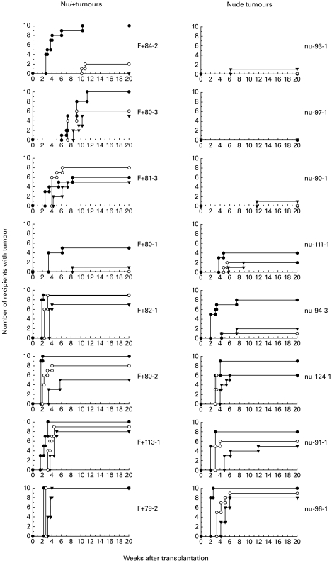
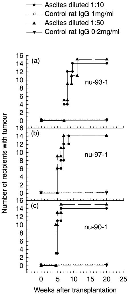
An attempt was made to identify the selection pressures put upon a growing tumour by CD8+ T cells. To this end tumours induced with 3-methylcholanthrene in T cell-deficient nude mice and in congenic T cell-competent nu/+ mice were transplanted to nu/+ recipients. The rejection rate of the sarcomas from nude mice was almost twice that of the sarcomas from nu/+ mice. Depletion of CD8+ T cells from nu/+ recipients prior to transplantation made them accept nude tumours that were consistently rejected by untreated nu/+ recipients. These findings suggest that a methylcholanthrene sarcoma during its growth in a T cell-competent host adapts to the T cell system through a selective elimination of highly immunogenic tumour cells that are susceptible to CD8+ T cell-mediated lysis.
Figures
References
-
- Nowell PC. The clonal evolution of tumor cell populations. Science. 1976;194:23–28. - PubMed
-
- Greenberg PD. Adoptive T cell therapy of tumors: mechanisms operative in the recognition and elimination of tumor cells. [Review] Adv Immunol. 1991;49:281–355. - PubMed
-
- Shimizu K, Shen FW. Role of different T cells sets in the rejection of syngenic chemically induced tumors. J Immunol. 1979;122:1162–5. - PubMed
-
- Ward PL, Schreiber H. MHC class I restricted T cells and immune surveillance against transplanted ultraviolet light-induced tumors. [Review] Seminars Cancer Biology. 1991;2:321–8. - PubMed
-
- van der Bruggen P, Traversari C, Chomez P, et al. A gene encoding an antigen recognized by cytolytic T lymphocytes on a human melanoma. Science. 1991;254:1643–7. - PubMed
Publication types
MeSH terms
Substances
LinkOut - more resources
Full Text Sources
Research Materials
