Comparative Study
. 2000 Aug 7;150(3):405-16.
doi: 10.1083/jcb.150.3.405.

Identification and characterization of SA/Scc3p subunits in the Xenopus and human cohesin complexes

Affiliations
Comparative Study

Identification and characterization of SA/Scc3p subunits in the Xenopus and human cohesin complexes

A Losada et al. J Cell Biol. .

Abstract

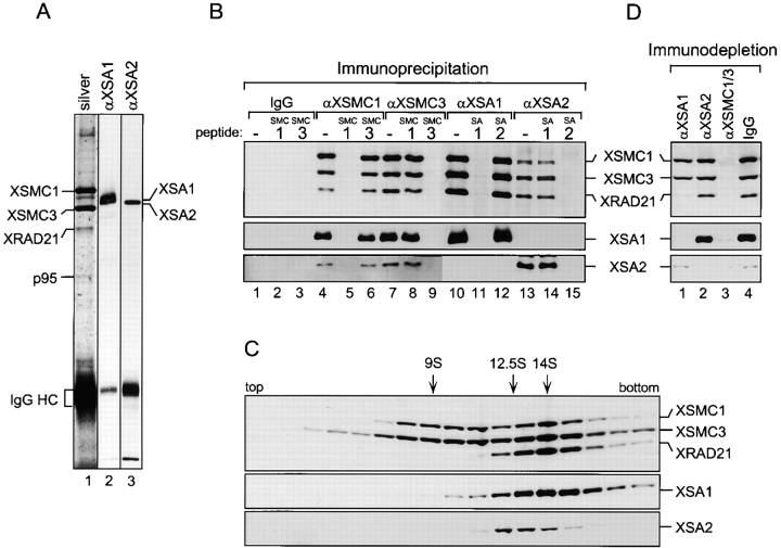
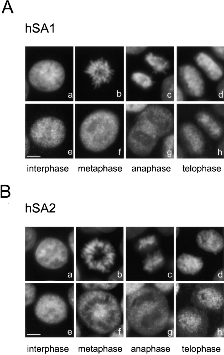
A multisubunit protein complex, termed cohesin, plays an essential role in sister chromatid cohesion in yeast and in Xenopus laevis cell-free extracts. We report here that two distinct cohesin complexes exist in Xenopus egg extracts. A 14S complex (x-cohesin(SA1)) contains XSMC1, XSMC3, XRAD21, and a newly identified subunit, XSA1. In a second 12.5S complex (x-cohesin(SA2)), XSMC1, XSMC3, and XRAD21 associate with a different subunit, XSA2. Both XSA1 and XSA2 belong to the SA family of mammalian proteins and exhibit similarity to Scc3p, a recently identified component of yeast cohesin. In Xenopus egg extracts, x-cohesin(SA1) is predominant, whereas x-cohesin(SA2) constitutes only a very minor population. Human cells have a similar pair of cohesin complexes, but the SA2-type is the dominant form in somatic tissue culture cells. Immunolocalization experiments suggest that chromatin association of cohesin(SA1) and cohesin(SA2) may be differentially regulated. Dissociation of x-cohesin(SA1) from chromatin correlates with phosphorylation of XSA1 in the cell-free extracts. Purified cdc2-cyclin B can phosphorylate XSA1 in vitro and reduce the ability of x-cohesin(SA1) to bind to DNA or chromatin. These results shed light on the mechanism by which sister chromatid cohesion is partially dissolved in early mitosis, far before the onset of anaphase, in vertebrate cells.


Figures

Figure 1
Figure 1
Sequence alignment of Xenopus and human SA1 and SA2. Identical amino acid residues are shaded. These sequence data are available from GenBank/EMBL/DDBJ under accession numbers AF255017 and AF255018.
Figure 3
Figure 3
Biochemical characterization of the human cohesin complexes. (A) Human cohesins were affinity-purified from HeLa cell nuclear extracts using anti-XSMC3 antibodies and analyzed by silver staining (lane 1) and by immunoblotting with anti-XSMC1 (lane 2), anti-XSMC3 (lane 3), anti-XRAD21 (lane 4), anti-XSA1 (lane 5), and anti-XSA2 (lane 6). (B) A HeLa cell nuclear extract was fractionated in a sucrose gradient as described in Fig. 2 C. The two peaks containing SMC subunits are indicated. (C) Immunoprecipitations from HeLa cell nuclear extracts were performed using control rabbit IgG (lane 1) or antibodies against XSMC3 (lanes 2–4), XSA1 (lanes 5–7) and XSA2 (lanes 8–10). The antigen peptides for XSMC1 (lanes 3), XSMC3 (lanes 4), XSA1 (lanes 6 and 9), or XSA2 (lanes 7 and 10) were added at 0.4 mg/ml to demonstrate the specificity of these reactions. The immunoprecipitates that were recovered on protein A agarose beads were analyzed by immunoblotting. (D) Aliquots of a HeLa cell nuclear extract were immunodepleted using control rabbit IgG (lane 1), anti-XSA1 (lane 2), anti-XSA2 (lane 3), or a mixture of anti-XSMC1 and anti-XSMC3 (lane 4), and analyzed by immunoblotting.
Figure 2
Figure 2
Biochemical characterization of Xenopus cohesin complexes containing XSA1 and XSA2. (A) Cohesins were immunoprecipitated from a Xenopus egg HSS using anti-XSMC3 antibodies and analyzed by silver staining (lane 1) and immunoblotting with anti-XSA1 (lane 2) or anti-XSA2 (lane 3). (B) Immunoprecipitations were performed using antibodies against XSMC1 (lanes 4–6), XSMC3 (lanes 7–9), XSA1 (lanes 10–12), XSA2 (lanes 13–15), or control rabbit IgG (lanes 1–3). The antigen peptides for XSMC1 (lanes 2, 5, and 8), XSMC3 (lanes 3, 6, and 9), XSA1 (lanes 11 and 14), or XSA2 (lanes 12 and 15) were added at 0.4 mg/ml to demonstrate the specificity of these reactions. The immunoprecipitates that were recovered on protein A agarose beads were analyzed by immunoblotting using the antibodies indicated. (C) An interphase HSS was fractionated in a 5–20% sucrose gradient centrifuged at 36,000 rpm for 15 h in an SW50.1 rotor (Beckman), and fractions were analyzed by immunoblotting. The peaks corresponding to x-cohesinSA1 (14S), x-cohesinSA2 (12.5S), and the XSMC1-XSMC3 heterodimer (9S) are indicated. (D) Aliquots of an HSS were immunodepleted using anti-XSA1 (lane 1), anti-XSA2 (lane 2), and a mixture of anti-XSMC1 and anti-XSMC3 (lane 3) or control rabbit IgG (lane 4), and analyzed by immunoblotting.
Figure 5
Figure 5
Localization of cohesin complexes in human tissue culture cells. HeLa cells were fixed with paraformaldehyde, and hSA1 (A) and hSA2 (B) were immunolocalized using anti-XSA1 and anti-XSA2, respectively. (a–d) DNA; (e–h) antibody staining. Bars, 5 μm.
Figure 4
Figure 4
Localization of cohesin complexes in nuclei and chromosomes assembled in Xenopus egg extracts. (A) Xenopus sperm chromatin was incubated in interphase or mitotic HSS. The assembled interphase chromatin (I) and mitotic chromosomes (M) were fixed, isolated through a glycerol cushion, and immunostained with anti-XSA1 (e and f, green) or anti-XSA2 (g and h, green). The DNA was counterstained with DAPI (a–d, red). Merged images are also shown (i–l). (B) Xenopus sperm chromatin was incubated at 22°C for 90 min in interphase LSS, where it was replicated. Half of this interphase mixture (I) was fixed and processed for immunostaining with anti-XSA1 (a, f, and k) or anti-XSA2 (d, i, and n). The other half was driven into mitosis (M) by the addition of a mitotic LSS, incubated for another 2 h, fixed, and immunostained. A mass of entangled chromosomes (b, g, l, e, j, and o) and an individual chromosome (c, h, and m) are shown. (a–e, red) DNA; (f–h, green) anti-XSA1; (i and j, green) anti-XSA2; (k–o) merged images. Stronger staining with anti-XSA1 was often observed on a bent region of the chromosome, most likely corresponding to the pericentromeric region (arrowhead). The images of antibody staining were captured using different exposure times to visualize weak signals on the mitotic chromosomes and, therefore, the label intensities cannot be compared quantitatively. (C) Xenopus sperm nuclei were incubated in an interphase LSS depleted of x-cohesins and supplemented with h-cohesinSA1 and h-cohesinSA2. After incubation at 22°C for 90 min, the nuclei were fixed, and hSA1 and hSA2 were detected with anti-XSA1 and anti-XSA2, respectively. (a and b, red) DNA; (c and d, green) antibody staining; (e and f) merged images. Bars, 5 μm.
Figure 7
Figure 7
Phosphorylation of x-cohesin by cdc2-cyclin B and its effect on DNA binding. (A) Xenopus cohesins were immunoprecipitated from an interphase HSS with anti-XSMC3, and incubated with purified cdc2-cyclin B (lanes 1 and 3) or no kinase (lanes 2 and 4) in the presence of γ-[32P]ATP. After washing, the immunoprecipitates were separated by SDS-PAGE and analyzed by autoradiography (lanes 1 and 2) or by immunoblotting (lanes 3 and 4). (B) Solid-phase chromatin was assembled on DNA-coupled magnetic beads in an interphase HSS, washed, and treated with cdc2-cyclin B (lanes 1 and 2) or no kinase (lanes 3 and 4) in the presence of γ-[32P]ATP. After incubation at 22°C for 1 h, proteins remaining bound to the chromatin beads (lanes 1 and 3; unreleased [U]) and those dissociated from the beads (lanes 2 and 4; released [R]) were analyzed by immunoblotting with antibodies against cohesin subunits (top) or by autoradiography (bottom). (C) Filter binding assay. Purified cohesins were first treated with no kinase (lane 2), cdc2-cyclin B (lane 3), or cdc2-cyclin B in the presence of the CDK inhibitor roscovitine (lane 4), and then incubated with plasmid DNA in a low salt buffer. Buffer alone (lane 1) or cdc2-cyclin B alone (lane 5) were also mixed with plasmid DNA. The binding reactions were passed through glass fiber filters, and washed sequentially with low salt buffer, high salt buffer, and buffer containing SDS. The ratio of bound DNA to total DNA was calculated as described previously (Kimura et al. 1999). The average of values from three independent experiments was plotted with error bars. (D) Bead-binding assay. Unphosphorylated (lanes 1, 3, and 5) or phosphorylated (lanes 2, 4, and 6) cohesins were mixed with no-DNA beads (lanes 1 and 2; control beads), DNA-coupled beads (lanes 3 and 4; DNA beads), or solid-phase chromatin beads assembled in a cohesin-depleted interphase HSS (lanes 5 and 6; chromatin beads). After incubation for 1 h, the beads were washed and proteins bound to the beads were analyzed by immunoblotting.
Figure 6
Figure 6
Regulation of cohesin by phosphorylation in the cell-free extracts. (A) DNA-coupled paramagnetic beads were incubated with mitotic (M) or interphase (I) HSS to assemble solid-phase chromatin (lanes 3 and 4). Uncoupled beads were treated in the same way as controls (lanes 1 and 2). After washing the beads, bound proteins were analyzed by immunoblotting with antibodies against cohesin (upper) and condensin subunits (lower). Alternatively, solid-phase chromatin was assembled in an interphase HSS, washed, and then placed in a mitotic HSS (lane 5; I > M) or a cohesin-depleted mitotic HSS (lane 6; I > ΔM). After incubation for 2 h, proteins bound to the beads were analyzed as above. The fraction released from the I > ΔM chromatin beads was immunoprecipitated with anti-XSMC3 and analyzed by immunoblotting (lane 7). (B) DNA-coupled beads were incubated with an interphase HSS (I) for 1 h (lane 1). Half of the reaction mixture was incubated for another 1 h after addition of okadaic acid to 1 μM (lane 2; OA). The DNA beads were also incubated in a mitotic HSS (M) without (lane 3) or with (lane 4) 5 mM 6-dimethylaminopurine (DMAP) for 2 h. The chromatin beads were separated from the extract and washed. Cohesin fractions, which were present in chromatin (top) or extracts (middle and bottom), were detected by immunoblotting with the indicated antibodies. (C) A Xenopus interphase LSS was immunodepleted of x-cohesins using anti-XSMC3. An aliquot of the depleted extract was saved to check the absence of x-cohesins (lane 1). The remainder was divided into two parts, and one of them was converted into a mitotic state by addition of cyclin BΔ90 (Glotzer et al. 1991). Both LSSs depleted of x-cohesins were supplemented with purified h-cohesinSA1 and h-cohesinSA2 (∼7.5 nM each). After incubation at 22°C for 90 min, aliquots were taken from the interphase (lane 2) and the mitotic (lane 3) extracts. The human cohesin complexes were recovered from the LSS by immunoprecipitation with anti-XSMC3, and treated with buffer alone (lane 4) or λ-phosphatase (lane 5) at 30°C for 1 h. The aliquots of the extracts and the immunoprecipitates were separated by SDS-PAGE, and hSA1 and hSA2 were detected by immunoblotting with anti-XSA1 (top) and anti-XSA2 (bottom), respectively.
Figure 8
Figure 8
Mitotic and meiotic cohesins in different organisms. The subunit composition of S. pombe mitotic cohesin and the three meiotic cohesin complexes are hypothetical. The Scc1p/Rad21/Rec8-like subunits are shown by white rectangles, whereas the Scc3p/Rec11/SA-like subunits are indicated by black ovals. For simplicity, no distinction is made between SMC1 and SMC3, and the heterodimers are represented as symmetrical V-shaped structures.

Similar articles

Cited by

References

    1. Bell S.P., Kobayashi R., Stillman B. Yeast origin recognition complex functions in transcription silencing and DNA replication. Science. 1993;262:1844–1849. - PubMed
    1. Biggins S., Murray A. Sister chromatid cohesion in mitosis. Curr. Opin. Cell Biol. 1999;9:230–236. - PubMed
    1. Birkenbihl R.P., Subramani S. The rad21 gene product of Schizosaccharomyces pombe is a nuclear, cell cycle-regulated phosphoprotein. J. Biol. Chem. 1995;270:7703–7711. - PubMed
    1. Blat Y., Kleckner N. Cohesins bind to preferential sites along yeast chromosome III, with differential regulation along arms versus the centric region. Cell. 1999;98:249–259. - PubMed
    1. Carramolino L., Lee B.C., Zaballos A., Peled A., Barthelemy I., Shav-Tal Y., Prieto I., Carmi P., Gothelf Y., González de Buitrago G. SA-1, a nuclear protein encoded by one member of a novel gene familymolecular cloning and detection in hemopoietic organs. Gene. 1997;195:151–159. - PubMed

Publication types

MeSH terms

Associated data