. 2000 Aug 7;150(3):417-32.
doi: 10.1083/jcb.150.3.417.

Tracking COL1A1 RNA in osteogenesis imperfecta. splice-defective transcripts initiate transport from the gene but are retained within the SC35 domain

Affiliations

Tracking COL1A1 RNA in osteogenesis imperfecta. splice-defective transcripts initiate transport from the gene but are retained within the SC35 domain

C Johnson et al. J Cell Biol. .

Abstract

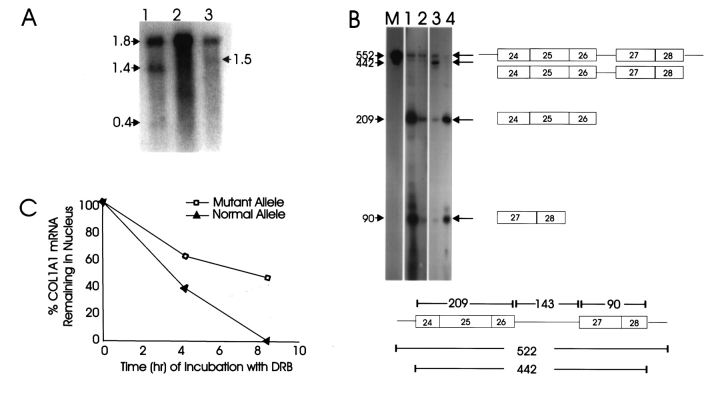
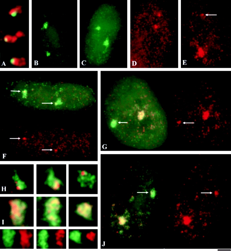
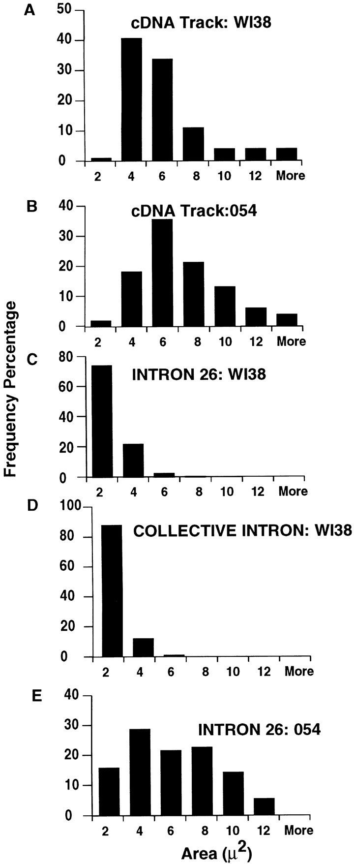
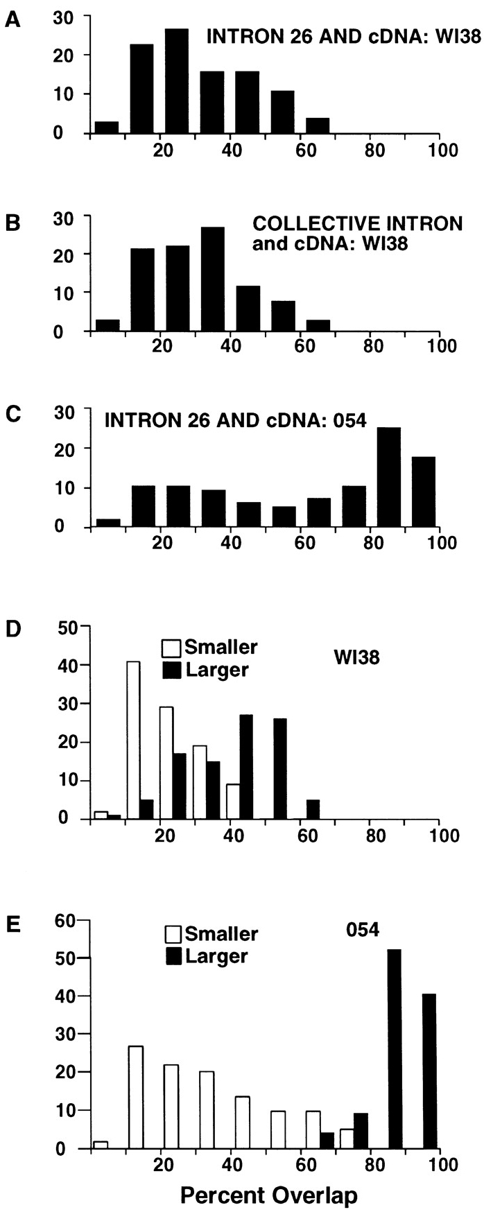
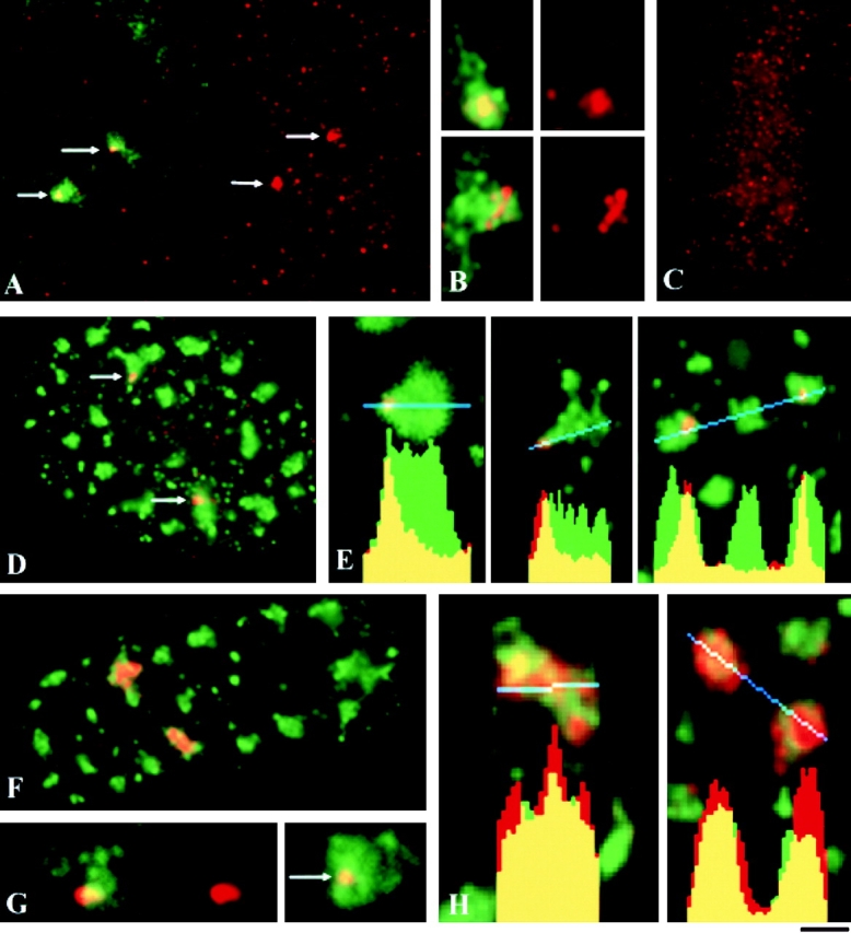
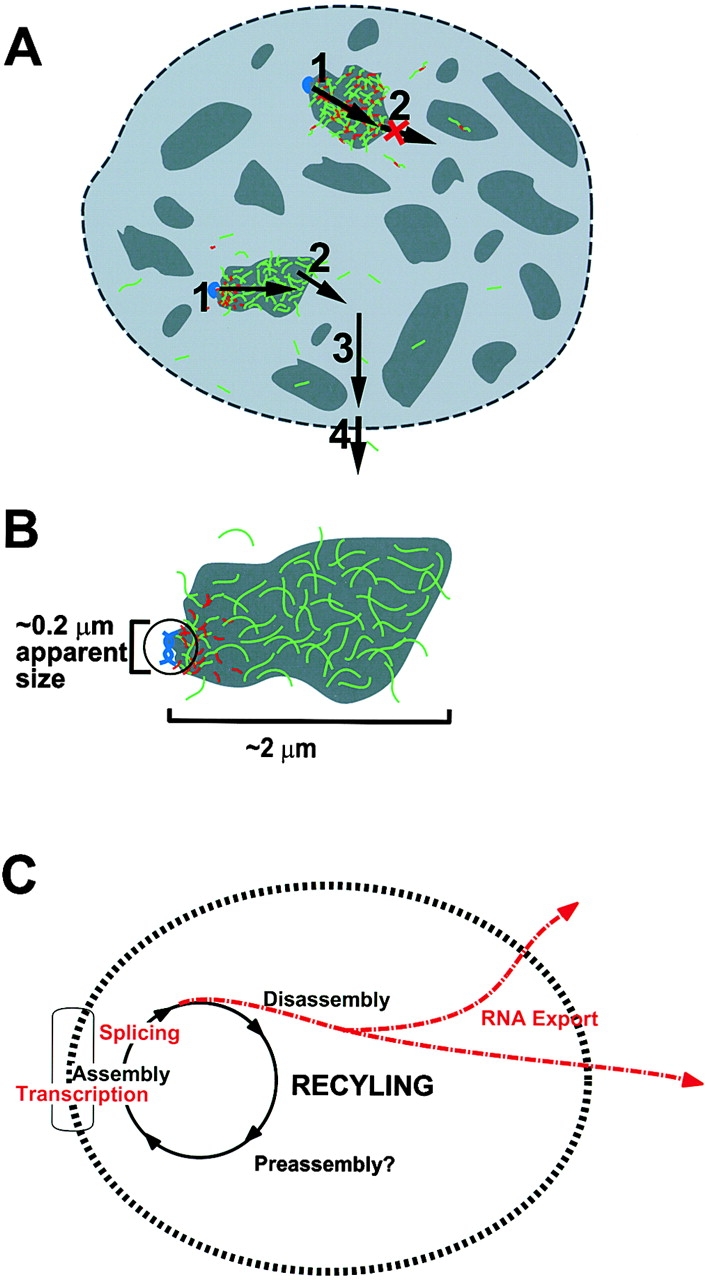
This study illuminates the intra-nuclear fate of COL1A1 RNA in osteogenesis imperfecta (OI) Type I. Patient fibroblasts were shown to carry a heterozygous defect in splicing of intron 26, blocking mRNA export. Both the normal and mutant allele associated with a nuclear RNA track, a localized accumulation of posttranscriptional RNA emanating to one side of the gene. Both tracks had slightly elongated or globular morphology, but mutant tracks were cytologically distinct in that they lacked the normal polar distribution of intron 26. Normal COL1A1 RNA tracks distribute throughout an SC-35 domain, from the gene at the periphery. Normally, almost all 50 COL1A1 introns are spliced at or adjacent to the gene, before mRNA transits thru the domain. Normal COL1A1 transcripts may undergo maturation needed for export within the domain such as removal of a slow-splicing intron (shown for intron 24), after which they may disperse. Splice-defective transcripts still distribute thru the SC-35 domain, moving approximately 1-3 micrometer from the gene. However, microfluorimetric analyses demonstrate mutant transcripts accumulate to abnormal levels within the track and domain. Hence, mutant transcripts initiate transport from the gene, but are impeded in exit from the SC-35 domain. This identifies a previously undefined step in mRNA export, involving movement through an SC-35 domain. A model is presented in which maturation and release for export of COL1A1 mRNA is linked to rapid cycling of metabolic complexes within the splicing factor domain, adjacent to the gene. This paradigm may apply to SC-35 domains more generally, which we suggest may be nucleated at sites of high demand and comprise factors being actively used to facilitate expression of associated loci.


Figures

Figure 1
Figure 1
(A) Indirect RNase protection by hybridization of cellular RNA with HF404, which encompasses COL1A1 exons 19–43. Nuclear mRNA from heterozygous patient 054 contains the primary 1.8-kb band, as well as a 1.4-kb and a weak 0.4-kb band, indicating the presence of an insertion (lane 1). Nuclear mRNA from a control patient (411), which lacks an intron retention mutation (lane 2). Total RNA from control cells (lane 3). Note the presence of a faint 1.5-kb band in normal cells suggesting late removal of intron 24 (see results). (B) Direct RNase protection. A 552-bp probe encompassing exons 24–28 and intron 26, was generated by RT/PCR for hybridization to nuclear (lanes 1 and 3) and cytoplasmic (lanes 2 and 4) RNA from control (lanes 1 and 2) and 054 (lanes 3 and 4) cells. Full protection of the 442-nt fragment was only seen in the nuclear compartment of cells from patient 054 (lane 3) which is consistent with the retention of intron 26. Even when the sample is loaded so that processed mRNA is abundantly detected in the nuclear fraction, the unprocessed intermediate is undetectable (lane 1). The smaller 209- and 90-nt bands represent the fully spliced exons 24–26 and 27, 28. (C) Nuclear retention of COL1A1 RNA. Transcription was inhibited in 054 cells using DRB and the proportion of normal and mutant COL1A1 transcripts remaining in the nuclear fraction was quantitated by direct RNase Protection, indicating a more stable population of mutant transcripts.
Figure 2
Figure 2
Fluorescent in situ hybridization using COL1A1 genomic, cDNA and intron 26 specific probes in normal and patient 054 fibroblasts. (A) Collagen transcripts (red) detected with a digoxigenin-labeled full-length genomic probe extend beyond and to the side of the gene, detected with the same probe sequence labeled with biotin (green). (B and C) Nuclear RNA tracks detected with a 1.8-kb 3′ end cDNA probe in (B) W138 and (C) 054 cells. (D and E) Intron 26 RNA in (D) W138 and (E) 054 cells. Arrow indicates small intron 26 signal associated with normal allele. (F, G, H, I, and J) Colocalization of intron 26 (red) and the cDNA track (green, overlap of red and green appears yellow). (F and H) Small intron 26 signal (arrows) localizes to the end of the cDNA track in W138 cells. An enlargement of the tracks (H) shows that intron 26 has been removed from a portion of the track. (G and J) In 054 cells, the normal cDNA track (arrow) is polar with respect to intron 26, whereas the other track is not, thus enabling identification of the mutant allele. (I) Higher magnification view of the retained intron 26 (red) completely colocalizing with the cDNA track from one allele in heterozygous mutant cells (green). (J) Illustrates patchy secondary signal sometimes observed in 054 cells, often near the mutant allele (see text). Bars, (A, H and I) 1.3 μm; (B, C, D, E, and F) 5 μm; (G and J) 4 μm.
Figure 3
Figure 3
Frequency distribution of COL1A1 RNA signal areas in normal WI38 and mutant 054 cells. Morphometric data quantitates the area of nuclear RNA accumulations in large cell samples, with a minimum of 50 WI38 and 054 cells measured. (A) cDNA in WI38 cells. (B) cDNA in heterozygous 054 cells. The distribution reflects equal contributions of the normal and mutant allele. (C) Intron 26 in WI38 cells. (D) Collective intron in WI38 cells representing hybridization to all 50 introns in the COL1A1 primary transcript using exon suppression hybridization. The distribution is remarkably similar to that seen for intron 26 alone (C). (E) Intron 26 in 054 cells. The mutant cells contain a population of intron 26 tracks which are much larger than seen in normal cells (C).
Figure 4
Figure 4
Frequency distribution showing the percent overlap of COL1A1 intron signal with cDNA tracks. Morphometric data was collected on over 50 normal W138 or mutant 054 cells by outlining on the computer each distinct signal in separate color channels. (A) Intron 26 overlap with cDNA in W138 cells. (B) Collective intron overlap with cDNA in W138 cells, note the distributions in A and B are very similar. (C) Intron 26 and cDNA overlap in heterozygous 054 cells. The 054 cells contain a population of tracks which have a much higher overlap than that seen in normal cells (A). (D and E) Overlap data from each individual cell was separated into the allele with the smaller (open bar) versus larger (solid bar) overlap. (D) Separated alleles from W138 cells. (E) Separated alleles from 054 cells. The clear bimodal distribution demonstrates the presence of two distinct populations of RNA tracks in 054 cells, with the higher overlap clearly reflecting the mutant allele.
Figure 5
Figure 5
Analysis of intron RNA distributions within COL1A1 RNA tracks and SC-35 domains, in normal WI38 cells. Exon-suppression hybridization was used to investigate the collective intron distribution (see Materials and Methods). (A and B) Colocalization of collective intron (red) and cDNA (green). Most of the introns colocalize in a tight focus at the edge of the cDNA track. Only occasionally is the intron signal elongated or diffuse (B, lower right). (C) cDNA tracks were completely competed away in control exon suppression experiments (compare with Fig. 2B, Fig. F, and Fig. H). Colocalization of collective intron (red, D and E) or cDNA (red, F and H) with SC35 domains (green). (D and E) The collective intron signal is concentrated at the edge of SC35 domains. Relatively little intron RNA is detected throughout the central portion of the domain despite the comparatively high concentration of SC35. Graphs show the fluorescence intensity for pixels along the line shown in blue (E). Note the sharp increase in SC-35 fluorescence at the boundary of an SC35 domain. (F and H) cDNA tracks (red) fill all or most of the SC35 domains with cDNA levels mirroring SC35 (H). (G) Colocalization of intron 26 (red) and intron 24 (green). Intron 24 is removed late, and is retained throughout much of the RNA accumulation or track from which intron 26 has already been removed. (I) Colocalization of intron 26 RNA (red) and the full-length COL1A1 gene (green). Signal from intron 26 RNA partially overlaps the gene, but in 20 randomly chosen examples, was consistently displaced to one side. Bar, (A, C, D, and I) 4 μm; (B and E, right most image) 1.3 μm; (E, left most image) 1 μm; (E, center image) 1.6 μm; (F and G) 3 μm; (H) 1 μm.
Figure 6
Figure 6
Early steps in nuclear transport of COL1A1 RNA, models A and B, summarize results presented here and their interpretation whereas Model C is more hypothetical. (A) Schematic of normal and mutant COL1A1RNA tracks (intron 26 in red and cDNA in green) and their progression through the nucleus. An early step in the export of both normal and mutant COL1A1 transcripts involves movement from the gene (blue) into an SC35 domain (dark gray, step 1). Both properly spliced (lower left track) and splice-defective RNA (upper track) accumulate within the domain, but the mutant transcripts fail to undergo the next step involving release from the domain and exit to the rest of the nucleoplasm (step 2). The fully spliced normal mRNA then traverses the nucleoplasm to the nuclear pore (step 3), which may involve more random dispersal of the RNA. A later or final step in nuclear export would involve RNA translocation through the nuclear pore (step 4). (B) Model showing the RNA track as a posttranscriptional accumulation of predominantly spliced transcripts in an early stage of export. The collective intron RNA (red) forms a focal signal at one end of the larger RNA track detected by cDNA (green), which in turn is much larger than the gene (blue) positioned at one end. The gene itself is at the limits of resolution, and thus may be smaller than the 0.2-μm fluorescent signal it generates. On a molecular scale, these transcripts have moved an appreciable distance relative to the gene. For COL1A1 RNA the track resides within an SC35 domain (not necessarily the case for all RNA tracks). (C) Hypothetical model showing a functional rationale for coupling the completion of mRNA maturation and release for mRNA export with the recycling/preassembly of factors within SC-35 domains. Because the RNA metabolic machinery for processing and transcription requires the interaction of such a large number of different factors, their concentration at specific sites and preassembly into components of a spliceosome or holoenzyme would facilitate recycling and maintenance of a high rate of splicing. The RNA template is synthesized at the periphery of the domain, and once released from factors within the domain would be released for export and the factors actively recycled at that site for rapid reuse. The closer the completion of processing occurs to putative sites of recycling the more efficient the total process. Further, if the time required to recycle splicing factors is long relative to the time they actually are engaged on the RNA, then sites of high splicing activity might be associated with large accumulations of factors not directly bound to unspliced RNA but engaged in dynamic recycling.

Similar articles

Cited by

References

    1. Ascoli C.A., Maul G.G. Identification of a novel nuclear domain. J. Cell Biol. 1991;112:785–795. - PMC - PubMed
    1. Barsh G.S., Roush C.L., Gelinas R.E. DNA and chromatin structure of the human alpha-1 (I) collagen gene. J. Biol. Chem. 1984;259:14906–14913. - PubMed
    1. Bauren G., Wieslander L. Splicing of Balbiani Ring 1 gene pre-mRNA occurs simultaneously with transcription. Cell. 1994;76:183–192. - PubMed
    1. Blencowe B.J., Nickerson J.A., Issner R., Penman S., Sharp P.A. Association of nuclear matrix antigens with exon-containing splicing complexes. J. Cell Biol. 1994;127:593–607. - PMC - PubMed
    1. Bridge E., Riedel K.U., Johansson B.M., Pettersson U. Spliced exons of adenovirus late RNAs colocalize with snRNP in a specific nuclear domain. J. Cell Biol. 1996;135:303–314. - PMC - PubMed

Publication types

MeSH terms