. 2000 Aug 7;150(3):433-46.
doi: 10.1083/jcb.150.3.433.

The dynamics of postmitotic reassembly of the nucleolus

Affiliations

The dynamics of postmitotic reassembly of the nucleolus

M Dundr et al. J Cell Biol. .

Abstract

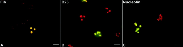
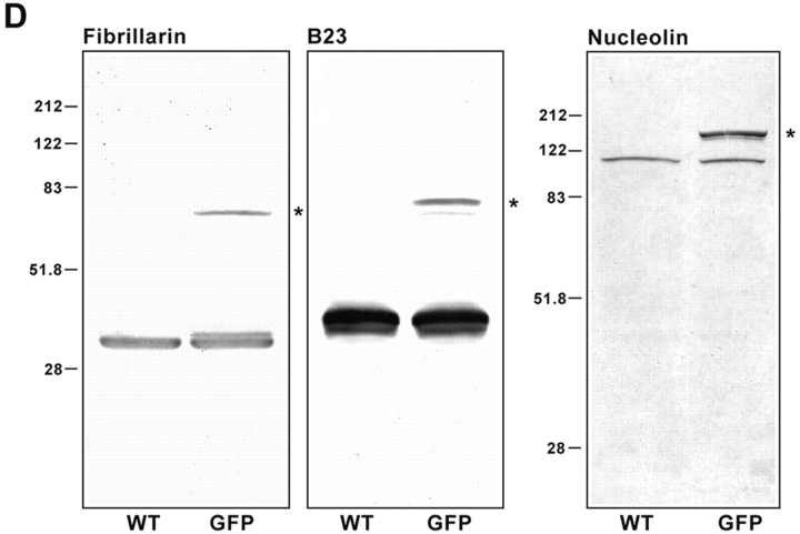
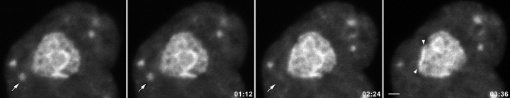
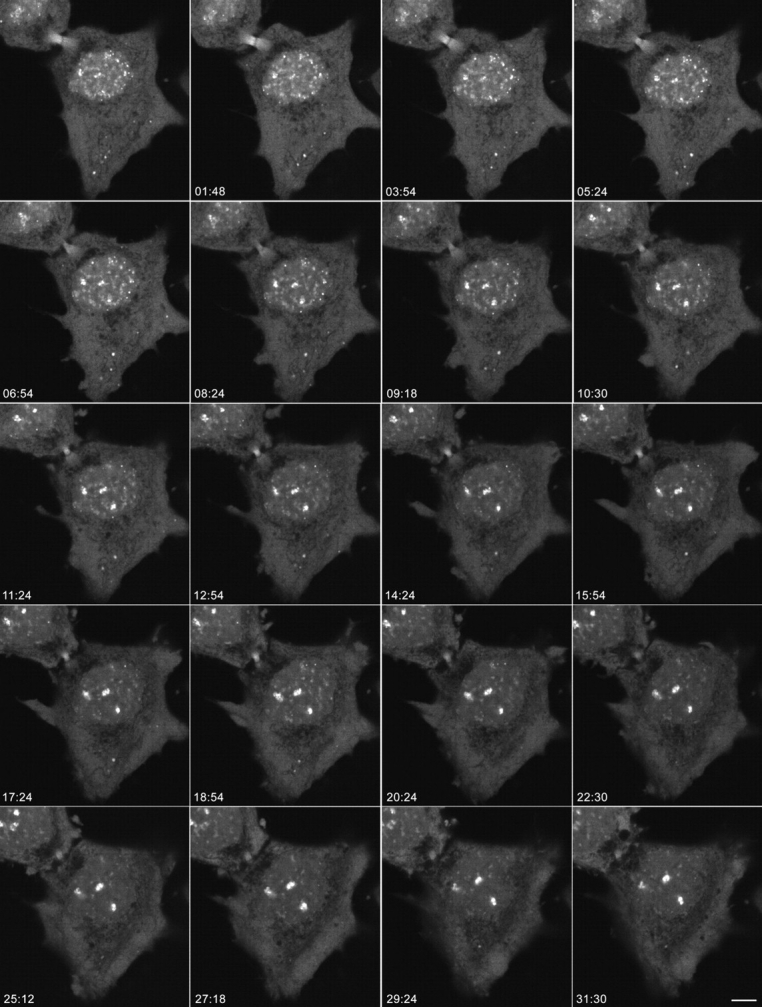
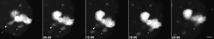
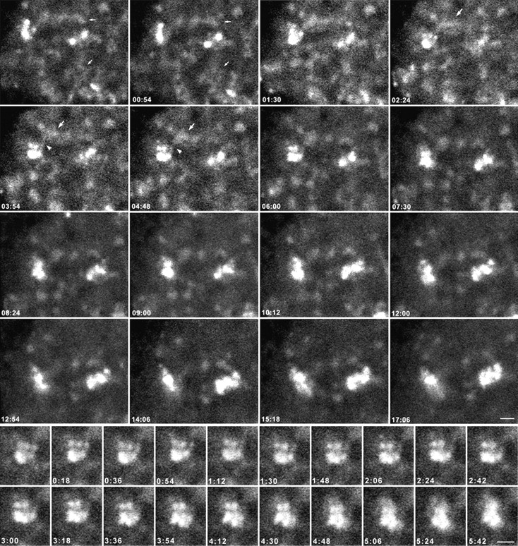
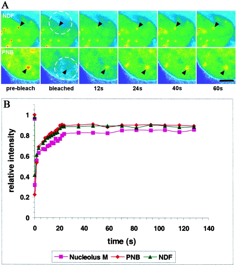
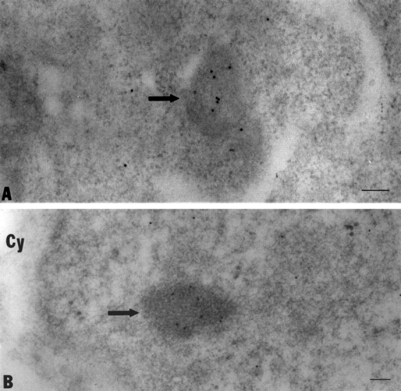
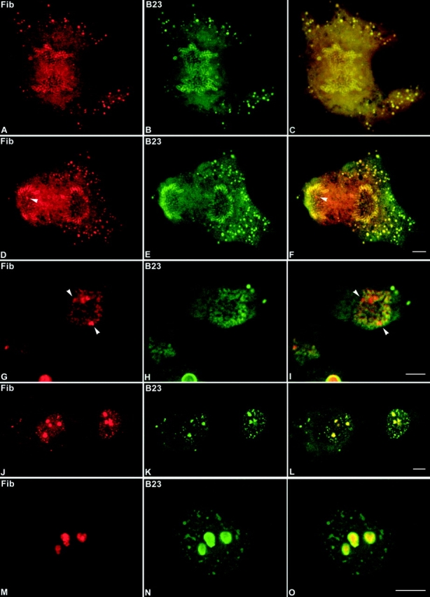
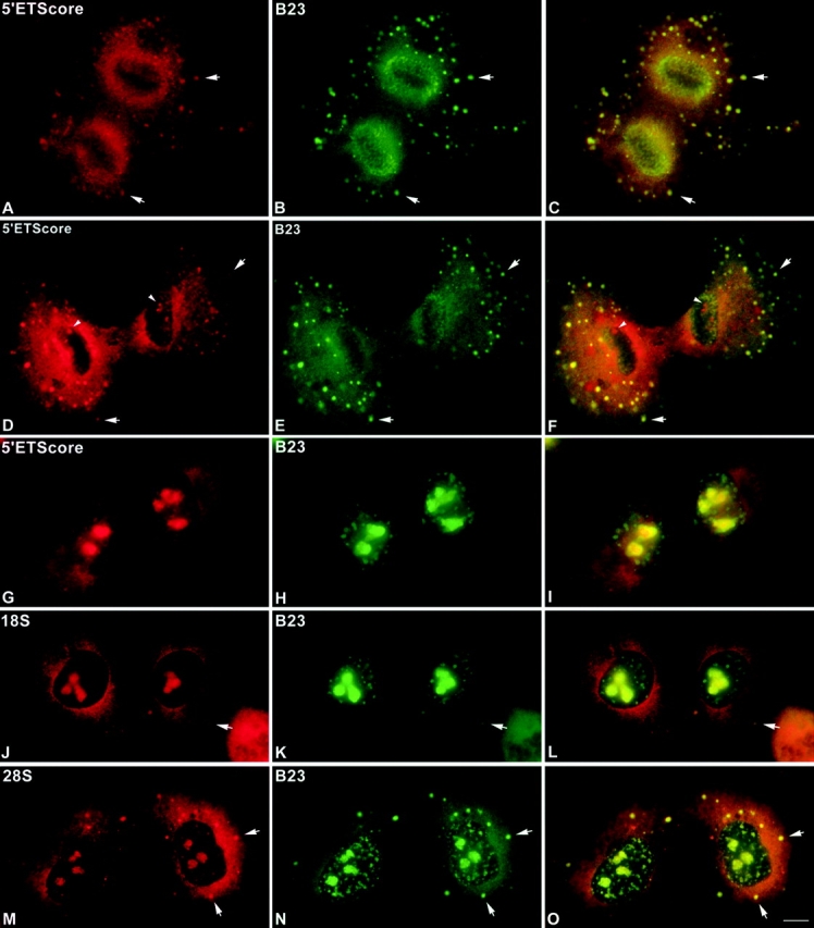
Mammalian cell nucleoli disassemble at the onset of M-phase and reassemble during telophase. Recent studies showed that partially processed preribosomal RNA (pre-rRNA) is preserved in association with processing components in the perichromosomal regions (PRs) and in particles called nucleolus-derived foci (NDF) during mitosis. Here, the dynamics of nucleolar reassembly were examined for the first time in living cells expressing fusions of the processing-related proteins fibrillarin, nucleolin, or B23 with green fluorescent protein (GFP). During telophase the NDF disappeared with a concomitant appearance of material in the reforming nuclei. Prenucleolar bodies (PNBs) appeared in nuclei in early telophase and gradually disappeared as nucleoli formed, strongly suggesting the transfer of PNB components to newly forming nucleoli. Fluorescence recovery after photobleaching (FRAP) showed that fibrillarin-GFP reassociates with the NDF and PNBs at rapid and similar rates. The reentry of processing complexes into telophase nuclei is suggested by the presence of pre-rRNA sequences in PNBs. Entry of specific proteins into the nucleolus approximately correlated with the timing of processing events. The mitotically preserved processing complexes may be essential for regulating the distribution of components to reassembling daughter cell nucleoli.


Figures

Figure 1
Figure 1
Fibrillarin-GFP, protein B23-GFP, and nucleolin-GFP chimeras colocalize with endogenous proteins in nuclear and nucleolar regions of CMT3 cells. The cells were transiently transfected with pEGFP-fibrillarin (A), pEGFP-protein B23 (B), or pEGFP-nucleolin (C) and fixed 24 h after transfection. The endogenous protein was detected by staining with the corresponding antibody followed by treatment with a specific secondary antibody conjugated with Texas red. The merged images show the colocalization of fibrillarin-GFP with endogenous fibrillarin in the dense fibrillar components (A, yellow). Protein B23-GFP (B, yellow) and nucleolin-GFP (C, yellow) were distributed throughout the nucleolus with higher accumulation in the nucleolar periphery and traces of signal in the nucleoplasm. Bars, 10 μm. (D) SDS-PAGE analysis of whole cell lysates from CMT3 cells transiently transfected with pEGFP-fibrillarin, pEGFP-protein B23 or pEGFP-nucleolin. Electrophoresis and immunoblotting with the corresponding antibody was done 40 h after transfection. The control lanes (WT) contain lysates of cells exposed to the FuGene 6 reagent without DNA. Each of the WT lanes shows only one band corresponding to the endogenous protein. In the samples from the transfected cells (GFP) both the wild-type protein and the GFP fusion protein (star), migrating at a slower rate, were detected. The molecular masses (in kD) of marker proteins are indicated on the left-hand sides of the blots.
Figure 1
Figure 1
Fibrillarin-GFP, protein B23-GFP, and nucleolin-GFP chimeras colocalize with endogenous proteins in nuclear and nucleolar regions of CMT3 cells. The cells were transiently transfected with pEGFP-fibrillarin (A), pEGFP-protein B23 (B), or pEGFP-nucleolin (C) and fixed 24 h after transfection. The endogenous protein was detected by staining with the corresponding antibody followed by treatment with a specific secondary antibody conjugated with Texas red. The merged images show the colocalization of fibrillarin-GFP with endogenous fibrillarin in the dense fibrillar components (A, yellow). Protein B23-GFP (B, yellow) and nucleolin-GFP (C, yellow) were distributed throughout the nucleolus with higher accumulation in the nucleolar periphery and traces of signal in the nucleoplasm. Bars, 10 μm. (D) SDS-PAGE analysis of whole cell lysates from CMT3 cells transiently transfected with pEGFP-fibrillarin, pEGFP-protein B23 or pEGFP-nucleolin. Electrophoresis and immunoblotting with the corresponding antibody was done 40 h after transfection. The control lanes (WT) contain lysates of cells exposed to the FuGene 6 reagent without DNA. Each of the WT lanes shows only one band corresponding to the endogenous protein. In the samples from the transfected cells (GFP) both the wild-type protein and the GFP fusion protein (star), migrating at a slower rate, were detected. The molecular masses (in kD) of marker proteins are indicated on the left-hand sides of the blots.
Figure 2
Figure 2
Behavior of NDF during telophase in living cells. The CMT3 cells were transiently transfected with the protein B23-GFP vector. After 24 h, cells in telophase were selected for observation by time-lapse confocal fluorescence microscopy. The images were collected every 18 s over a 90-min period. See also video 1 available at http://www.jcb.org/cgi/content/full/150/3/433/DC1. Protein B23-GFP was present in several NDF in the cytoplasm and distributed throughout the nucleoplasm and in newly forming nucleoli. The disappearance of the NDF in the proximity of the nuclear envelope (arrows) was accompanied by a concomitant increase in the fluorescence of the adjacent nuclear interior (arrowheads). Bar, 2 μm.
Figure 3
Figure 3
Dynamics of nucleolar reassembly in telophase. CMT3 cells in telophase, which were transiently expressing the fibrillarin-GFP protein, were subjected to time-lapse confocal fluorescence microscopy. The images were collected every 18 s over a 60-min period. See also video 2 available at http://www.jcb.org/cgi/content/full/150/3/433/DC1. The series of frames shows the progression of the cell from early to late telophase with transfer of material from PNBs to the newly forming nucleoli (the three brightest spots in the nuclei). Note the gradual disappearance of the PNBs in the nucleoplasm and the NDF in the cytoplasm with a concomitant increase in the fluorescent signal in the nucleoli. Bar, 10 μm.
Figure 5
Figure 5
Behavior of nucleolin-GFP during telophase. CMT3 cells in telophase, which were transiently expressing the nucleolin-GFP fusion protein were subjected to time-lapse confocal fluorescence microscopy. The images were collected every 18 s over a 30-min period. See also video 4 available at http://www.jcb.org/cgi/content/full/150/3/433/DC1. In the time-lapse sequence, material from two PNBs (arrowheads) is incorporated into nucleoli. In frame 0 a stream of small particles appears between the PNB and the assembling nucleolus (large arrowhead). By 18 min the PNB as well as the connecting stream have disappeared. The second PNB (small arrowhead) approaches the nucleolus in the 6 min frame, forming a connection at 12 min and nearly disappears by 24 min. Bar, 2 μm.
Figure 4
Figure 4
Higher magnification views of the nucleoplasm of the fibrillarin-GFP–expressing telophase cell in Fig. 3. The upper panels show that the PNBs are distributed throughout the nucleoplasm in a network-like organization during the early time points (small arrows). For the upper panels, see also video 3 available at http://www.jcb.org/cgi/content/full/150/3/433/DC1. As the PNBs (large arrows) move close to growing nucleoli, material appears to transfer between PNBs and nucleoli through streams of small particles (arrowhead). In the later time points the PNBs become distinct and are diminished in size and number. The lower panels show enlargements of images containing a single assembling nucleolus at early time points in telophase. Areas from the upper left-hand corners of the frames in the upper panels were enlarged to show a single nucleolus as it is formed. Each of the three distinct spots above the major spot resembles a dense fibrillar component (DFC) surrounding a fibrillar center (FC); i.e., a relatively bright ring of signal around a tiny open area. These units grow in size and brightness over the time of observation and eventually fuse with the main body of the nucleolus (see frame 5:42). Bars, 2 μm.
Figure 6
Figure 6
FRAP on fibrillarin-GFP in NDF, PNB and nucleoli during telophase. An area in a telophase cell including either an NDF or a PNB was bleached for 0.5 s using the 488-nm laser line of a confocal microscope at high power. The cells were then observed under normal fluorescence microscopy conditions, with images collected at 2-s intervals and the kinetics of recovery measured. (A) Images before and immediately after the bleach pulse and during recovery were taken at the indicated times. The bleached area is indicated by a dashed circle and the positions of the bleached NDF or PNB are indicated by arrowheads. Bar, 4 μm. (B) Quantitative measurements of fluorescence recovery. Values represent the relative recovery relative to total cellular fluorescence. The values were averages of determinations from six different cells for each subcellular compartment.
Figure 7
Figure 7
The ultrastructures of the NDF and PNBs have similar features. The CMT3 cells were synchronized and mitotic cells were harvested and embedded in Lowicryl. After sectioning and immunogold labeling with an anti-B23 polyclonal antibody the sections were viewed under the electron microscope. The NDF (A) in the cell plasms of anaphase cells identified by the 10-nm immunogold particle labeling (arrow) showed the same general fibrogranular structure as seen in the PNBs (B) in the nuclei of telophase cells (arrow). Bars, 100 nm.
Figure 8
Figure 8
Fibrillarin and protein B23 enter reassembling nucleoli at different times during mitosis. The CMT3 cells were double-labeled with anti-fibrillarin autoimmune serum (red) and mouse monoclonal antibody against protein B23 (green). Fibrillarin and protein B23 colocalized in the perichromosomal region and in numerous NDF (arrowheads) during early (A–C) and late (D–F) anaphase. From late anaphase (D) to early telophase (G) fibrillarin is detectable in tiny newly formed nucleoli (arrowheads), which are negative for protein B23 (E and H). In late telophase fibrillarin (J) and protein B23 (K) colocalized in PNBs and postmitotic nucleoli (L). In early G1 phase fibrillarin (M) is exclusively localized in postmitotic nucleoli in contrast to protein B23 (N), which is still detectable in persisting PNBs (O). Bars, 10 μm.
Figure 9
Figure 9
Preribosomal RNA sequences are present in telophase nuclei. CMT3 cells were subjected to fluorescence in situ hybridization (FISH) to probe for various segments of pre-rRNA including sequences in the 5′ETS core region and in 28S rRNA. During anaphase the 5′ETS core sequence was detected in the perichromosomal regions (PRs) and in numerous NDF (arrows) where it colocalized with protein B23 (A–C). The distribution of this sequence in early telophase (D–F) was similar to that seen in anaphase, with the signal present in NDF (arrows) and in the vicinity of the PRs (arrowheads). The 5′ETS core sequence was also seen in newly forming nucleoli (D and F, arrowheads). Protein B23 did not colocalize with the 5′ETS core sequence in nucleoli in cells at this stage of telophase (E and F). In late telophase/early G1 cells the 5′ETS core sequence was present primarily in newly forming nucleoli, with no detectable signal for this sequence in the PNBs labeled by anti-B23 antibody (G–I). The signal for the 18S rRNA sequence was seen in newly forming nucleoli of late telophase/early G1 cells, but not in the PNBs labeled by anti-B23 antibody (J–L). In late telophase cells the FISH signal for a 28S rRNA sequence was clearly visible in PNBs (M), where it colocalized with protein B23 (N and O). Bar, 10 μm.

References

    1. Aris J.P., Blobel G. cDNA cloning and sequencing of human fibrillarin, a conserved nucleolar protein recognized by autoimmune antisera. Proc. Natl. Acad. Sci. USA. 1991;88:931–935. - PMC - PubMed
    1. Azum-Gélade M.C., Noaillac-Depeyre J., Caizergues-Ferrer M., Gas N. Cell cycle redistribution of U3 snRNA and fibrillarin. Presence in the cytoplasmic nucleolus remnant and in the prenucleolar bodies at telophase. J. Cell Sci. 1994;107:463–475. - PubMed
    1. Bell P., Scheer U. Prenucleolar bodies contain coilin and are assembled in Xenopus egg extract depleted of specific nucleolar proteins and U3 RNA. J. Cell Sci. 1996;109:43–54. - PubMed
    1. Bell P., Dabauvalle M.-C., Scheer U. In vitro assembly of prenucleolar bodies in Xenopus egg extract. J. Cell Biol. 1992;118:1297–1304. - PMC - PubMed
    1. Benavente R., Rose K.M., Reimer G., Hügle-Dörr B., Scheer U. Inhibition of nucleolar reformation after microinjection of antibodies to RNA polymerase I into mitotic cells. J. Cell Biol. 1987;105:1483–1491. - PMC - PubMed

Publication types

MeSH terms